Review
. 2022 May 21;1(5):720-732.
doi: 10.1016/j.gastha.2022.05.004. eCollection 2022.

Examining the Role of Type 2 Inflammation in Eosinophilic Esophagitis

Affiliations
Review

Examining the Role of Type 2 Inflammation in Eosinophilic Esophagitis

Mirna Chehade et al. Gastro Hep Adv. .

Abstract

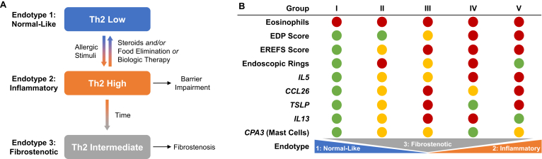
Eosinophilic esophagitis (EoE) is a chronic type 2 inflammatory disease characterized by an eosinophilic inflammatory infiltrate in the esophagus, leading to remodeling, stricture formation, and fibrosis. Triggered by food and aeroallergens, type 2 cytokines interleukin (IL)-4, IL-13, IL-5 produced by CD4+ T helper 2 cells (Th2), eosinophils, mast cells, basophils, and type 2 innate lymphoid cells alter the esophageal epithelial barrier and increase inflammatory cell tissue infiltration. Clustering analysis based on the expression of type 2 inflammatory genes demonstrated the diversity of EoE endotypes. Despite the availability of treatment options for patients with EoE, which include dietary restriction, proton pump inhibitors, swallowed topical steroids, and esophageal dilation, there are still no Food and Drug Administration-approved medications for this disease; as such, there are clear unmet medical needs for these patients. A number of novel biologic therapies currently in clinical trials represent a promising avenue for targeted therapeutic approaches in EoE. This review summarizes our current knowledge on the role of type 2 inflammatory cells and mediators in EoE disease pathogenesis, as well as the future treatment landscape targeting underlying inflammation in EoE.

Keywords: Endotypes; Eosinophilic Esophagitis; Eosinophils; Type 2 Inflammation.


Figures

Figure
Figure
Endotypes of eosinophilic esophagitis and their characteristic features. (A) Model depicting patient progression from Th2-low phenotype (endotype 1) to a Th2-high phenotype (endotype 2) following allergic or inflammatory insult. On steroid treatment, food elimination, or biologic therapy the Th2-gene expression decreases and patients either resolve inflammation by reverting to a Th2-low phenotype or develop a fibrostenotic (endotype 3) signature. (B) Five subgroups of patients with active EoE were identified based on a variety of criteria, including expression of IL5, IL13, CCL26, TSLP, and CPA3. Relative levels of each criterion are reported in red (high), yellow (intermediate), or green (low). The 5 groups differed in the EoE endotypes spanned, but not in eosinophil levels, which were universally high. Group V patients had the highest expression of IL5, TSLP, CCL26, and genes associated with tissue remodeling. Groups II and III (which exhibited intermediate expression of IL5 and CPA3) were differentiated by high TSLP and IL13 in group III. CCL26, C-C motif chemokine ligand 26; EDP, eosinophilic esophagitis diagnostic panel; EREFS, endoscopic reference score; Th2, T helper cell type 2; TSLP, thymic stromal lymphopoietin (Adapted from J Allergy Clin Immunol: 2020;145:1629–1640.e4.).

References

    1. Dellon E.S., Liacouras C.A., Molina-Infante J., et al. Updated international consensus diagnostic criteria for eosinophilic esophagitis: proceedings of the AGREE conference. Gastroenterology. 2018;155:1022–1033.e10. - PMC - PubMed
    1. Greuter T., Hirano I., Dellon E.S. Emerging therapies for eosinophilic esophagitis. J Allergy Clin Immunol. 2020;145:38–45. - PMC - PubMed
    1. Gonsalves N.P., Aceves S.S. Diagnosis and treatment of eosinophilic esophagitis. J Allergy Clin Immunol. 2020;145:1–7. - PMC - PubMed
    1. Davis B.P., Rothenberg M.E. Mechanisms of disease of eosinophilic esophagitis. Annu Rev Pathol. 2016;11:365–393. - PMC - PubMed
    1. Aceves S.S., Chen D., Newbury R.O., et al. Mast cells infiltrate the esophageal smooth muscle in patients with eosinophilic esophagitis, express TGF-β1, and increase esophageal smooth muscle contraction. J Allergy Clin Immunol. 2010;126:1198–1204.e4. - PubMed

LinkOut - more resources