. 2024 Jan 31;3(2):101259.
doi: 10.1016/j.jscai.2023.101259. eCollection 2024 Feb.

SCAI Expert Consensus Statement on the Management of Calcified Coronary Lesions

Affiliations

SCAI Expert Consensus Statement on the Management of Calcified Coronary Lesions

Robert F Riley et al. J Soc Cardiovasc Angiogr Interv. .

Abstract

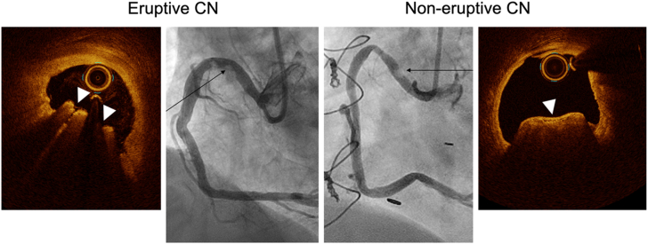
The prevalence of calcification in obstructive coronary artery disease is on the rise. Percutaneous coronary intervention of these calcified lesions is associated with increased short-term and long-term risks. To optimize percutaneous coronary intervention results, there is an expanding array of treatment modalities geared toward calcium modification prior to stent implantation. The Society for Cardiovascular Angiography and Interventions, herein, puts forth an expert consensus document regarding methods to identify types of calcified coronary lesions, a central algorithm to help guide use of the various calcium modification strategies, tips for when using each treatment modality, and a look at future studies and trials for treating this challenging lesion subset.

Keywords: calcium; coronary artery disease; percutaneous coronary intervention.


Figures

Figure 1
Figure 1
Criteria for coronary calcium modification (group consensus). Angiographic criteria: fluoroscopic radiopacities noted without cardiac motion before contrast injection involving both sides of the arterial wall in at least 1 location and total length of calcium of at least 15 mm. Intravascular imaging criteria: By both intravascular ultrasound (IVUS) and optical coherence tomography (OCT), the total amount of calcium (calcium arc, thickness, and length) and negative remodeling or small vessel size are associated with stent expansion. OCT can evaluate thickness of calcium and the minimum thickness of calcium of <0.5 mm should be associated with the creation of calcium fracture without calcium modification. IVUS (A) and OCT (F) showed 360° of calcium. (B, G) Calcified nodules. (C) Calcium arc measured is 310°. (D) Frame with visible external elastic lamina (EEL) adjacent to (C), and the EEL diameter measured 3.2 mm. (E) further distal frame and EEL diameter measured 3.6 mm. Proximal EEL diameter smaller than distal EEL diameter in (D) indicates vessel negative remodeling. (H,J) OCT shows similar findings but with added calcium minimum thickness measured 0.38 mm.
Figure 2
Figure 2
Eruptive vs noneruptive calcified nodule (CN). Optical coherence tomography (OCT) images illustrate an eruptive CN and noneruptive CN. The eruptive CN is a protruding calcium mass with an irregular shape and strong signal attenuation. The noneruptive CN has a smooth fibrous cap overlying a protruding calcium mass with strong signal attenuation. In both cases, the corresponding coronary angiography demonstrates a radiolucent mass (black arrows).
Figure 3
Figure 3
Calcium distribution in a long lesion. Preintervention coronary angiography and optical coherence tomography (OCT) images after 3 mm noncompliant balloon dilatation at 20 atm. (A) Circumferential calcium without fracture requiring further preparation. Because the minimum thickness of calcium measured 0.43 mm, additional angioplasty was performed with a 3.5 mm noncompliant balloon at 20 atm. (B) 180° of calcium with a large dissection in the fibrous plaque (arrowhead). (C) Thin calcium at the site of the most obstructive portion of the lesion, with 3 calcium fractures (white arrows in C′). (D) 90° of thick calcium with dissection on both sides of the calcium (arrowhead). Poststent OCT demonstrated calcium fracture with optimal stent expansion (minimum stent area of 6.5 mm2). In long lesions, all calcium segments should be confirmed to be fractured on intravascular imaging or yielded to allow full expansion in 2 angiographic views with a 1:1 NC balloon inflation prior to stenting. (A′-D′) Same images as (A-D) with annotation. White shaded areas in (A′-D′) indicate calcification.
Figure 4
Figure 4
Balloon-uncrossable chronic total occlusion lesion treated with rotational atherectomy and intravascular lithotripsy. Coronary angiogram shows calcification in the occluded segment (white arrow) of a chronic total occlusion in the proximal left anterior descending artery. After successful antegrade guide wire escalation, no device was able to cross. Rotational atherectomy (RA) was performed, followed by intravascular lithotripsy (IVL). Intravascular ultrasound (IVUS) after RA demonstrated circumferential calcium with reverberation (equidistant white circles, indicated by green arrows). A post-IVL IVUS image shows 2 calcium fractures (blue arrows). A poststent IVUS image shows good stent expansion (stent area of 6.5 mm2).
Figure 5
Figure 5
Stent failure with calcified plaque. (A) Optical coherence tomography (OCT) image of a stent implanted 5 weeks prior. A protruding mass with strong attenuation indicates a calcified nodule (CN). Because neoatherosclerosis would not accrue in such a short time span, it was interpreted as reprotrusion of a CN through the recently implanted stent. (B) CN found in the stent placed many years prior was interpreted as calcified neoatherosclerosis with a CN. (C) Thick calcium within and outside of an old stent (white area) diagnosed as neoatherocalcification. Because of the thick calcium, calcium modification is necessary before stenting. (D) Old underexpanded stent with limited neointimal hyperplasia causing restenosis and thick, circumferential calcium outside the stent. (A′-D′) are the same images as (A-D), with annotation. White areas indicate calcium; yellow areas indicate a CN, and the green lines indicate stent struts.
Figure 6
Figure 6
Treatment algorithm for calcified CAD. Ca, calcium; EEL, external elastic lamina; OCT, optical coherence tomography; NC, noncompliant; PCI, percutaneous coronary intervention. ∗Criteria for calcium modification shown on left.

Similar articles

Cited by

References

    1. Szerlip M., Al-Azizi K., Alasnag M., et al. SCAI publications committee manual of standard operating procedures: 2022 update. J Soc Cardiovasc Angiogr Interv. 2022;1(4) doi: 10.1016/j.jscai.2022.100389. - DOI - PubMed
    1. Sutton N.R., Malhotra R., St. Hilaire C., et al. Molecular mechanisms of vascular health: insights from vascular aging and calcification. Arterioscler Thromb Vasc Biol. 2023;43(1):15–29. doi: 10.1161/ATVBAHA.122.317332. - DOI - PMC - PubMed
    1. McClelland R.L., Chung H., Detrano R., Post W., Kronmal R.A. Distribution of coronary artery calcium by race, gender, and age: results from the multi-ethnic study of atherosclerosis (MESA) Circulation. 2006;113(1):30–37. doi: 10.1161/CIRCULATIONAHA.105.580696. - DOI - PubMed
    1. Kronmal R.A., McClelland R.L., Detrano R., et al. Risk factors for the progression of coronary artery calcification in asymptomatic subjects: results from the multi-ethnic study of atherosclerosis (MESA) Circulation. 2007;115(21):2722–2730. doi: 10.1161/CIRCULATIONAHA.106.674143. - DOI - PubMed
    1. Chellan B., Sutton N.R., Hofmann Bowman M.A. S100/RAGE-mediated inflammation and modified cholesterol lipoproteins as mediators of osteoblastic differentiation of vascular smooth muscle cells. Front Cardiovasc Med. 2018;5:163. doi: 10.3389/fcvm.2018.00163. - DOI - PMC - PubMed

LinkOut - more resources