Review
. 2022 Sep 16;1(6):100452.
doi: 10.1016/j.jscai.2022.100452. eCollection 2022 Nov-Dec.

Transcatheter Pulmonary Valve Replacement: A Review of Current Valve Technologies

Affiliations
Review

Transcatheter Pulmonary Valve Replacement: A Review of Current Valve Technologies

Neil D Patel et al. J Soc Cardiovasc Angiogr Interv. .

Abstract

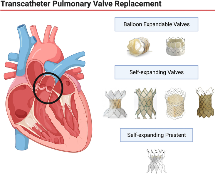
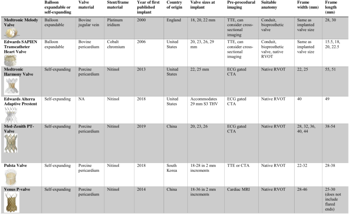
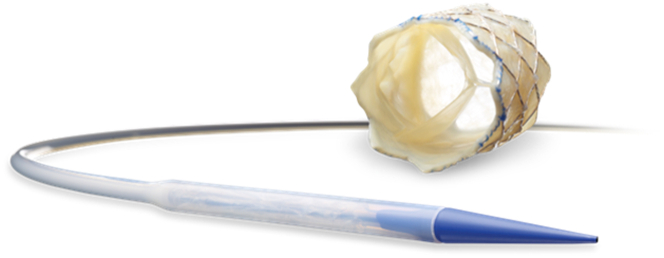
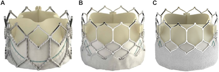
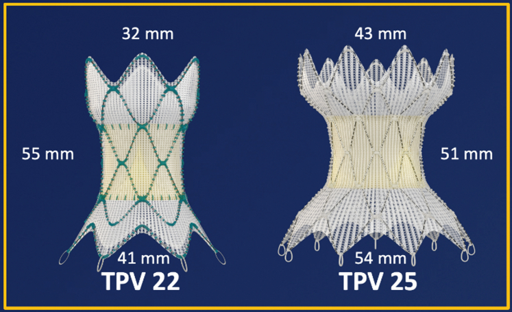
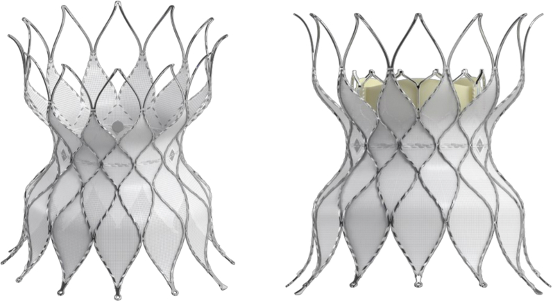
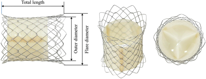
Transcatheter pulmonary valve replacement was first performed by Dr Philip Bonhoeffer, who implanted a Medtronic Melody valve in a human in 2000. Over the past 2 decades, there have been many advances in transcatheter pulmonary valve technology. This includes the use of the SAPIEN transcatheter heart valve in the pulmonary position, modifications and refinements to valve implantation procedures, and development of self-expanding valves and prestents to treat large diameter native or patched right ventricular outflow tracts. This article reviews the current transcatheter pulmonary valve technologies with a focus on valve design, screening process, implant procedure, and clinical outcomes.

Keywords: pulmonary valve replacement; tetralogy of Fallot; transcatheter.


Figures

None
Graphical abstract
Central Illustration
Transcatheter pulmonary valve replacement has evolved significantly over the last 22 years. Current technologies include balloon expandable valves, self-expanding valves, and self-expanding prestents.
Figure 1
Figure 1
Comparison of valve and prestent types. TTE, transthoracic echo; RVOT, right ventricular outflow tract; NA, not applicable; S3 THV, Sapien 3 Transcatheter Heart Valve
Figure 2
Figure 2
Melody Valve and Ensemble delivery system.
Figure 3
Figure 3
Edwards SAPIEN THV iterations. (A) SAPIEN XT valve, (B) SAPIEN 3 valve, (C) SAPIEN 3 Ultra valve.
Figure 4
Figure 4
Harmony TPV 22 and TPV25. TPV, transcatheter pulmonary valve.
Figure 5
Figure 5
Harmony screening report. The report contains a (A) perimeter plot in systole and diastole, (B) virtual implantation, and (C) estimated fluoroscopic viewing projections with landing zone.
Figure 6
Figure 6
Alterra Adaptiveprestent. The Alterra Adaptive prestent is shown with the SAPIEN 3 THV. The proximal inflow portion of the pre-stent is covered, while the distal outflow portion has an open-cell design.
Figure 7
Figure 7
Alterra Prestent delivery system. (A) Schematic diagram showing the Alterra Prestent delivery system components. (B) Enlarged image of the distal end of the delivery system with approximately 50% of the Alterra Prestent deployed. At this point, recovering, recapture, and subsequent redeployment are possible. (C) With the outer shaft completely withdrawn, the Alterra Prestent is completely deployed.
Figure 8
Figure 8
Edwards Pulmonicdeliverysystem. The system features a covered balloon assembly that protects the SAPIEN 3 THV and cardiac structures from one another during navigation through the right side of the heart. THV, transcatheter heart valve.
Figure 9
Figure 9
Med-Zenith PT valve. (A) Med-Zenith PT-valve with a (B) schematic diagram. 1, nitinol stent; 2, leaflets; 3, seal membrane; 4, markers; 5, anchors.
Figure 10
Figure 10
Med-Zenith PT-valvescreening report. Perimeter plots in (A) diastole and (B) systole are generated from an ECG-gated CTA. CTA, computed tomography angiography; ECG, electrocardiogram.
Figure 11
Figure 11
Pulsta valve. The valve diameter ranges from 18 to 32 mm with 2-mm increments. Both ends of the valve are flared 4 mm wider than the outer diameter and are uncovered.
Figure 12
Figure 12
Venus P-valve. Self-expanding nitinol stent with a trileaflet porcine pericardial valve.

References

    1. Bonhoeffer P., Boudjemline Y., Saliba Z., et al. Percutaneous replacement of pulmonary valve in a right-ventricle to pulmonary-artery prosthetic conduit with valve dysfunction. Lancet. 2000;356(9239):1403–1405. doi: 10.1016/S0140-6736(00)02844-0. - DOI - PubMed
    1. Zahn E.M., Hellenbrand W.E., Lock J.E., McElhinney D.B. Implantation of the melody transcatheter pulmonary valve in patients with a dysfunctional right ventricular outflow tract conduit early results from the U.S. clinical trial. J Am Coll Cardiol. 2009;54(18):1722–1729. doi: 10.1016/j.jacc.2009.06.034. - DOI - PubMed
    1. Martin M.H., Meadows J., McElhinney D.B., et al. Safety and feasibility of melody transcatheter pulmonary valve replacement in the native right ventricular outflow tract: a multicenter pediatric heart network scholar study. JACC Cardiovasc Interv. 2018;11(16):1642–1650. doi: 10.1016/j.jcin.2018.05.051. - DOI - PubMed
    1. Cheatham S.L., Holzer R.J., Chisolm J.L., Cheatham J.P. The Medtronic Melody® transcatheter pulmonary valve implanted at 24-mm diameter – it works. Catheter Cardiovasc Interv. 2013;82(5):816–823. doi: 10.1002/ccd.24821. - DOI - PubMed
    1. Berman D.P., McElhinney D.B., Vincent J.A., Hellenbrand W.E., Zahn E.M. Feasibility and short-term outcomes of percutaneous transcatheter pulmonary valve replacement in small (<30 kg) children with dysfunctional right ventricular outflow tract conduits. Circ Cardiovasc Interv. 2014;7(2):142–148. doi: 10.1161/CIRCINTERVENTIONS.113.000881. - DOI - PubMed

LinkOut - more resources