Review
. 2024 Feb 18;3(5):101337.
doi: 10.1016/j.jscai.2024.101337. eCollection 2024 May.

The Intra-aortic Balloon Pump: A Focused Review of Physiology, Transport Logistics, Mechanics, and Complications

Affiliations
Review

The Intra-aortic Balloon Pump: A Focused Review of Physiology, Transport Logistics, Mechanics, and Complications

Lauren E Gillespie et al. J Soc Cardiovasc Angiogr Interv. .

Abstract

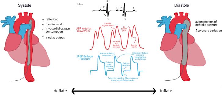
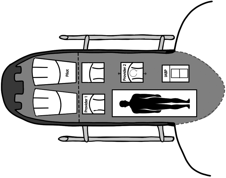
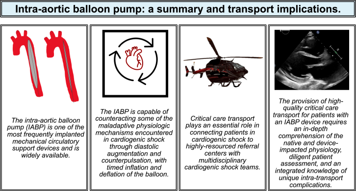
Critical care transport medicine (CCTM) teams are playing an increasing role in the care of patients in cardiogenic shock requiring mechanical circulatory support devices. Hence, it is important that CCTM providers are familiar with the pathophysiology of cardiogenic shock, the role of mechanical circulatory support, and the management of these devices in the transport environment. The intra-aortic balloon pump is a widely used and accessible cardiac support device capable of increasing cardiac output and reducing work on the left ventricle through diastolic augmentation and counterpulsation. This article reviews essential CCTM-based considerations for patients supported by intra-aortic balloon pump, including indications for placement, mechanics and physiology, potential issues during transport, and associated complications.

Keywords: cardiogenic shock; heart failure; mechanical circulatory support.


Figures

Figure 1
Figure 1
SCAI classification of cardiogenic shock incorporating the 2022 revisions., BNP, brain natriuretic peptide; CPR, cardiopulmonary resuscitation; CVP, central venous pressure; JVP, jugular venous pressure; LFT, liver function test; MAP, mean arterial pressure; PCWP, pulmonary capillary wedge pressure; SBP, systolic blood pressure.
Figure 2
Figure 2
Pressure-volume loop in normal cardiac physiology. The PV loop demonstrates normal cardiac physiology and the relationships between volume and pressure. Diastolic filling of the ventricle (D to A) is followed by early systole and isovolumetric contraction (A to B), during which the pressure generated by the ventricle results in opening of the semilunar valves, closure of the atrioventricular valves, and forward ejection of blood. Systolic ejection (B to C) is followed by ventricular isovolumetric relaxation (C to D) during which ventricular pressures drop below the diastolic aortic and pulmonary pressure leading to semilunar valve closure and the beginning of diastole. EDPVR, end-diastolic pressure-volume relationship; ESPVR, end-systolic pressure-volume relationship; LV, left ventricle.
Figure 3
Figure 3
Neurohumoral activation and LV dysfunction impact the PV loop. The physiologic adaptations found in cardiogenic shock with acute left ventricular dysfunction are demonstrated by changes in the PV loop. With a downward and rightward shift in the acute LV dysfunction loop, filling pressures (LV volume) are significantly elevated, with lower overall generated systolic pressures, and reduced contractility as portrayed by the shift in ESPVR. Neurohumoral activation releases endogenous catecholamines resulting in increased myocardial oxygen demands, centralization of blood volume leading to pulmonary congestion, and further elevation of filling pressures. LV, left ventricle; PV, pressure-volume.
Figure 4
Figure 4
IABP impact on the PV loop. The IABP has multiple favorable effects on cardiac function. While inflated, the IAB displaces intra-aortic blood during diastole, propagating forward movement of blood into systemic circulation and ideally, increasing coronary blood flow. Owing to the reduction of end-diastolic intra-aortic blood volume, the pressure required of the LV to initiate aortic valve opening is reduced (afterload reduction, seen at point B), leading to reduced LV work and myocardial oxygen demand. Before systole, the balloon then deflates, which also contributes to reduced LV afterload and aortic pressures due to negative pressure mechanics. The time spent in isovolumetric contraction is reduced, leading to reduced myocardial demand. SV increases (EDV, point A; ESV, point C), resulting in modest increases in CO. EDV, end-diastolic volume; IAB, intra-aortic balloon; IABP, intra-aortic balloon pump; LV, left ventricle.
Figure 5
Figure 5
IABP console configuration. The IABP console consists of a foldable screen, where settings may be entered and adjusted, waveforms are represented, and alarms display; a connection panel, for fiberoptic (top right port), ECG (green, top left), pressure/arterial line (second from top left, red), and helium extender tubing (donut port, central) cables; 2 lithium ion batteries (pictured below front-facing vents); and a helium tank system (not pictured, located on the lower back-side of the console). ECG, electrocardiogram; IABP, intra-aortic balloon pump.
Figure 6
Figure 6
Pictorial representation of IABP in systole and diastole with ECG tracing, arterial augmented waveforms and corresponding IABP tracings. The IABP functions to inflate during diastole and deflate during systole. This can be accomplished by timing relative to ECG or pressure waveforms, pictured above, to accurately inflate during the appropriate portion of the cardiac cycle. The IAB waveform, pictured in blue, is timed to correlate vertically with diastole in the arterial and ECG tracings. ECG, electrocardiogram; IAB, intra-aortic balloon; IABP, intra-aortic balloon pump.
Figure 7
Figure 7
Normal and abnormal IABP waveforms and their impact on cardiac physiology. IABP monitor interface demonstrating waveform changes in the setting of (A) correct inflation timing, (B) early inflation, (C) early deflation, (D) late inflation, and (E) late deflation. The arterial waveform in normal IABP inflation timing results in augmentation of diastole correlating with balloon inflation. The balloon pressure waveform illustrates rapid inflation of the balloon, followed by the plateau at maximal inflation, and subsequently rapid deflation before returning to baseline in preparation for the next cycle. In early inflation (B), afterload increases, and the aortic valve undergoes premature closure, which increases the end-systolic volume and reduces CO. In early deflation (C), there is reduced overall impact and efficacy of the IABP on cardiac function; there is minimal diastolic augmentation and a lack of reduction in myocardial oxygen demand and LV afterload. In late inflation (D), there is significantly reduced diastolic augmentation given the delayed timing; there still may be a small impact on improved coronary perfusion, although it is suboptimal compared with the potential of an adequately timed device. Finally, in late deflation (E), there is an increase, rather than decrease, in LV afterload, which translates into increased myocardial oxygen demand. CO, cardiac output; IABP, intra-aortic balloon pressure.
Figure 8
Figure 8
Chest x-ray demonstrating correct positioning of the IAB. The IAB is highlighted by the radiopaque marker at the distal point of the balloon (superior aspect of the yellow-highlighted portion). There is also a radiopaque midline marker within the balloon itself, which is faintly visible. Additional support devices pictured include bilateral chest tubes, an endotracheal tube, and a right internal jugular approach Swan-Ganz catheter. IAB, intra-aortic balloon.
Figure 9
Figure 9
Top-down view of the configuration of an EC145 helicopter with a patient supported by an IABP. Pictured is the proposed, commonly encountered configuration for rotor-wing transport of a patient with IABP support. Dotted lines represent closed doors, with clamshell doors depicted in the open position.
Central Illustration
The intra-aortic balloon pump is a frequently encountered mechanical support device. Critical care providers must be expertly trained in caring for patients supported by the IABP, given the essential role of transport providers in connecting patients to highly resourced specialty centers for cardiogenic shock. IABP, intra-aortic balloon pressure.
Supplemental Figure S1
Supplemental Figure S2

Similar articles

Cited by

References

    1. Moghaddam N., van Diepen S., So D., Lawler P.R., Fordyce C.B. Cardiogenic shock teams and centres: a contemporary review of multidisciplinary care for cardiogenic shock. ESC Heart Fail. 2021;8(2):988–998. doi: 10.1002/ehf2.13180. - DOI - PMC - PubMed
    1. Reynolds H.R., Hochman J.S. Cardiogenic shock: current concepts and improving outcomes. Circulation. 2008;117(5):686–697. doi: 10.1161/circulationaha.106.613596. - DOI - PubMed
    1. Sinha S.S., Rosner C.M., Tehrani B.N., et al. Cardiogenic shock from heart failure versus acute myocardial infarction: clinical characteristics, hospital course, and 1-year outcomes. Circ Heart Fail. 2022;15(6) doi: 10.1161/circheartfailure.121.009279. - DOI - PMC - PubMed
    1. Abraham J., Blumer V., Burkhoff D., et al. Heart failure-related cardiogenic shock: pathophysiology, evaluation and management considerations: review of heart failure-related cardiogenic shock. J Card Fail. 2021;27(10):1126–1140. doi: 10.1016/j.cardfail.2021.08.010. - DOI - PubMed
    1. Kolte D., Khera S., Aronow W.S., et al. Trends in incidence, management, and outcomes of cardiogenic shock complicating ST-elevation myocardial infarction in the United States. J Am Heart Assoc. 2014;3(1) doi: 10.1161/jaha.113.000590. - DOI - PMC - PubMed

LinkOut - more resources