[Preprint]. 2024 Jul 30:2024.07.27.24310296.
doi: 10.1101/2024.07.27.24310296.

Reemergence of Oropouche virus between 2023 and 2024 in Brazil

Affiliations

Reemergence of Oropouche virus between 2023 and 2024 in Brazil

Gabriel C Scachetti et al. medRxiv. .

Update in

  • Re-emergence of Oropouche virus between 2023 and 2024 in Brazil: an observational epidemiological study.
    Scachetti GC, Forato J, Claro IM, Hua X, Salgado BB, Vieira A, Simeoni CL, Barbosa ARC, Rosa IL, de Souza GF, Fernandes LCN, de Sena ACH, Oliveira SC, Singh CML, de Lima STS, de Jesus R, Costa MA, Kato RB, Rocha JF, Santos LC, Rodrigues JT, Cunha MP, Sabino EC, Faria NR, Weaver SC, Romano CM, Lalwani P, Proenca-Modena JL, de Souza WM. Scachetti GC, et al. Lancet Infect Dis. 2025 Feb;25(2):166-175. doi: 10.1016/S1473-3099(24)00619-4. Epub 2024 Oct 16. Lancet Infect Dis. 2025. PMID: 39423838 Free PMC article.

Abstract

Background: Oropouche virus (OROV; species Orthobunyavirus oropoucheense) is an arthropod-borne virus that has caused outbreaks of Oropouche fever in Central and South America since the 1950s. This study investigates virological factors contributing to the reemergence of Oropouche fever in Brazil between 2023 and 2024.

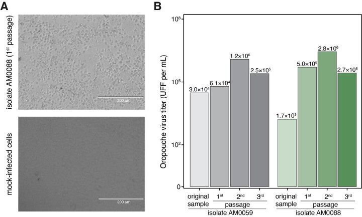
Methods: In this study, we combined OROV genomic, molecular, and serological data from Brazil from 1 January 2015 to 29 June 2024, along with in vitro and in vivo characterization. Molecular screening data included 93 patients with febrile illness between January 2023 and February 2024 from the Amazonas State. Genomic data comprised two genomic OROV sequences from patients. Serological data were obtained from neutralizing antibody tests comparing the prototype OROV strain BeAn 19991 and the 2024 epidemic strain. Epidemiological data included aggregated cases reported to the Brazilian Ministry of Health from 1 January 2014 to 29 June 2024.

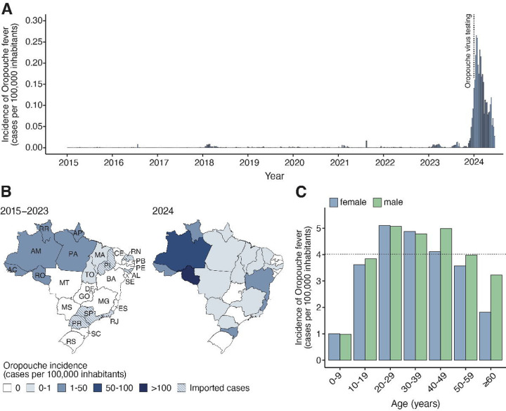
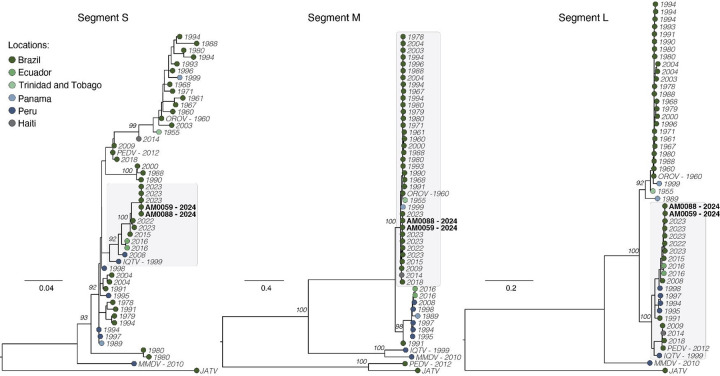
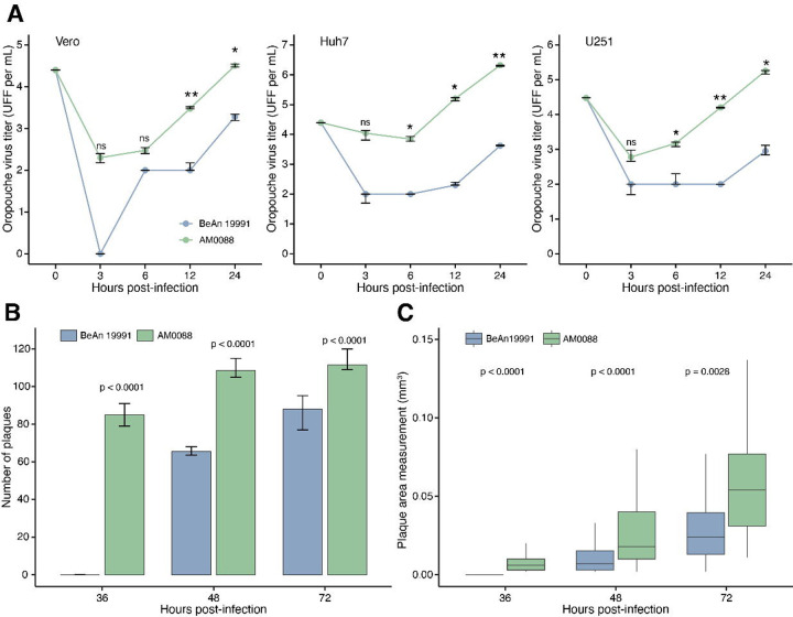
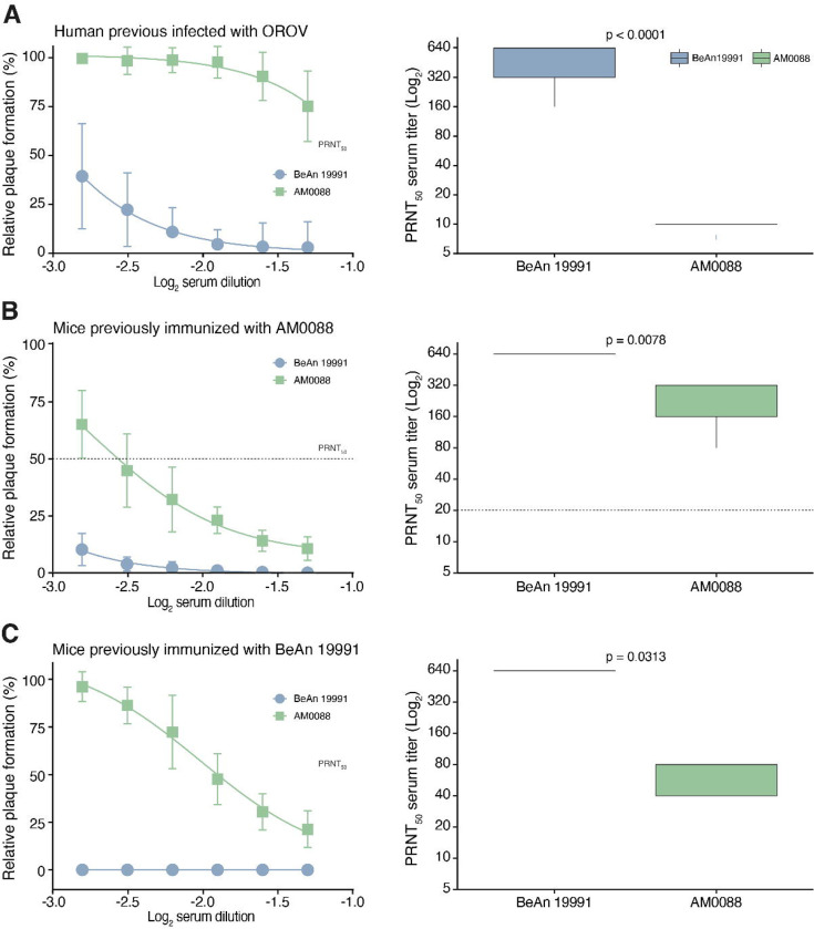
Findings: In 2024, autochthonous OROV infections were detected in previously non-endemic areas across all five Brazilian regions. Cases were reported in 19 of 27 federal units, with 83.2% (6,895 of 8,284) of infections in Northern Brazil and a nearly 200-fold increase in incidence compared to reported cases over the last decade. We detected OROV RNA in 10.8% (10 of 93) of patients with febrile illness between December 2023 and May 2024 in Amazonas. We demonstrate that the 2023-2024 epidemic was caused by a novel OROV reassortant that replicated approximately 100-fold higher titers in mammalian cells compared to the prototype strain. The 2023-2024 OROV reassortant displayed plaques earlier than the prototype, produced 1.7 times more plaques, and plaque sizes were 2.5 larger compared to the prototype. Furthermore, serum collected in 2016 from previously OROV-infected individuals showed at least a 32-fold reduction in neutralizing capacity against the reassortment strain compared to the prototype.

Interpretation: These findings provide a comprehensive assessment of Oropouche fever in Brazil and contribute to a better understanding of the 2023-2024 OROV reemergence. The recent increased incidence may be related to a higher replication efficiency of a new reassortant virus that also evades previous immunity.


Conflict of interest statement

Declaration of interests The authors declare no competing interests.

Figures

Figure 1.
Figure 1.. Spatial-temporal dynamics of Oropouche fever between 2015 and 2024 in Brazil.
(A) Incidence of laboratory-confirmed Oropouche fever cases per epidemiological week in all 26 Brazilian States and the Federal District, from epidemiological week 1 of 2015 (04 to 10 January) to epidemiological week 26 of 2024 (23 to 29 June). The dashed line indicates the implementation of OROV diagnosis across all Central Public Health Laboratories in Brazil. (B) Maps were colored according to the incidence of laboratory-confirmed Oropouche fever cases by federal unit between January 2015 and December 2023 (left) and January to June 2024 (right). AC=Acre. AL=Alagoas. AM=Amazonas. AP=Amapá. BA=Bahia. CE=Ceará. ES=Espírito Santo. DF=Distrito Federal (Federal District). GO=Goiás. MA=Maranhão. MG=Minas Gerais. MS=Mato Grosso do Sul. MT=Mato Grosso. PA=Pará. PB=Paraíba. PE=Pernambuco. PI=Piauí. PR=Paraná. RJ=Rio de Janeiro. RN=Rio Grande do Norte. RO=Rondônia. RR=Roraima. RS=Rio Grande do Sul. SC=Santa Catarina. SE=Sergipe. SP=São Paulo. TO=Tocantins. (C) Oropouche fever incidence based on the age-sex distribution of cases from 2015 to 2024.
Figure 2.
Figure 2.. Oropouche virus from the 2024 epidemic (AM0059 and AM0088) isolated from serum samples of Oropouche fever patients from Manaus City, Amazonas, Brazil.
(A) Isolation of the 2024 epidemic of OROV was performed in Vero CCL81 cells, and a typical cytopathic effect was observed approximately 30 hours post-infection in inoculated cells in comparison with uninfected cells. (B) The isolation was also confirmed by focus forming assay, where it is possible to observe an increase in viral load. Images were obtained in an EVOS inverted microscope kindly provided by Thermo Fisher Scientific.
Figure 3.
Figure 3.. Phylogenetic analysis of the Oropouche virus.
Maximum likelihood phylogenetic tree of 48 representative OROV genomes, including two new genomes (black bold) from Manaus City generated in this study. Phylogenetic trees are shown for the S segment (left), M segment (center), and L segment (right). Tips are colored according to the location country of each sample. Phylogenies were midpoint rooted for clarity of presentation. Scale bar indicates the evolutionary distance of substitutions per nucleotide site. Bootstrap values based on 1,000 replicates are shown on principal nodes. The GenBank accession numbers of sequences used in this figure are presented in Appendix Table S4.
Figure 4.
Figure 4.. Characterization in vitro of 2024 Oropouche virus reassortment strain.
(A) Viral replication properties of OROV strain AM0088 and OROV strain BeAn 19991 in Vero CCL81 (left), Huh7 (center), and U-251 (right) cell lines. Cells were infected at a multiplicity of infection of 0.1. At indicated time points, samples were harvested, and titers were determined by focus-forming assay on Vero CCL81 cells. Statistical analyses were performed using the paired t-test. The median is presented by the middle line, and the upper and lower limits represent the 75th and 25th percentiles (whiskers). Statistical significance is ***p < 0.001, **p < 0.01, and *p < 0.05; ns, not significant. (B) Number of plaques formation by OROV strains AM0088 and BeAn 19991 at 36-, 48-, and 72- hpi in Vero CCL8 cells (n=12 wells). (C) Size of plaques by OROV strains AM0088 and BeAn 19991 at 36-, 48-, and 72- hpi in Vero CCL81 cells (n≥ 118 plaques).
Figure 5.
Figure 5.. Neutralization of Oropouche virus strains BeAn 19991 and AM0088 by PRNT50.
(A) Serum samples were tested by PRNT50 in Vero CCL81 cells after incubation with OROV strain BeAn 19991 or AM0088. PRNT50 represents the sample dilution that showed a 50% reduction in plaque formation compared with a control well inoculated with OROV alone (without serum), after linear regression analysis. Each data point represents the mean of all serum samples for each group at each dilution level (shown as log2 serum dilution), and error bars represent SD (left). PRNT50 titer that is defined as the reciprocal value of the serum dilution that reduced cytopathic effects by 50% against cytopathic effects (right). (A). Serum samples individuals previously infected with OROV in Coari, Amazonas State, Brazil (n=22). (B). Serum samples were collected from C57BL/6 mice 28 days after infection with OROV strain AM0088 (n=7). (C). Serum samples were collected from C57BL/6 mice 28 days after infection with OROV strain BeAn 19991 (n=5).

References

    1. Travassos da Rosa JF, de Souza WM, Pinheiro FP, et al. Oropouche Virus: Clinical, Epidemiological, and Molecular Aspects of a Neglected Orthobunyavirus. Am J Trop Med Hyg 2017; 96(5): 1019–30. - PMC - PubMed
    1. Wesselmann KM, Postigo-Hidalgo I, Pezzi L, et al. Emergence of Oropouche fever in Latin America: a narrative review. Lancet Infect Dis 2024. - PubMed
    1. Bastos MeS Figueiredo LT, Naveca FG, et al. Identification of Oropouche Orthobunyavirus in the cerebrospinal fluid of three patients in the Amazonas, Brazil. Am J Trop Med Hyg 2012; 86(4): 732–5. - PMC - PubMed
    1. Vernal S, Martini CCR, da Fonseca BAL. Oropouche Virus-Associated Aseptic Meningoencephalitis, Southeastern Brazil. Emerg Infect Dis 2019; 25(2): 380–2. - PMC - PubMed
    1. Pinheiro FP, Travassos da Rosa AP, Gomes ML, LeDuc JW, Hoch AL. Transmission of Oropouche virus from man to hamster by the midge Culicoides paraensis. Science 1982; 215(4537): 1251–3. - PubMed

Publication types

LinkOut - more resources