. 2024 Jan-Dec;16(1):2388801.
doi: 10.1080/19490976.2024.2388801. Epub 2024 Aug 12.

Porphyromonas gingivalis fuels colorectal cancer through CHI3L1-mediated iNKT cell-driven immune evasion

Affiliations

Porphyromonas gingivalis fuels colorectal cancer through CHI3L1-mediated iNKT cell-driven immune evasion

Angélica Díaz-Basabe et al. Gut Microbes. 2024 Jan-Dec.

Abstract

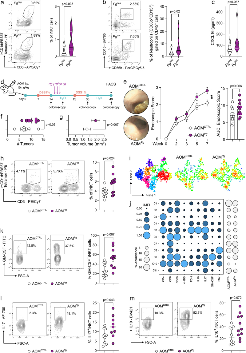
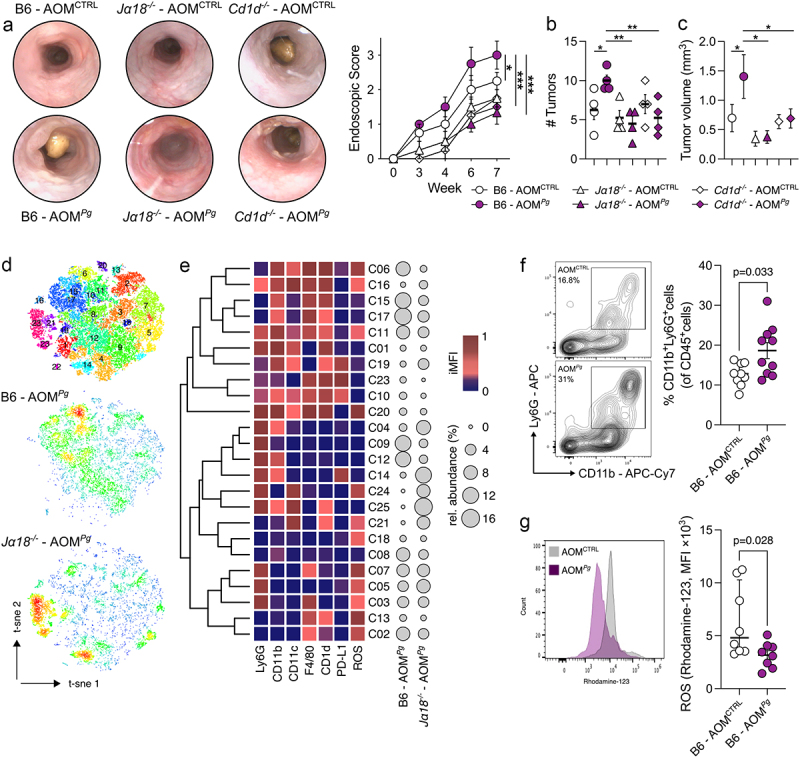
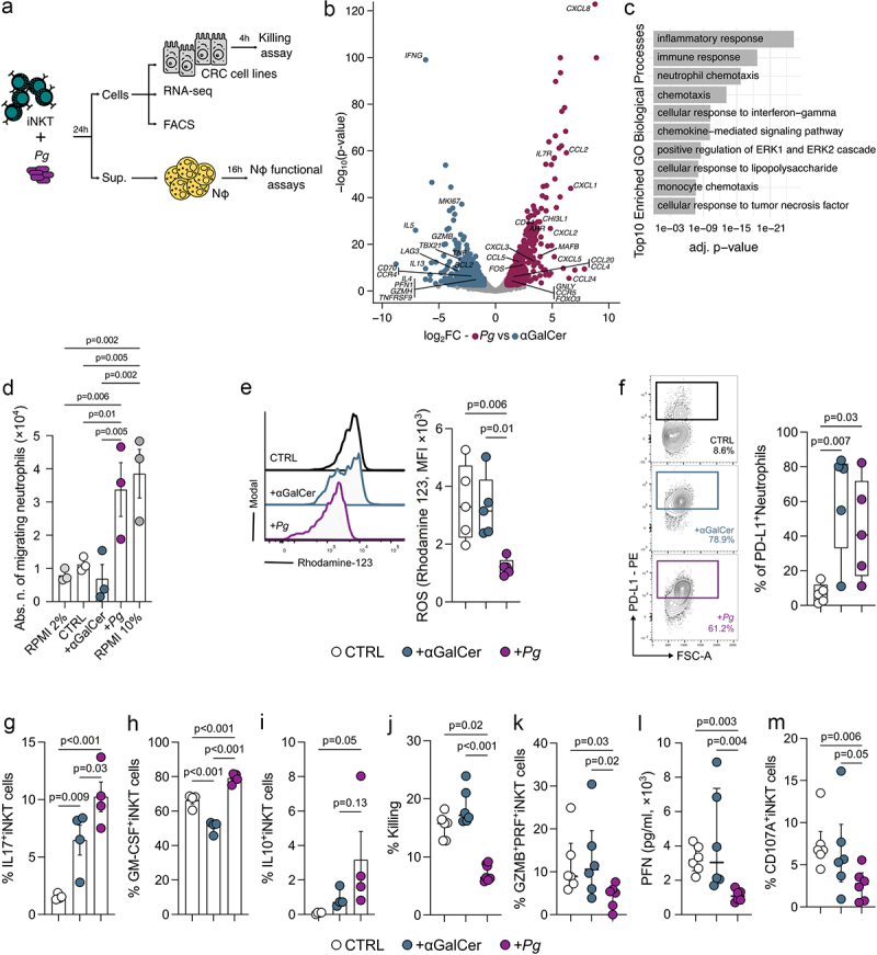
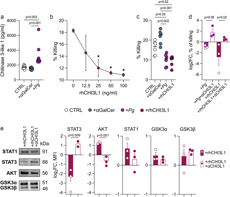
The interaction between the gut microbiota and invariant Natural Killer T (iNKT) cells plays a pivotal role in colorectal cancer (CRC). The pathobiont Fusobacterium nucleatum influences the anti-tumor functions of CRC-infiltrating iNKT cells. However, the impact of other bacteria associated with CRC, like Porphyromonas gingivalis, on their activation status remains unexplored. In this study, we demonstrate that mucosa-associated P. gingivalis induces a protumour phenotype in iNKT cells, subsequently influencing the composition of mononuclear-phagocyte cells within the tumor microenvironment. Mechanistically, in vivo and in vitro experiments showed that P. gingivalis reduces the cytotoxic functions of iNKT cells, hampering the iNKT cell lytic machinery through increased expression of chitinase 3-like-1 protein (CHI3L1). Neutralization of CHI3L1 effectively restores iNKT cell cytotoxic functions suggesting a therapeutic potential to reactivate iNKT cell-mediated antitumour immunity. In conclusion, our data demonstrate how P. gingivalis accelerates CRC progression by inducing the upregulation of CHI3L1 in iNKT cells, thus impairing their cytotoxic functions and promoting host tumor immune evasion.

Keywords: CHI3L1; CRC; Porphyromonas gingivalis; iNKT cells.


Conflict of interest statement

No potential conflict of interest was reported by the author(s).

Figures

Figure 1.
Figure 1.
P. gingivalis promotes colorectal tumorigenesis by modulating iNKT cell functions. A) frequency of tumor-infiltrating iNKT cells and B) tumor-associated neutrophils in CRC patients (n = 31) positive (Pgpos; n = 16) or negative (Pgneg; n = 15) for P. gingivalis in their mucosa-associated microbiota with representative dot plots. C) CXCL16 concentration from tissue lysates (200 µg of total protein). D) schematic representation of the AOM-DSS experimental plan. E) tumour endoscopic score, AUC and representative endoscopic pictures, F) number and G) volume of tumors from AOM-DSS treated C57BL/6 animals orally gavaged with PBS (AOMCTRL) or 109 CFUs of P. gingivalis (AOMPg). H) frequency of tumor-infiltrating iNKT cells in AOMCTRL and AOMPg C57BL/6 mice with representative plots. I) t-sne map of iNKT cells based on phenograph metaclustering analysis of AOMCTRL and AOMPg tumor samples. J) balloon plot of the scaled integrated mean fluorescent intensity (iMFI) of phenograph clusters generated in panel J. K-M) frequency of tumor-infiltrating K) GM-CSF+ L) IL17+ and M) IL10+ iNKT cells in AOMCTRL and AOMPg C57BL/6 mice with representative plots. Data (n = 10, AOMCTRL; n = 11, AOMPg) from two pooled independent experiments representative of at least three.
Figure 2.
Figure 2.
iNKT cells are essential to promote P. gingivalis-elicited colorectal tumorigenesis. A) Tumour endoscopic score and representative endoscopic pictures, B) number and C) volume of tumors from AOM-DSS treated B6, Traj18−/− (Jα18−/−) and Cd1d−/− mice orally gavaged with PBS (AOMCTRL) or 109 CFUs of P. gingivalis (AOMPg); data (n = 4) from one representative experiment. D) t-sne map of intratumour myeloid cells based on phenograph clustering from AOM-DSS, P. gingivalis treated C57BL/6 (B6 - AOMPg) and Traj18−/− mice (Jα18−/− - AOMPg). E) heatmap of scaled integrated MFI data from phenograph clustering analysis; relative abundance of the identified clusters is also shown. F) frequency of CD11b+Ly6G+ and G) respiratory burst quantification of tumor-associated neutrophils from AOM-DSS treated C57BL/6 mice orally gavaged with PBS (AOMCTRL) or 109 CFUs of P. gingivalis (AOMPg), with representative dot plots. Data (n = 8-10) from two pooled independent experiments representative of at least three.
Figure 3.
Figure 3.
P. gingivalis promotes the iNKT cell-mediated recruitment of TANs while impairing iNKT cell cytotoxicity. A) schematic representation of the experimental plan. B) volcano plot representing the differentially expressed genes (DEGs) in Pg- vs αGalCer-primed iNKT cells; the volcano plot shows for each gene (dots) the differential expression [log2fold-change (log2FC)] and its associated statistical significance (log10p-value). Dots indicate those genes with an fdr-corrected p < 0.05 and log2FC > |1|. C) gene ontology (GO) analysis of differentially expressed genes (Bonferroni-corrected p < 0.05 and log2FC >2). D) absolute numbers of migrating neutrophils upon exposure to RPMI + 2%FBS (negative control), RPMI + 10%FBS (positive control), unloaded moDC (CTRL), αGalCer- and pg-primed (+Pg) iNKT cell supernatants. Results are representative of three (n = 3) independent experiments. p < 0.05 (*), p < 0.01 (**), one-way ANOVA. E) respiratory burst assay quantification and F) frequency of PD-L1+ cells from neutrophils exposed to the culture supernatants of unloaded moDC (CTRL), αGalCer- and Pg-primed (+Pg) iNKT cells with representative plots. G-I) frequency of G) IL17+ H) GM-CSF+ and I) IL10+ iNKT cells upon stimulation with P. gingivalis. J) percentage of killed tumor cells by unloaded moDC (CTRL), αGalCer- and Pg-primed (+Pg) iNKT cells. K) frequency of GZMB+PFN+iNKT cells. L) perforin concentration in the culture supernatant of unloaded moDC (CTRL), αGalCer- and Pg-primed (+Pg) iNKT cells. M) frequency of CD107a+iNKT cells. Data are representative of at least three independent experiments.
Figure 4.
Figure 4.
P. gingivalis exerts its effect on iNKT cells through CHI3L1. A) CHI3L1 concentration in the culture supernatant of Pg-primed (+Pg) iNKT cells and unloaded moDC (CTRL). B) percentage of killed tumor cells by iNKT cells treated with increasing doses of rhCHI3L1. C) percentage of killed tumor cells by iNKT cells basal activated (CTRL), primed with 109 CFUs of P. gingivalis (+Pg) or treated with rhCHI3L1 (100 ng/ml). D) Log2 fold-change of the percentage of killed tumor cells by iNKT cells primed with 109 CFUs of P. gingivalis (+Pg) or treated with rhCHI3L1 (100 ng/ml) upon neutralization with anti-CHI3L1 antibody (full bars); data have been normalized vs iNKT cells primed with unloaded moDC. E) western blot analysis; MFI is expressed as log2 fold-change normalized vs iNKT cells primed with unloaded moDC. Data are representative of at least three independent experiments.

References

    1. Deo SVS, Sharma J, Kumar SG.. Report on global cancer burden: challenges and opportunities for surgical oncologists. Ann Surg Oncol. 2022;2020;29(11):6497–17. doi:10.1245/s10434-022-12151-6. - DOI - PubMed
    1. Schmitt M, Greten FR. The inflammatory pathogenesis of colorectal cancer. Nat Rev Immunol. 2021;21(10):653–667. doi:10.1038/s41577-021-00534-x. - DOI - PubMed
    1. Chen J, Pitmon E, Wang K. Microbiome, inflammation and colorectal cancer. Semin Immunol. 2017;32:43–53. doi:10.1016/j.smim.2017.09.006. - DOI - PubMed
    1. Ternes D, Karta J, Tsenkova M, Wilmes P, Haan S, Letellier E. Microbiome in colorectal cancer: how to get from meta-omics to mechanism? Trends Microbiol. 2020;28(5):401–423. doi:10.1016/j.tim.2020.01.001. - DOI - PubMed
    1. Hajishengallis G. Periodontitis: from microbial immune subversion to systemic inflammation. Nat Rev Immunol. 2015;15(1):30–44. doi:10.1038/nri3785. - DOI - PMC - PubMed

MeSH terms

Substances