

Prevalence of S-methyl-5'-thioadenosine Phosphorylase (MTAP) Deficiency in Human Cancer: A Tissue Microarray Study on 13,067 Tumors From 149 Different Tumor Types
- PMID: 39132873
- PMCID: PMC11404761
- DOI: 10.1097/PAS.0000000000002297
Prevalence of S-methyl-5'-thioadenosine Phosphorylase (MTAP) Deficiency in Human Cancer: A Tissue Microarray Study on 13,067 Tumors From 149 Different Tumor Types
Abstract
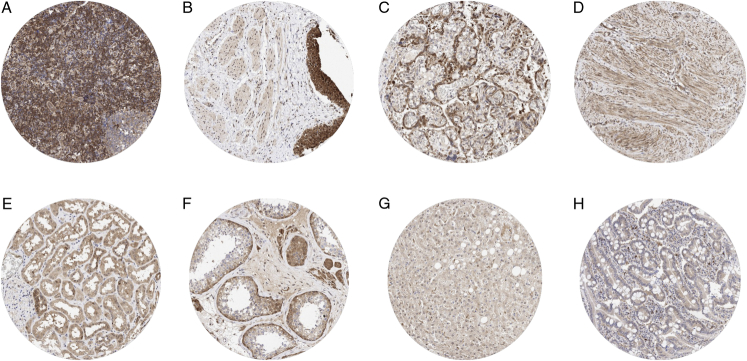
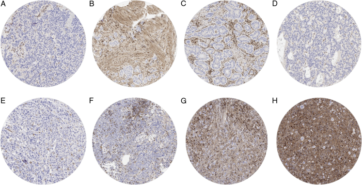
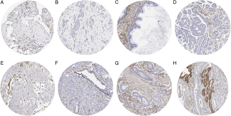
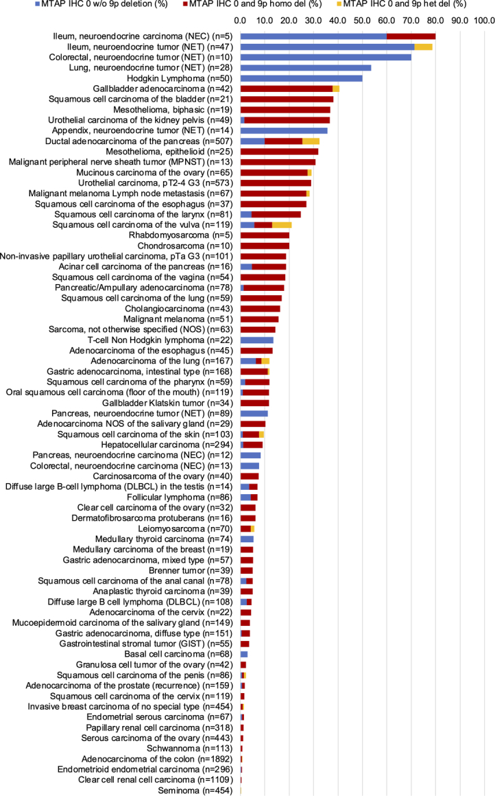
Loss of S-methyl-5'-thioadenosine phosphorylase (MTAP) expression is a common event in cancer leading to a critical vulnerability of cancer cells towards anti-cancer drugs. Homozygous MTAP deletions result in a complete expression loss that can be detected by immunohistochemistry (IHC). In this study, a tissue microarray containing 17,078 samples from 149 different tumor entities was analyzed by IHC, and complete MTAP loss was validated by fluorescence in situ hybridization. MTAP loss was observed in 83 of 149 tumor categories, including neuroendocrine neoplasms (up to 80%), Hodgkin lymphoma (50.0%), mesothelioma (32.0% to 36.8%), gastro-intestinal adenocarcinoma (4.0% to 40.5%), urothelial neoplasms (10.5% to 36.7%), squamous cell carcinomas (up to 38%), and various types of sarcomas (up to 20%) and non-Hodgkin lymphomas (up to 14%). Homozygous MTAP deletion was found in 90% to 100% of cases with MTAP expression loss in most tumor categories. However, neuroendocrine tumors, Hodgkin lymphomas, and other lymphomas lacked MTAP deletions. MTAP deficiency was significantly linked to unfavorable tumor phenotype in selected tumor entities and the presence of PD-L1 expression on tumor cells, absence of PD-L1 expression on immune cells, and a low density of CD8 + lymphocytes. In summary, MTAP deficiency can occur in various tumor entities and is linked to unfavorable tumor phenotype and noninflamed tumor microenvironment, but is not always related to deletions. MTAP IHC is of considerable diagnostic value for the detection of neoplastic transformation in multiple different applications.
Copyright © 2024 The Author(s). Published by Wolters Kluwer Health, Inc.
Conflict of interest statement
Conflicts of Interest and Source of Funding: The recombinant rabbit monoclonal antibody, clone MSVA-741R was provided from MS Validated Antibodies GmbH, Hamburg, Germany (owned by a family member of G.S.). For the remaining authors none were declared.
Figures
References
-
- Della Ragione F, Carteni-Farina M, Gragnaniello V, et al. Purification and characterization of 5’-deoxy-5’-methylthioadenosine phosphorylase from human placenta. J Biol Chem. 1986;261:12324–12329. - PubMed
-
- Zhang H, Chen ZH, Savarese TM. Codeletion of the genes for p16INK4, methylthioadenosine phosphorylase, interferon-alpha1, interferon-beta1, and other 9p21 markers in human malignant cell lines. Cancer Genet Cytogenet. 1996;86:22–28. - PubMed
MeSH terms
Substances
LinkOut - more resources
Full Text Sources
Medical
Research Materials
