. 2024 Sep 19;206(9):e0010724.
doi: 10.1128/jb.00107-24. Epub 2024 Aug 12.

Regulation of potassium uptake in Caulobacter crescentus

Affiliations

Regulation of potassium uptake in Caulobacter crescentus

Alex Quintero-Yanes et al. J Bacteriol. .

Abstract

Potassium (K+) is an essential physiological element determining membrane potential, intracellular pH, osmotic/turgor pressure, and protein synthesis in cells. Here, we describe the regulation of potassium uptake systems in the oligotrophic α-proteobacterium Caulobacter crescentus known as a model for asymmetric cell division. We show that C. crescentus can grow in concentrations from the micromolar to the millimolar range by mainly using two K+ transporters to maintain potassium homeostasis, the low-affinity Kup and the high-affinity Kdp uptake systems. When K+ is not limiting, we found that the kup gene is essential while kdp inactivation does not impact the growth. In contrast, kdp becomes critical but not essential and kup dispensable for growth in K+-limited environments. However, in the absence of kdp, mutations in kup were selected to improve growth in K+-depleted conditions, likely by increasing the affinity of Kup for K+. In addition, mutations in the KdpDE two-component system, which regulates kdpABCDE expression, suggest that the inner membrane sensor regulatory component KdpD mainly works as a phosphatase to limit the growth when cells reach late exponential phase. Our data therefore suggest that KdpE is phosphorylated by another non-cognate histidine kinase. On top of this, we determined the KdpE-dependent and independent K+ transcriptome. Together, our work illustrates how an oligotrophic bacterium responds to fluctuation in K+ availability.IMPORTANCEPotassium (K+) is a key metal ion involved in many essential cellular processes. Here, we show that the oligotroph Caulobacter crescentus can support growth at micromolar concentrations of K+ by mainly using two K+ uptake systems, the low-affinity Kup and the high-affinity Kdp. Using genome-wide approaches, we also determined the entire set of genes required for C. crescentus to survive at low K+ concentration as well as the full K+-dependent regulon. Finally, we found that the transcriptional regulation mediated by the KdpDE two-component system is unconventional since unlike Escherichia coli, the inner membrane sensor regulatory component KdpD seems to work rather as a phosphatase on the phosphorylated response regulator KdpE~P.

Keywords: KdpD; KdpE; Kup; potassium transport; two-component system.


Conflict of interest statement

The authors declare no conflict of interest.

Figures

Fig 1
Fig 1
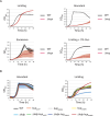
Impact of K+ on C. crescentus growth. Growth of WT in M2G-K supplemented with different K+ concentrations using K2HPO4 + KH2PO4 (A, B) and KCl (C, D) as K+ source. Data represent average, n = 3, and error bars = ±SD. Other concentrations tested are presented in Fig. S1.
Fig 2
Fig 2
Deletion of K+-related genes impact C. crescentus growth. Growth of WT, ΔkefB, ΔkefC, ΔCCNA_01688, and ΔkdpABCDE mutants in minimal medium M2G-K supplemented with (A) limiting (0.025 mM), (B) abundant (0.5 mM), and (C) excessive (50 mM) K+ concentrations. Data represent average, n = 3, and error bars = ±SD.
Fig 3
Fig 3
Tn-seq profile of C. crescentus cells in limiting K+ conditions. (A) Diagram of the Tn-seq experiment. As explained in detail in Materials and Methods section, E. coli cells were used to deliver a mini Tn5 vector into C. crescentus by conjugation. Mating spots were washed twice in either M2G or M2G-K. Cells were plated in either M2G or M2G-K square plates supplemented with kanamycin (Km) and aztreonam (Az) to counter select C. crescentus cells with Tn5 insertions (green cells) over C. crescentus without insertions and E. coli cells. Cells were collected to complete >3 × 10⁵ clones. Thereafter, gDNA extraction and Tn5-directed sequencing were performed to determine the frequency of insertions for each gene in C. crescentus genome and determine genes that are essential, high fitness cost, or non-essential in M2G and M2G-K plates. (B) Growth of WT and Δkdp cells in M2G-K plates without or supplemented with K+. (C) Number of Transposon insertions (#Tn insertions) against the length (#bp) of 4,186 genes in M2G and M2G-K media. Non-essential genes are highlighted in gray, high fitness cost genes in light blue, and essential in dark blue. (D) Representation of genes that changed in their fitness cost categories according to analysis done in M2G and M2G-K (C). (C, D) The percentage of genes in each category is indicated following the color code.
Fig 4
Fig 4
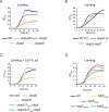
Impact of deletion and overexpression of the kup gene. (A) Growth of WT and Δkup mutant in minimal medium M2G-K supplemented with limiting (0.025 mM), abundant (0.5 mM), excessive (50 mM) K+ concentrations or limiting (0.025 mM) K+ concentrations and 3% sucrose. (B) Growth of WT, kupA87P, kupG253S, and kupS456R in an otherwise WT or Δkdp background, in abundant and limiting K+ conditions. Data represent average, n = 3, and error bars = ±SD.
Fig 5
Fig 5
Molecular dynamics-highlighted structural changes of the putative K+ binding sites of Kup mutants. (A) Snapshot of Kup WT after 1 µs of simulation. The protein is represented in cyan as cartoon, the considered mutation sites are colored in magenta and indicated by arrows, and the residues composing the KimA-based K+ binding cavity are highlighted as sticks in dark blue. (B) Snapshot of the KimA-based K+ binding site of KupA87P, (C) KupG253S, and (D) KupS456R after 1 µs of simulation. On each of these panels, WT Kup (cyan) and mutated Kup (magenta) aligned to KimA (PDB entry: 6S3K; light gray) are shown as cartoon, conserved residues between Kup and KimA constitutive of the K+ binding pocket are represented as sticks in the corresponding color, and K+ cations as orange spheres. The displayed amino acid numbering only corresponds to Kup sequence.
Fig 6
Fig 6
K+- and KdpE-dependent regulon. (A, B) Volcano plots representing the relation between the log2 FC and −log FDR on gene expression between (A) WT in limiting vs abundant K+ condition, and (B) ΔkdpE vs WT in limiting K+ condition. Genes identified are presented as dots. Significant downregulated and upregulated genes are presented as blue (A) and purple (B) dots, while genes with no significant alterations are presented as black dots. Top 10 downregulated and upregulated genes are highlighted for each analysis.
Fig 7
Fig 7
KdpD downregulates KdpE to control growth in low K+. (A) Growth of WT, ΔkdpABC, ΔkdpD, ΔkdpE, and ΔkdpDE. (B) Growth of WT and ΔkdpD carrying either pMR10 empty vector (EV) or pMR10 Pkdp::kdpD. (C) Growth of WT, ΔkdpE, and ΔkdpDE expressing kdpE from the xylX locus (PxylX::kdpE). (D) Growth of kdpD and kdpE catalytic point mutants. Growth was done in minimal medium M2G-K supplemented with limiting (0.025 mM) K+ concentrations (A–D), and tetracycline for plasmid selection (B) or with 0.01% xylose for the induction of kdpE expression (C). Data represent average, n = 3, and error bars = ±SD.
Fig 8
Fig 8
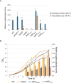
Regulation of kdp expression. (A) Relative β-galactosidase activities of WT and kdp mutants carrying a transcriptional Pkdp::lacZ fusion grown in M2G-K supplemented with limiting (0.025 mM) or abundant (0.5 mM) K+ concentrations. The data were normalized to the WT grown in limiting K+ conditions (blue bars). (B) Relative β-galactosidase activities of WT and kdp mutants carrying a transcriptional Pkdp::lacZ fusion along the growth in M2G-K supplemented with limiting (0.025 mM). The data were normalized to the WT grown in limiting K+ conditions (orange bars) after 20 h of growth. Data represent average, n = 3, and error bars = ±SD. Means were statistically compared using a two-way analysis of variance, followed by Tukey’s multiple comparisons test; not significant (NS), P < 0.05 (*), P < 0.01 (**), P < 0.001 (***), P < 0.0001 (****).

Similar articles

References

    1. Danchin A, Nikel PI. 2019. Why nature chose potassium. J Mol Evol 87:271–288. doi:10.1007/s00239-019-09915-2 - DOI - PubMed
    1. Epstein W. 2003. The roles and regulation of potassium in bacteria. Prog Nucleic Acid Res Mol Biol 75:293–320. doi:10.1016/s0079-6603(03)75008-9 - DOI - PubMed
    1. Bakker EP, Mangerich WE. 1981. Interconversion of components of the bacterial proton motive force by electrogenic potassium transport. J Bacteriol 147:820–826. doi:10.1128/jb.147.3.820-826.1981 - DOI - PMC - PubMed
    1. Booth IR. 1999. The regulation of intracellular pH in bacteria. Novartis Found Symp 221:19–28. doi:10.1002/9780470515631.ch3 - DOI - PubMed
    1. Ochrombel I, Ott L, Krämer R, Burkovski A, Marin K. 2011. Impact of improved potassium accumulation on pH homeostasis, membrane potential adjustment and survival of Corynebacterium glutamicum. Biochim Biophys Acta 1807:444–450. doi:10.1016/j.bbabio.2011.01.008 - DOI - PubMed

Publication types

MeSH terms