. 2024 Aug 12;15(1):346.
doi: 10.1007/s12672-024-01178-8.

Trimethylamine N-oxide promotes the proliferation and migration of hepatocellular carcinoma cell through the MAPK pathway

Affiliations

Trimethylamine N-oxide promotes the proliferation and migration of hepatocellular carcinoma cell through the MAPK pathway

Chunfang Zhou et al. Discov Oncol. .

Abstract

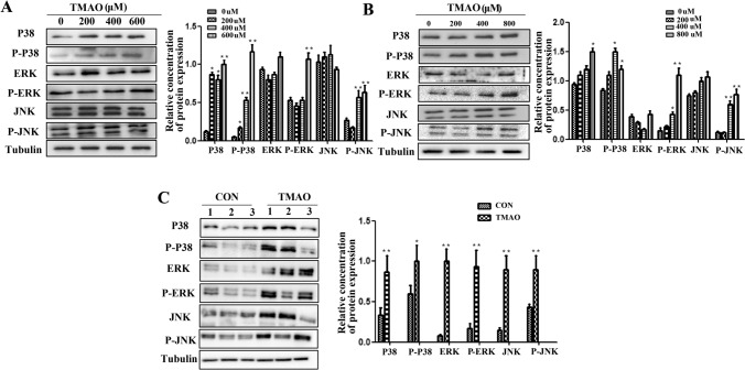
Trimethylamine-n-oxide (TMAO) is a metabolite of intestinal flora following the consumption of phosphatidylcholine-rich foods. Clinical cohort studies have shown that plasma TMAO may be a risk factor for cancer development, including hepatocellular carcinoma (HCC), but fundamental research data supporting this hypothesis are lacking. In this study, HCC cells were treated with TMAO in vivo and in vitro to evaluate the effect on some indicators related to the malignancy degree of HCC, and the relevant molecular mechanisms were explored. In vitro, TMAO promoted the proliferation and migration of HCC cells and significantly upregulated the expression of proteins related to epithelial-mesenchymal transformation (EMT). In vivo, after HCC cells were inoculated subcutaneously in nude mice given water containing TMAO, the tumors grew faster and larger than those in the mice given ordinary water. The immunohistochemistry analysis showed that proliferation, migration and EMT-related proteins in the tumor tissues were significantly upregulated by TMAO. Furthermore, TMAO obviously enhanced the phosphorylation of MAPK signaling molecules in vivo and in vitro. In conclusion, TMAO promotes the proliferation, migration and EMT of HCC cells by activating the MAPK pathway.

Keywords: Hepatocellular carcinoma; MAPK; Migration; Proliferation; Trimethylamine N-oxide.


Conflict of interest statement

The authors declare no competing interests.

Figures

Fig. 1
Fig. 1
TMAO promotes human hepatocellular carcinoma (HCC) cell growth and colony formation. A Huh-7 cells were treated with different concentrations of TMAO, and cell proliferation was measured with EdU assays (a) and RTCA assays (b). B The proliferation of HepG2 cells treated with TMAO was measured with an EdU assay (a) and RTCA (b). C Colony formation assay of HepG2 and Huh-7. All experiments were performed in triplicate. TMAO-treated vs. PBS-treated; *P < 0.05, **P < 0.01
Fig. 2
Fig. 2
TMAO promotes HCC cell migration. A Detection of Huh-7 migration speed by a scratch assay (a), quantitative analysis of migration speed by migration index (b); detection of the migration ability of Huh-7 cells by a Transwell assay (c), statistical analysis of the number of Huh-7 cells passing through the chambers (d). B The migration ability of HepG2 cells was detected by a scratch assay (a), the migration index was quantitatively analyzed (b), the migration ability of HepG2 cells was detected with a Transwell assay (c), and the number of HepG2 cells passing through the chambers was quantitatively analyzed (d). All experiments were performed in triplicate. TMAO-treated vs. PBS-treated; *P < 0.05, **P < 0.01
Fig. 3
Fig. 3
Effects of TMAO on the expression of proliferation- and migration-related proteins in hepatocellular carcinoma cells. A The expression of VEGF and MMP9 in HepG2 cells after TMAO treatment was detected by immunofluorescence. B Western blot analysis of PCNA, MMP9 and VEGF expression in Huh-7 cells treated with TMAO. C Western blot analysis was used to detect the expression of PCNA, Ki67 and VEGF in HepG2 cells treated with TMAO. A few strips were clipped before color development because ladder staining strongly affected the display of target proteins. All experiments were performed in triplicate. TMAO-treated vs. PBS-treated; *P < 0.05, **P < 0.01
Fig. 4
Fig. 4
Effects of TMAO on the epithelial mesenchymal transformation of hepatocellular carcinoma cells. A Observation of the morphology of HCC cells after treatment with TMAO. B Immunofluorescence detection of vimentin expression in HepG2 cells treated with TMAO. C Western blot analysis was used to detect the expression of EMT-related proteins in Huh-7 cells treated with different concentrations of TMAO. D Western blot analysis of EMT-related protein expression in HepG2 cells treated with different concentrations of TMAO. E Western blot analysis of EMT-related protein expression in Huh-7 cells treated with 600 μmol/L TMAO over time. F Western blot analysis of EMT-related protein expression in HepG2 cells treated with 800 μmol/L TMAO over time. A few strips were clipped before color development because ladder staining strongly affected the display of target proteins. All experiments were performed in triplicate. TMAO-treated vs. PBS-treated; *P < 0.05, **P < 0.01
Fig. 5
Fig. 5
TMAO promotes tumor growth in xenograft mice. A Twenty-five days after HCC cell injection, the mice were euthanized, and the xenograft tumors were collected. The image shows the tumors generated in two different treatment groups (a). Xenograft volumes were monitored at the indicated time (b). Tumor weight was analyzed (c). B PCNA, vimentin, MMP9, VEGF and E-cadherin expression in subcutaneous tumors detected by IHC. C PCNA and EMT-related protein expression in subcutaneous tumors detected by a Western blot analysis. *P < 0.05, **P < 0.01, compared with control
Fig. 6
Fig. 6
MAPK pathway was activated by TMAO in HCC cells. A The protein levels of p-p38, p38, p-ERK, ERK, p-JNK, and JNK were detected by a Western blot analysis after Huh-7 cells were exposed to 0, 200, 400 and 600 μmol/L TMAO for 48 h. B The protein levels of the MAPK pathway were detected by a Western blot analysis after HepG2 cells were exposed to 0, 200, 400 and 800 μmol/L TMAO for 48 h. C Western blot analysis of MAPK pathway protein expression in subcutaneous tumors from xenograft mice. All experiments were performed in triplicate. *P < 0.05, **P < 0.01, compared with control

Similar articles

Cited by

References

    1. Sung H, Ferlay J, Siegel RL, Laversanne M, Soerjomataram I, Jemal A, et al. Global Cancer Statistics 2020: GLOBOCAN estimates of incidence and mortality worldwide for 36 cancers in 185 countries. CA Cancer J Clin. 2021;71(3):209–49. 10.3322/caac.21660. 10.3322/caac.21660 - DOI - PubMed
    1. Sayiner M, Golabi P, Younossi ZM. Disease burden of hepatocellular carcinoma: a global perspective. Dig Dis Sci. 2019;64(4):910–7. 10.1007/s10620-019-05537-2. 10.1007/s10620-019-05537-2 - DOI - PubMed
    1. Gnyawali B, Pusateri A, Nickerson A, Jalil S, Mumtaz K. Epidemiologic and socioeconomic factors impacting hepatitis B virus and related hepatocellular carcinoma. World J Gastroenterol. 2022;28(29):3793–802. 10.3748/wjg.v28.i29.3793. 10.3748/wjg.v28.i29.3793 - DOI - PMC - PubMed
    1. Vogel A, Meyer T, Sapisochin G, Salem R, Saborowski A. Hepatocellular carcinoma. Lancet (London, England). 2022;400(10360):1345–62. 10.1016/S0140-6736(22)01200-4. 10.1016/S0140-6736(22)01200-4 - DOI - PubMed
    1. Ayares G, Idalsoaga F, Díaz LA, Arnold J, Arab JP. Current medical treatment for alcohol-associated liver disease. J Clin Exp Hepatol. 2022;12(5):1333–48. 10.1016/j.jceh.2022.02.001. 10.1016/j.jceh.2022.02.001 - DOI - PMC - PubMed

LinkOut - more resources