

Epidemiology and Risk Factors of Actinic Keratosis. What is New for The Management for Sun-Damaged Skin
- PMID: 39133637
- PMCID: PMC11566825
- DOI: 10.5826/dpc.1403S1a146S
Epidemiology and Risk Factors of Actinic Keratosis. What is New for The Management for Sun-Damaged Skin
Abstract
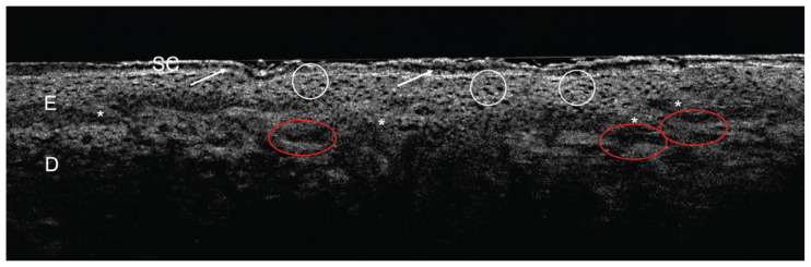
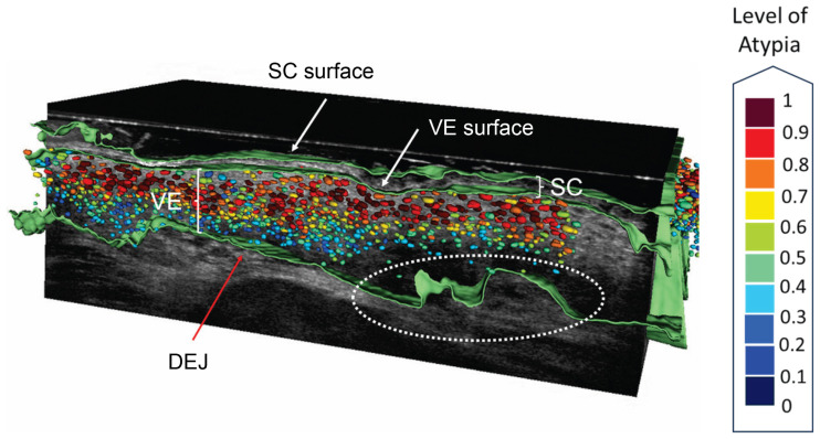
Actinic keratosis (AK) is considered a chronic skin disease mostly caused by long-term exposure to UV radiation and other risk factors such as immunosuppression, leading to an individual susceptibility for skin cancer manifestation. The treatment of AK is laborious and costly, and the incidence of skin cancer is forecasted to double until the year 2030 in an aging society.Risk factors in AK for malignant transformation in cutaneous squamous cell carcinoma (cSCC) are not fully understood, but studies suggest that histological features, such as atypia in the basal epidermal third and basal proliferation (PRO score) in AK play a pivotal role for development of malignancy. As the clinical appearance of AK does not correlate with the risk for malignancy, guidelines suggest treating every single AK lesion upon diagnosis. Skin imaging techniques, such as line-field confocal optical coherence tomography (LC-OCT) can help to provide an individual holistic follow-up for AK lesions by non-invasive visualization of atypia and basal proliferation. A follow-up for patients with AK may be critical for treatment success in terms of strengthening therapy adherence. When AK presents therapy refractory, cSCC manifests in nearly 30% of the cases after several years. Patients with AK suffering from field cancerization and immunosuppression are susceptible for a severe course of disease including metastasis and high mortality rates. Those vulnerable subgroups benefit from close skin cancer screening, early adequate treatment and chemoprevention, such as niacinamide or acitretin. Skin cancer prevention is substantial. Primary prevention should include chemical and physical UV-light protection and avoidance of indoor tanning. Secondary prevention is essential in high-risk populations, such as fair skin type elderly men and STORs. Tertiary prevention should comprise adequate treatment strategies to prevent therapy resistance, reoccurrence and cSCC, especially when field cancerization and immunosuppression are present.
Conflict of interest statement
Figures
References
-
- Thamm JR, Welzel J, Schuh S. Diagnosis and therapy of actinic keratosis. J Dtsch Dermatol Ges. 2024 - PubMed
-
- Schafer I, Reusch M, Siebert J, Spehr C, Augustin M. Health care characteristics of basal cell carcinoma in Germany: the role of insurance status and socio-demographic factors. J Dtsch Dermatol Ges. 2014;12(9):803–11. - PubMed
-
- Marks R, Rennie G, Selwood TS. Malignant transformation of solar keratoses to squamous cell carcinoma. Lancet. 1988;1(8589):795–7. - PubMed
Publication types
LinkOut - more resources
Full Text Sources
Research Materials