

Use of sedation-awakening electroencephalography in dogs with epilepsy
- PMID: 39133769
- PMCID: PMC11423447
- DOI: 10.1111/jvim.17153
Use of sedation-awakening electroencephalography in dogs with epilepsy
Abstract
Background: Electroencephalography (EEG) recording protocols have been standardized for humans. Although the utilization of techniques in veterinary medicine is increasing, a standard protocol has not yet been established.
Hypothesis: Assessment of a sedation-awakening EEG protocol in dogs.
Animals: Electroencephalography examination was performed in a research colony of 6 nonepileptic dogs (control [C]) and 12 dogs with epilepsy admitted to the clinic because of the epileptic seizures.
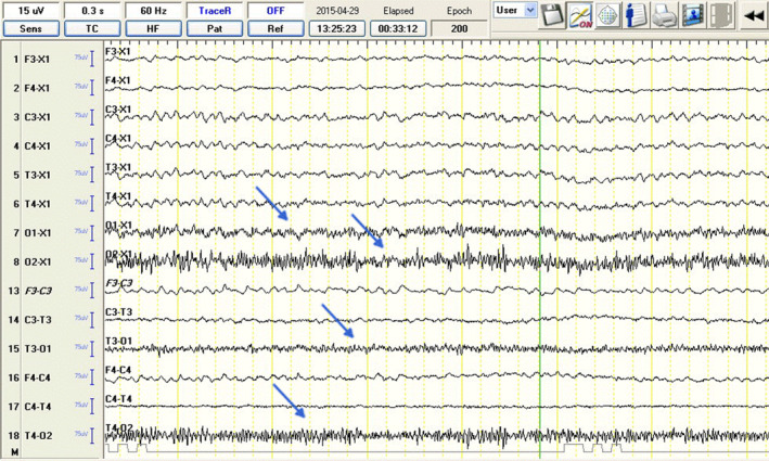
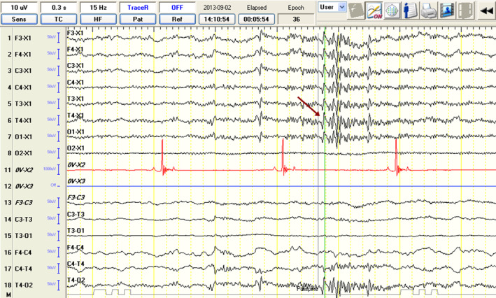
Methods: It was a prospective study with retrospective control. Dogs with epilepsy were divided into 2 equal groups, wherein EEG acquisition was performed using a "sedation" protocol (IE-S, n = 6) and a "sedation-awakening" protocol (IE-SA, n = 6). All animals were sedated using medetomidine. In IE-SA group, sedation was reversed 5 minutes after commencing the EEG recording by injecting atipamezole IM. Type of background activity (BGA) and presence of EEG-defined epileptiform discharges (EDs) were evaluated blindly. Statistical significance was set at P > 0.05.
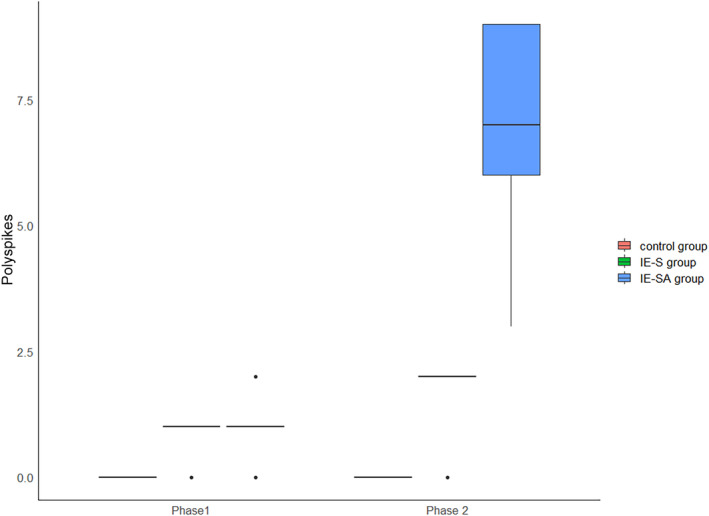
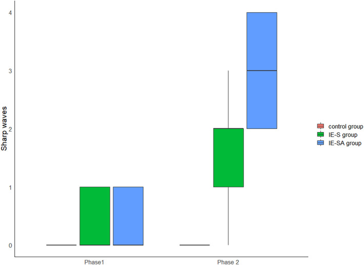
Results: Epileptiform discharges were found in 1 of 6 of the dogs in group C, 4 of 6 of the dogs in IE-S group, and 5 of 6 of the dogs in IE-SA group. A significantly greater number of EDs (spikes, P = .0109; polyspikes, P = .0109; sharp waves, P = .01) were detected in Phase 2 in animals subjected to the "sedation-awakening" protocol, whereas there was no statistically significant greater number of discharges in sedated animals.
Conclusions and clinical importance: A "sedation-awakening" EEG protocol could be of value for ambulatory use if repeated EEG recordings and monitoring of epilepsy in dogs is needed.
Keywords: ambulatory EEG; canine electroencephalography; canine epilepsy diagnosis; epilepsy; paroxysmal; sedation; seizure.
© 2024 The Author(s). Journal of Veterinary Internal Medicine published by Wiley Periodicals LLC on behalf of American College of Veterinary Internal Medicine.
Conflict of interest statement
Authors declare no conflict of interest.
Figures


References
-
- Falco‐Walter J. Epilepsy‐definition, classification, pathophysiology, and epidemiology. Semin Neurol. 2020;40:617‐623. - PubMed
-
- Koutroumanidis M, Smith S. Use and abuse of EEG in the diagnosis of idiopathic generalized epilepsies. Epilepsia. 2005;46:96‐107. - PubMed
-
- Sinha SR, Sullivan L, Sabau D, et al. American clinical neurophysiology society guideline 1: minimum technical requirements for performing clinical electroencephalography. J Clin Neurophysiol. 2016;33:303‐307. - PubMed
