. 2024 Nov 1;327(5):R486-R496.
doi: 10.1152/ajpregu.00227.2023. Epub 2024 Aug 12.

Spontaneous and evoked angiotensin II sniffer cell activity in the lamina terminalis in vitro

Affiliations

Spontaneous and evoked angiotensin II sniffer cell activity in the lamina terminalis in vitro

George E Farmer et al. Am J Physiol Regul Integr Comp Physiol. .

Abstract

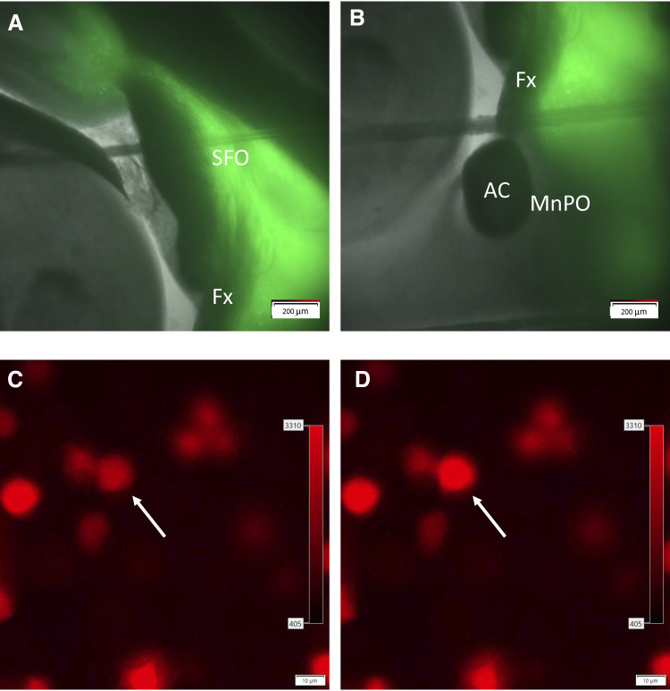
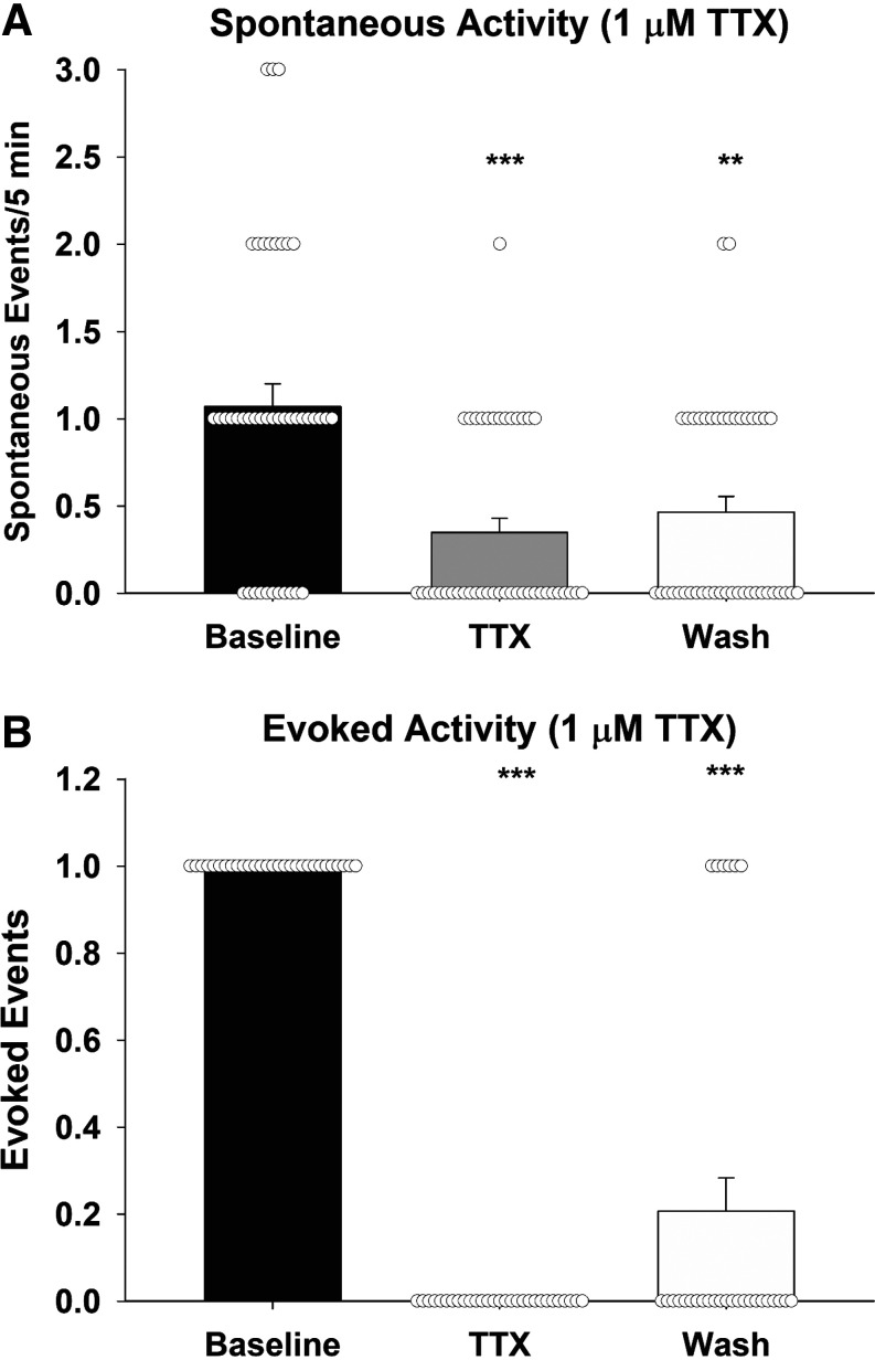
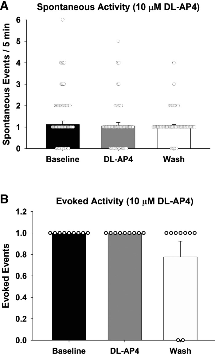
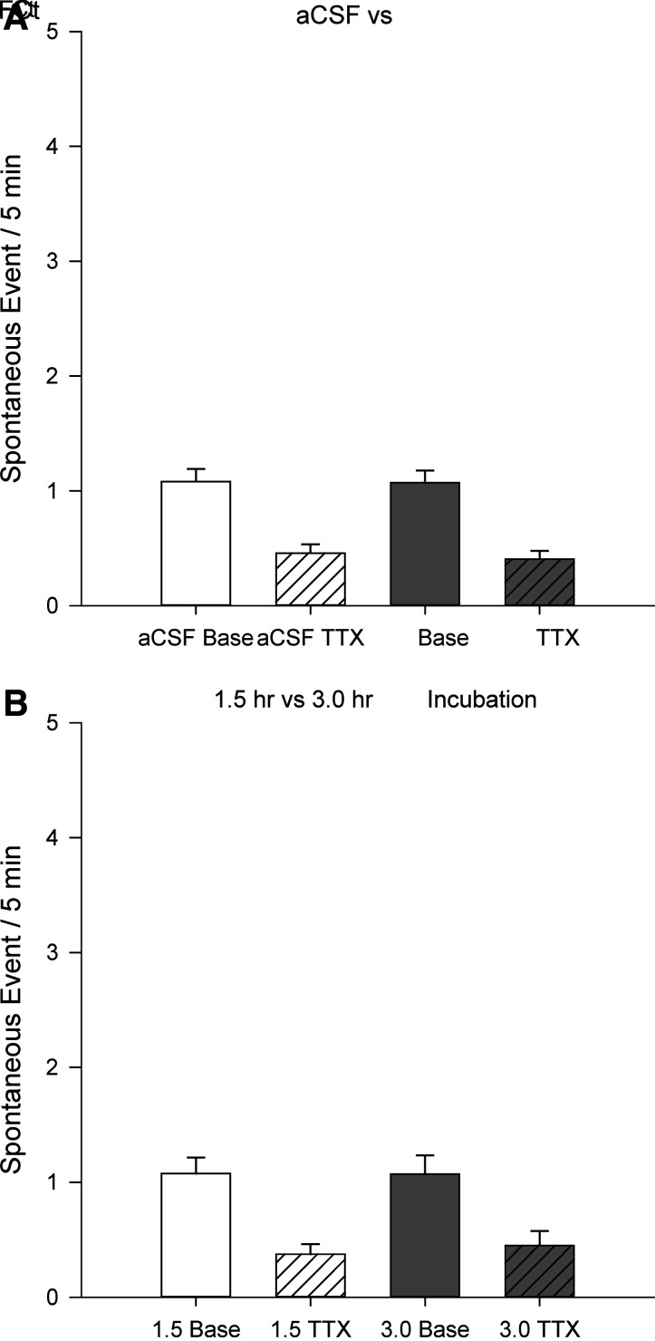
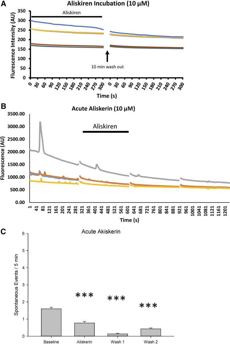
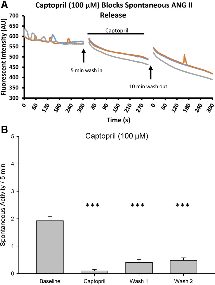
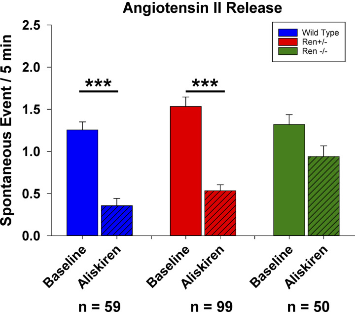
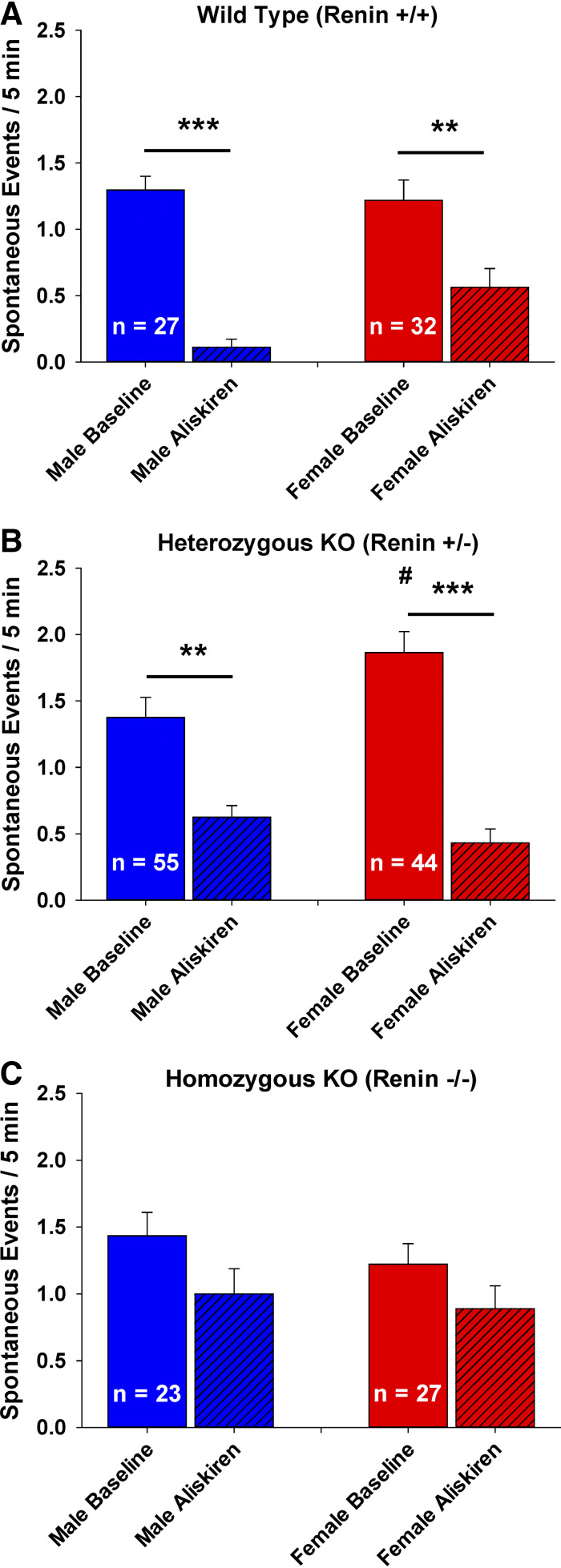
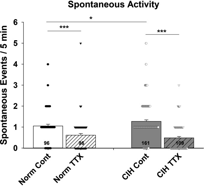
Angiotensin II (ANG II) has been shown to have central nervous system effects. Although tissue renin-angiotensin systems (RAS) have been demonstrated in multiple tissues, the existence of a brain RAS is still a matter of debate. These studies test for angiotensin release from brain slices prepared from adult male Sprague-Dawley rats and male and female renin knock-out rats using Chinese hamster ovary cells modified to express both the angiotensin II type 1 receptor and a fluorescent calcium indicator. Sniffer cells were placed on the slices and calcium transients were measured from those located on or adjacent to the median preoptic nucleus with and without stimulation of the subfornical organ. Bath application of tetrodotoxin (1 µM) significantly attenuated spontaneous events while abolishing evoked sniffer cell activity. Bath application of dl-AP4 (10 µM, glutamatergic antagonist) did not affect either spontaneous or evoked release. Incubating the slices with fluorocitrate to inactive astrocytes did not influence sniffer cell activity in the MnPO. Pharmacological experiments indicate that ANG II release is largely both renin (aliskiren 10 µM) and ACE-1 (captopril 100 µM) dependent. However, experiments with brain slices prepared from male and female Renin knock-out rats suggest that alternative synthetic pathways may exist. Finally, these studies demonstrate that increases in ANG II release are observed following 7 days of chronic intermittent hypoxia. These studies suggest the existence of a tissue-specific RAS in the brain that involves canonical and alternative ANG II synthetic pathways and is upregulated in an animal model of sleep apnea.NEW & NOTEWORTHY These studies used Chinese hamster ovary cells that were cloned to express an angiotensin receptor (At1ra) and a calcium indicator (R-GECO) to detect the release of angiotensin from brain slices containing the lamina terminalis of rats. Some of the experiments use tissue from renin knockout rats. The results support the existence of an angiotensin system in the brain that may involve alternative synthetic pathways and is upregulated by intermittent hypoxia.

Keywords: angtiotensin; circumventricular organ; hypertension; hypothalamus; peptide.


Conflict of interest statement

No conflicts of interest, financial or otherwise, are declared by the authors.

Figures

Figure 1.
Figure 1.
Expression of channel rhodopsin in the SFO (A) projections to the MnPO (B). Sniffer cells placed on the MnPO (C) show an increase in fluorescence in the presence of ANG II (D). AC, anterior commissure; ANG II, angiotensin II; Fx, fornix; MnPO, median preoptic nucleus; SFO, subfornical organ.
Figure 2.
Figure 2.
Release of ANG II is activity dependent. Spontaneous sniffer cell responses are reduced in the presence of 1 µM TTX (A). TTX completely blocks evoked sniffer cell responses (B). ***P < 0.001, **P < 0.01 compared with baseline. ANG II, angiotensin II; TTX, tetrodotoxin.
Figure 3.
Figure 3.
Both spontaneous (A) and evoked (B) sniffer cell responses are not influenced by broad-spectrum blockade of glutamatergic signaling. dl-AP4, dl-2-amino-4-phosphonobutyric acid.
Figure 4.
Figure 4.
Inactivation of astrocytes does not influence baseline spontaneous sniffer cell activity or TTX-mediated reductions in sniffer cell activity (A). There are no time-dependent effects of astrocyte inactivation on spontaneous sniffer cell activity (B). aCSF, artificial cerebrospinal fluid; FCt, fluorocitrate; TTX, tetrodotoxin.
Figure 5.
Figure 5.
Renin inhibition reduces spontaneous sniffer cell activity. Prolonged exposure to aliskerin completely abolishes spontaneous sniffer cell activity in MnPO slices (A). Acute application of aliskerin produces a delayed, but marked, decrease in spontaneous sniffer cell activity (B). A summary of the time-dependent reduction in sniffer cell activity is presented in C. ***P < 0.001 compared with baseline. MnPO, median preoptic nucleus.
Figure 6.
Figure 6.
Inhibition of ACE1 inhibits spontaneous sniffer cell activity. Bath application of captopril reduces the spontaneous sniffer cell activity (A). Spontaneous sniffer cell activity recovers following washout of captopril (B). ACE1, angiotensin-converting enzyme-1; ANG, angiotenisin II. ***P < 0.001 compared with baseline.
Figure 7.
Figure 7.
Renin inhibition reduces spontaneous sniffer cell activity in wild-type and heterozygous renin knockout rats but does not reduce spontaneous sniffer cell activity in homozygous renin knockout rats. ***P < 0.001 compared with baseline.
Figure 8.
Figure 8.
Sex differences in the role of renin on spontaneous sniffer cell activity are observed in MnPO slices. Males exhibit a more pronounced decrease in sniffer cell activity following renin inhibition than females in wild-type rats (A). In heterozygous renin KO rats, females exhibit a greater basal sniffer cell activity than males but both males and females show similar sniffer cell activity in response to renin inhibition (B). Male and female homozygous renin KO rats show similar basal levels of sniffer cell activity that is insensitive to renin inhibition (C). ***P < 0.001, **P < 0.01 compared with baseline. #P < 0.05 compared with the baseline from males. KO, knockout rats; MnPO, median preoptic nucleus.
Figure 9.
Figure 9.
Chronic intermittent hypoxia increases basal spontaneous sniffer cell activity in the MnPO. The increase in basal sniffer cell activity following CIH is due to an increase in activity-dependent release of ANG II. ***P < 0.001, *P < 0.05. ANG II, angiotensin II; CIH, chronic intermittent hypoxia; MnPO, median preoptic nucleus; TTX, tetrodotoxin.

Similar articles

References

    1. Nwia SM, Leite APO, Li XC, Zhuo JL. Sex differences in the renin-angiotensin-aldosterone system and its roles in hypertension, cardiovascular, and kidney diseases. Front Cardiovasc Med 10: 1198090, 2023. doi:10.3389/fcvm.2023.1198090. - DOI - PMC - PubMed
    1. Patel S, Rauf A, Khan H, Abu-Izneid T. Renin-angiotensin-aldosterone (RAAS): The ubiquitous system for homeostasis and pathologies. Biomed Pharmacother 94: 317–325, 2017. doi:10.1016/j.biopha.2017.07.091. - DOI - PubMed
    1. Hall JE. Historical perspective of the renin-angiotensin system. Mol Biotechnol 24: 27–39, 2003. doi:10.1385/MB:24:1:27. - DOI - PubMed
    1. Paul M, Poyan Mehr A, Kreutz R. Physiology of local renin-angiotensin systems. Physiol Rev 86: 747–803, 2006. doi:10.1152/physrev.00036.2005. - DOI - PubMed
    1. Ahmad H, Khan H, Haque S, Ahmad S, Srivastava N, Khan A. Angiotensin-converting enzyme and hypertension: a systemic analysis of various ACE inhibitors, their side effects, and bioactive peptides as a putative therapy for hypertension. J Renin Angiotensin Aldosterone Syst 2023: 7890188, 2023. doi:10.1155/2023/7890188. - DOI - PMC - PubMed

LinkOut - more resources