. 2024 Oct 1;327(4):R410-R422.
doi: 10.1152/ajpregu.00083.2024. Epub 2024 Aug 12.

Liraglutide ameliorates inflammation and fibrosis by downregulating the TLR4/MyD88/NF-κB pathway in diabetic kidney disease

Affiliations

Liraglutide ameliorates inflammation and fibrosis by downregulating the TLR4/MyD88/NF-κB pathway in diabetic kidney disease

Linjing Huang et al. Am J Physiol Regul Integr Comp Physiol. .

Abstract

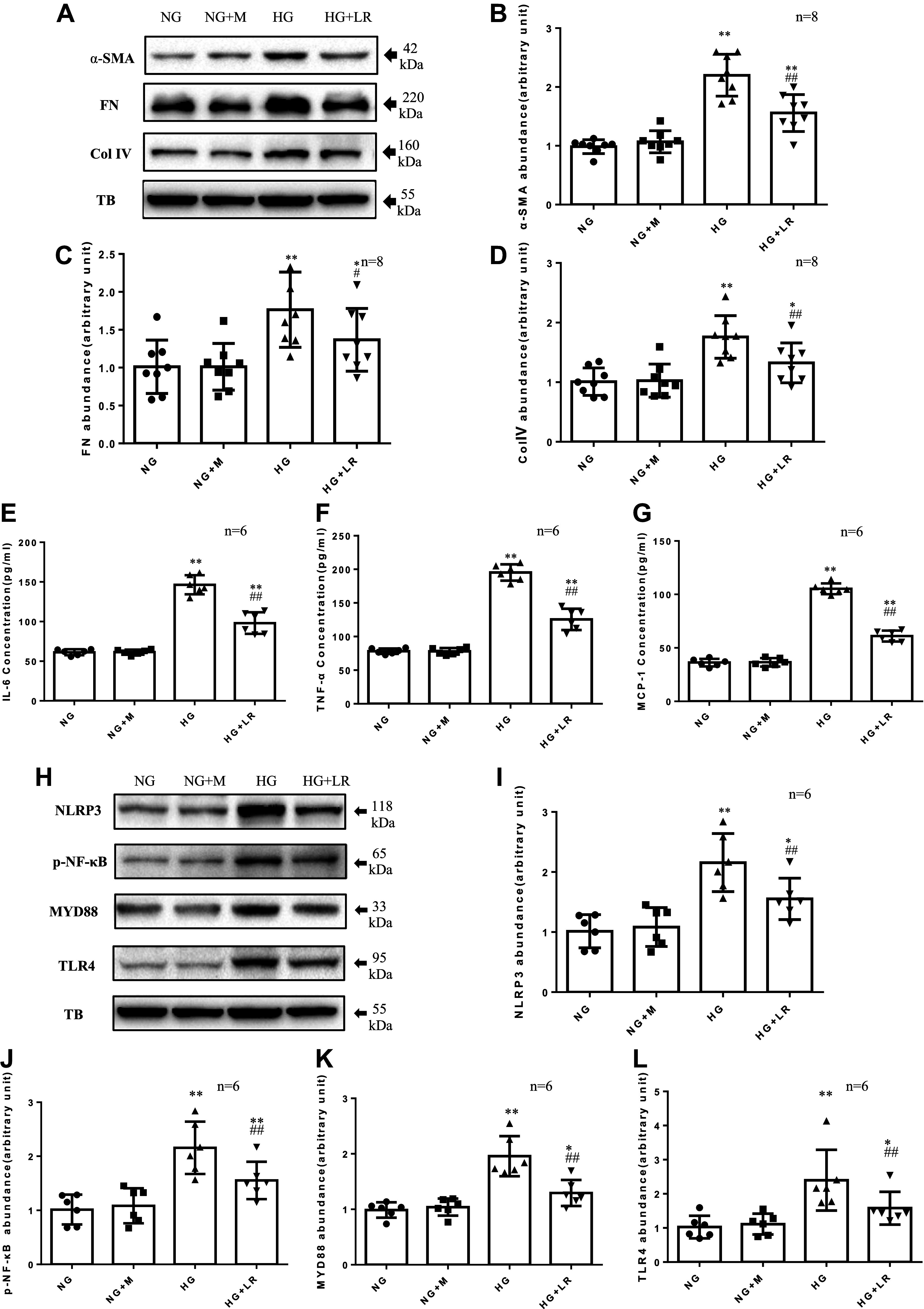
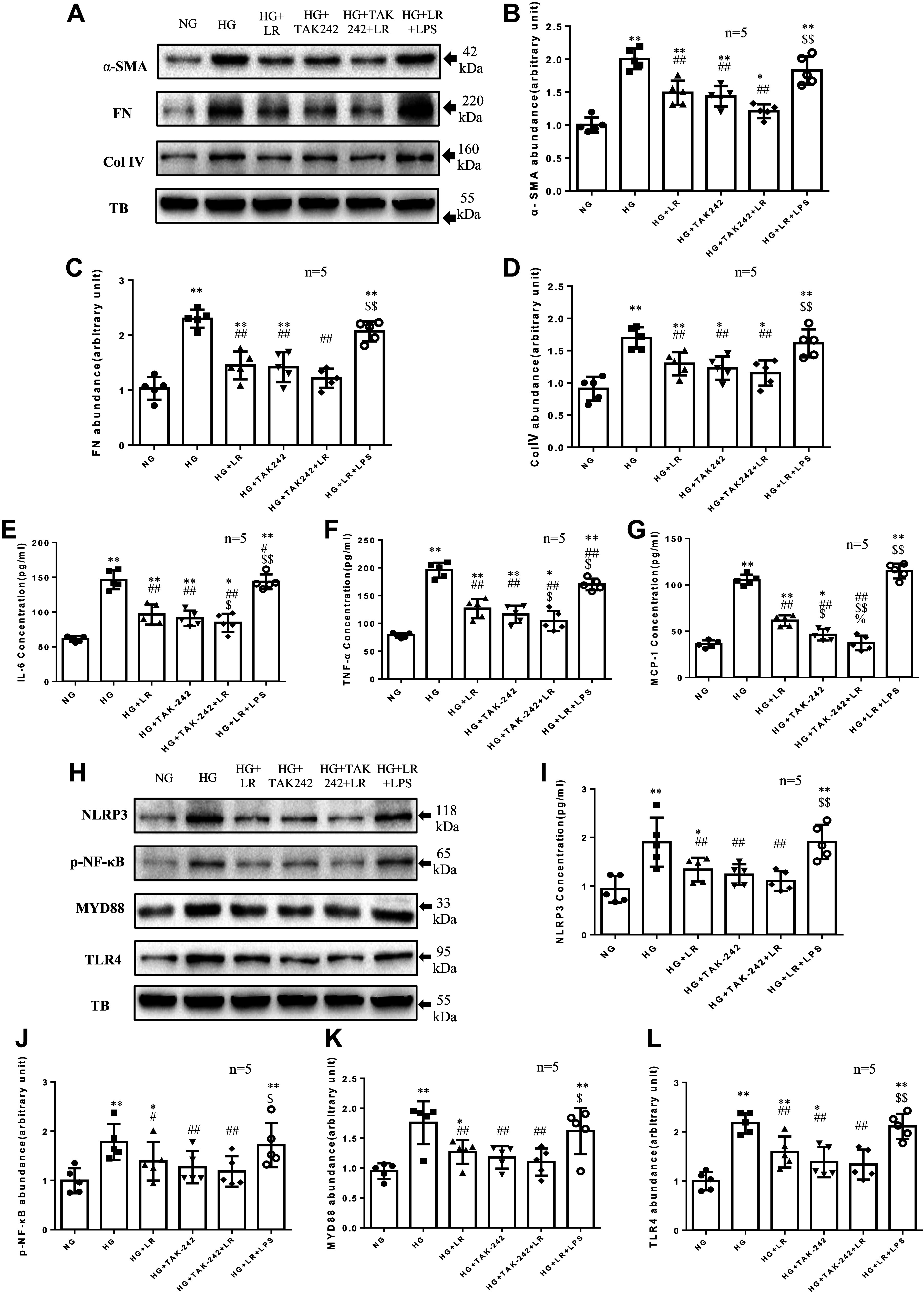
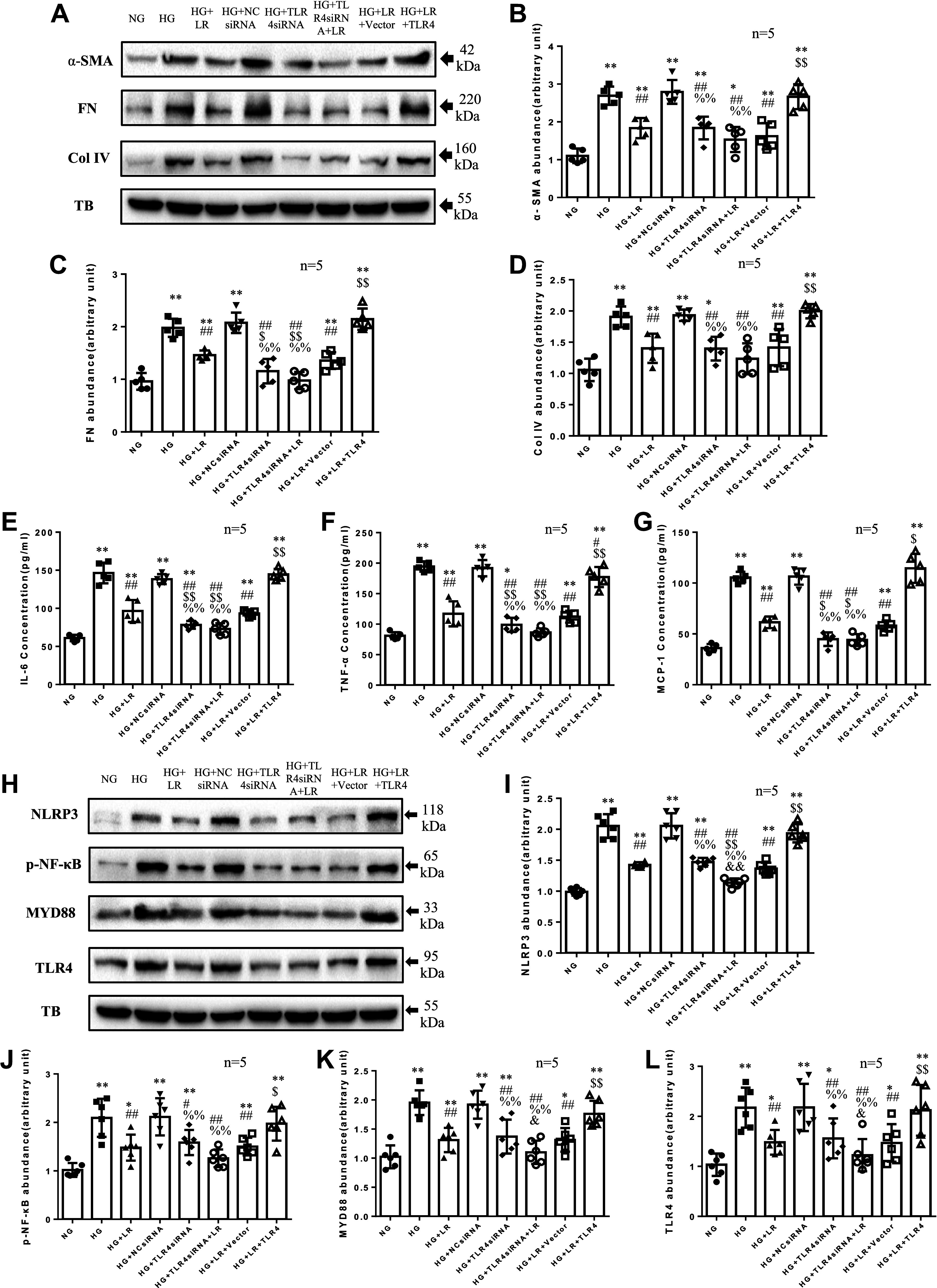
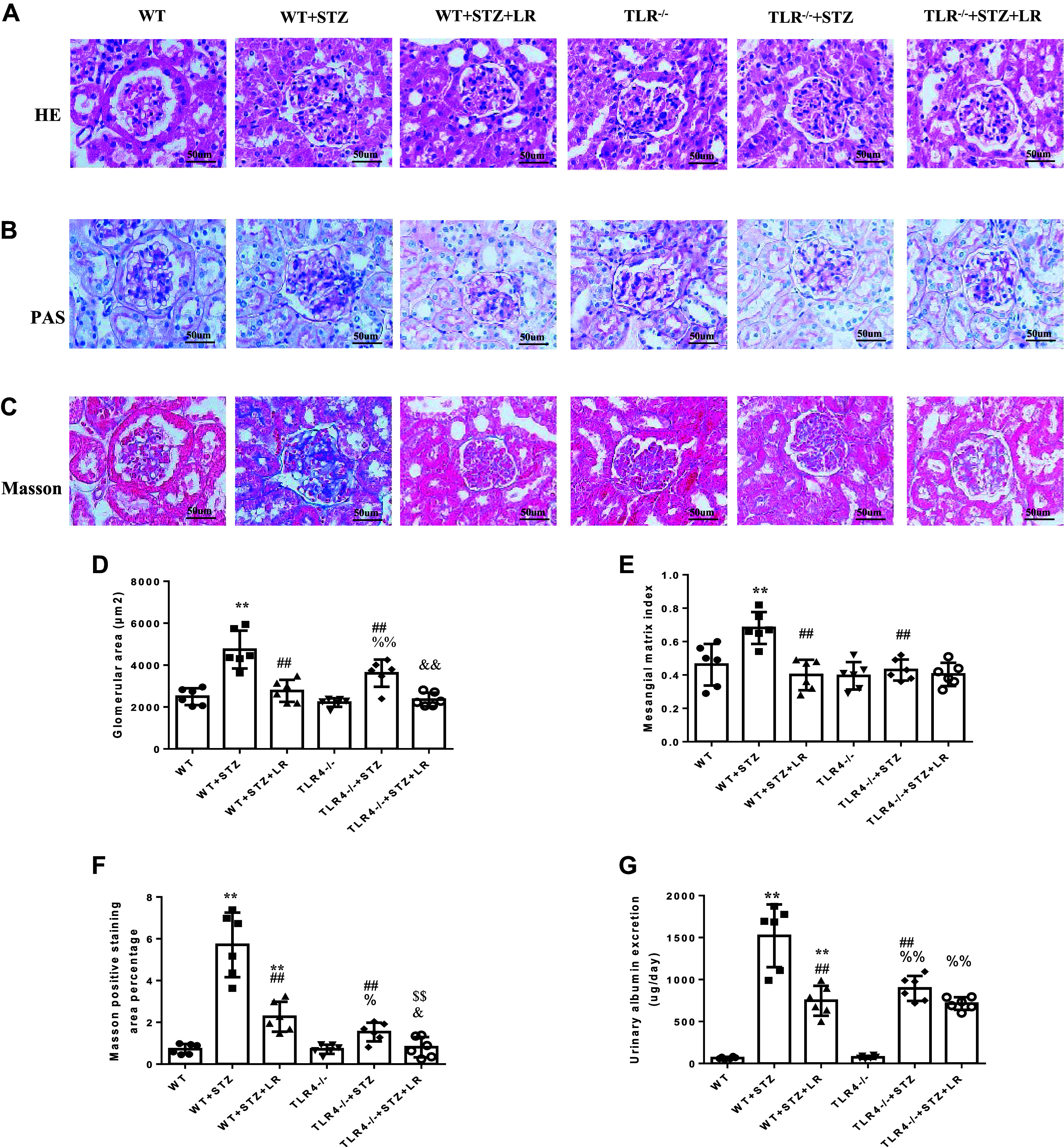
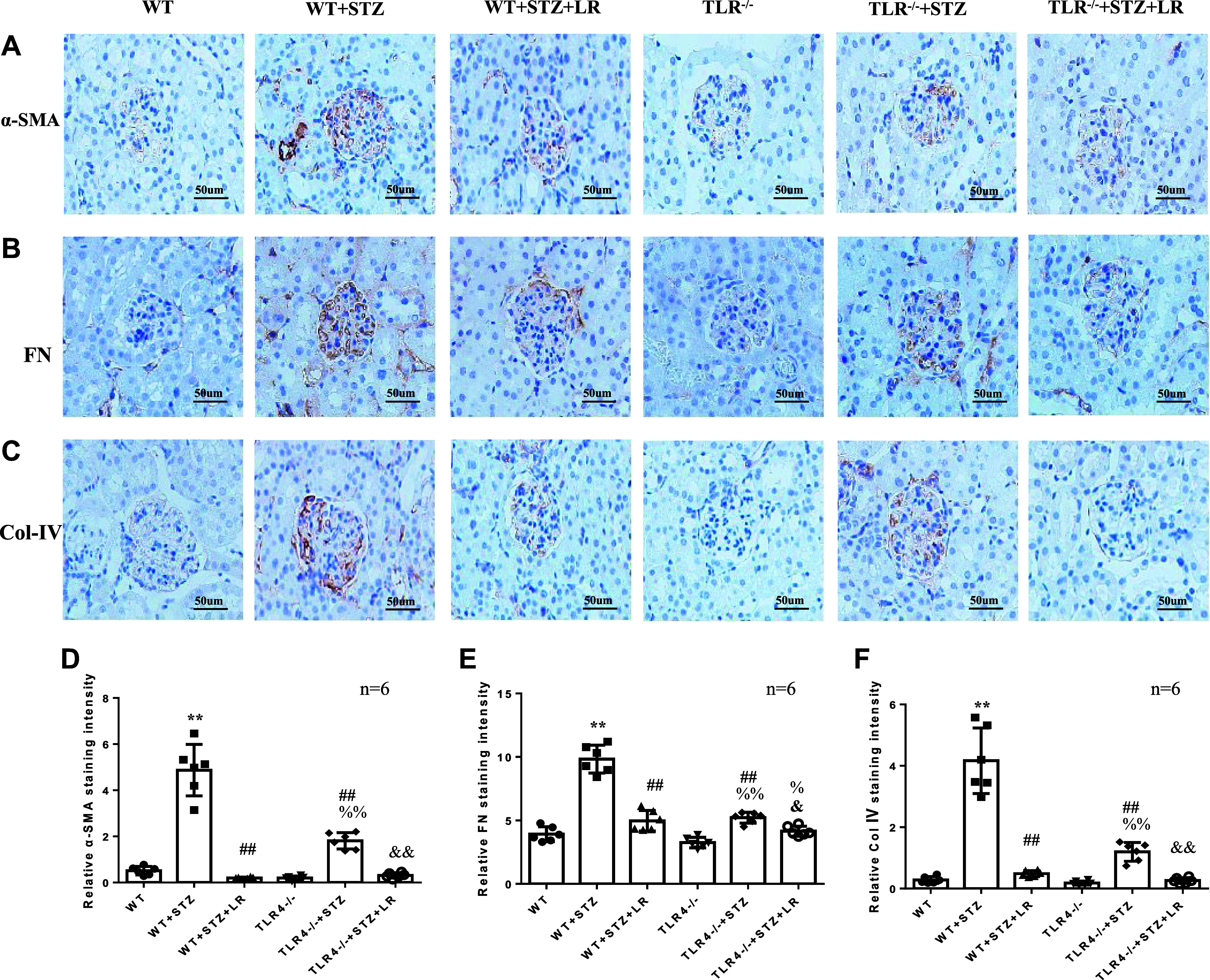
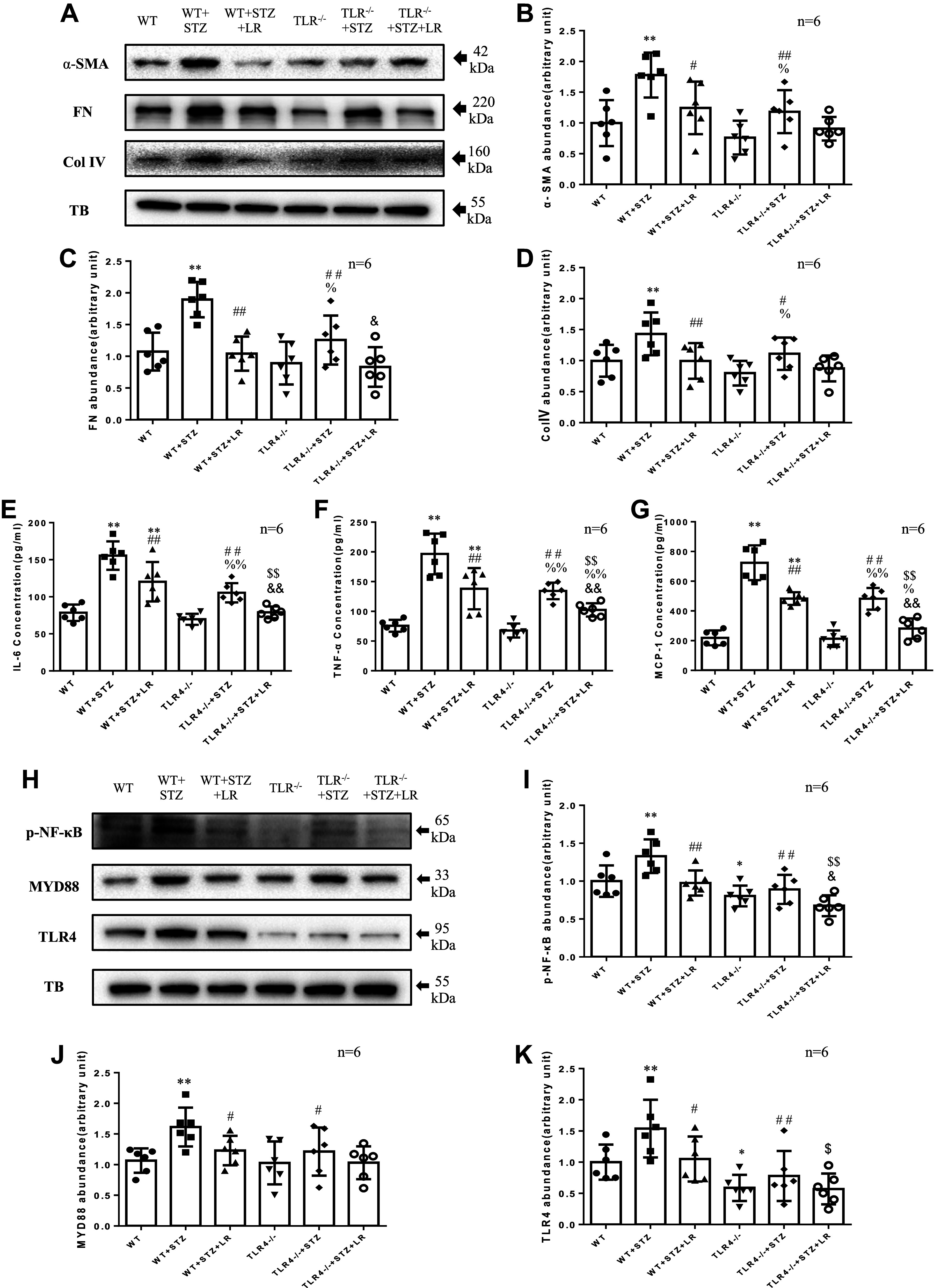
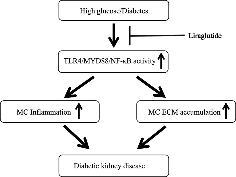
Inflammation and fibrosis play important roles in diabetic kidney disease (DKD). Previous studies have shown that glucagon-like peptide-1 receptor (GLP-1R) agonists had renal protective effects. However, the mechanisms are not clear. The present study explored the effect of liraglutide (LR), a GLP-1R agonist, on the downregulation of glomerular inflammation and fibrosis in DKD by regulating the Toll-like receptor (TLR)4/myeloid differentiation marker 88 (MyD88)/nuclear factor κB (NF-κB) signaling pathway in mesangial cells (MCs). In vitro, rat MCs were cultured in high glucose (HG). We found that liraglutide treatment significantly reduced the HG-mediated activation of the TLR4/MYD88/NF-κB signaling pathway, extracellular matrix (ECM)-related proteins, and inflammatory factors. A combination of TLR4 inhibitor (TAK242) and liraglutide did not synergistically inhibit inflammatory factors and ECM proteins. Furthermore, in the presence of TLR4 siRNA, liraglutide significantly blunted HG-induced expression of fibronectin protein and inflammatory factors. Importantly, TLR4 selective agonist LPS or TLR4 overexpression eliminated the improvement effects of liraglutide on the HG-induced response. In vivo, administration of liraglutide for 8 wk significantly improved the glomerular damage in streptozotocin-induced diabetic mice and reduced the expression of TLR4/MYD88/NF-κB signaling proteins, ECM protein, and inflammatory factors in renal cortex. TLR4-/- diabetic mice showed significant amelioration in urine protein excretion rate, glomerular pathological damage, inflammation, and fibrosis. Liraglutide attenuated glomerular hypertrophy, renal fibrosis, and inflammatory response in TLR4-/- diabetic mice. Taken together, our findings suggest that TLR4/MYD88/NF-κB signaling is involved in the regulation of inflammatory response and ECM protein proliferation in DKD. Liraglutide alleviates inflammation and fibrosis by downregulating the TLR4/MYD88/NF-κB signaling pathway in MCs.NEW & NOTEWORTHY Liraglutide, a glucagon-like peptide-1 receptor agonist (GLP-1RA), has renoprotective effect in diabetic kidney disease (DKD). In DKD, TLR4/MYD88/NF-κB signaling is involved in the regulation of inflammatory responses and extracellular matrix (ECM) protein proliferation. Liraglutide attenuates renal inflammation and overexpression of ECM proteins by inhibiting TLR4/MYD88/NF-κB signaling pathway. Therefore, we have identified a new mechanism that contributes to the renal protection of GLP-1RA, thus helping to design innovative treatment strategies for diabetic patients with various complications.

Keywords: TLR4/MyD88/NF-κB; diabetic kidney disease; extracellular matrix; inflammation; liraglutide.


Conflict of interest statement

No conflicts of interest, financial or otherwise, are declared by the authors.

Figures

Figure 1.
Figure 1.
Liraglutide reduced high glucose (HG)-induced increases of Toll-like receptor (TLR)4/myeloid differentiation marker 88 (MyD88)/nuclear factor κB (NF-κB) pathway, extracellular matrix (ECM), and inflammatory factors in rat glomerular mesangial cell lines (RMCs). RMCs were incubated in media containing 5.6 mM d-glucose (normal glucose, NG) or 5.6 mM d-glucose + 20 mM mannitol (NG + M) or 25 mM d-glucose (HG) with or without 100 nM liraglutide (LR). The protein levels of α-smooth muscle actin (α-SMA), fibronectin (FN), collagen type IV (Col IV), NOD-like receptor family pyrin domain containing 3 (NLRP3), NF-κB, MyD88, and TLR4 were measured by Western blot (AD, HL). The protein levels of interleukin-6 (IL-6), tumor necrosis factor-α (TNF-α), and monocyte chemotactic protein-1 (MCP-1) were measured by ELISA (EG). TB, α-tubulin. *P < 0.05, **P < 0.01 compared with both NG and NG + M groups. #P < 0.05, ##P < 0.01 vs. HG group. n, no. of independent experiments.
Figure 2.
Figure 2.
Toll-like receptor (TLR)4 agonist and inhibitor influence the effects of liraglutide (LR) on high glucose (HG)-induced extracellular matrix (ECM) protein abundance and inflammatory factors production in rat glomerular mesangial cell lines (RMCs). RMCs were incubated in media containing 5.5 mM glucose (normal glucose, NG) or HG in the absence or presence of 100 nM liraglutide (LR) or TAK242 (1 µM) or lipopolysaccharides (LPS, 1 µg/mL). The protein levels of α-smooth muscle actin (α-SMA), fibronectin (FN), collagen type IV (Col IV), NOD-like receptor family pyrin domain containing 3 (NLRP3), nuclear factor κB (NF-κB), myeloid differentiation marker 88 (MyD88), and TLR4 were measured by Western blot (AD, HL). The protein levels of interleukin-6 (IL-6), tumor necrosis factor-α (TNF-α), and monocyte chemotactic protein-1 (MCP-1) were measured by ELISA (EG). TB, α-tubulin. *P < 0.05, **P < 0.01 compared with NG groups. #P < 0.05, ##P < 0.01 vs. HG group. $P < 0.05, $$P < 0.01 vs. HG + LR group. %P < 0.05. n, no. of independent experiments.
Figure 3.
Figure 3.
Effects of liraglutide (LR) with or without Toll-like receptor (TLR)4 silence or overexpression on high glucose (HG)-induced extracellular matrix (ECM) protein abundance and inflammatory factors production in rat glomerular mesangial cell lines (RMCs). RMCs were incubated in media containing 5.5 mM glucose (normal glucose, NG) or HG in the absence or presence of 100 nM liraglutide (LR), without transfection or transfected with normal control siRNA (NCsiRNA) or TLR4 siRNA or empty pcDNA3.1 vector (Vector) or TLR4 expression plasmid pcDNA3.1-TLR4 (TLR4). The protein levels of α-smooth muscle actin (α-SMA), fibronectin (FN), collagen type IV (Col IV), NOD-like receptor family pyrin domain containing 3 (NLRP3), nuclear factor κB (NF-κB), myeloid differentiation marker 88 (MyD88), and TLR4 were measured by Western blot (AD, HL). The protein levels of interleukin-6 (IL-6), tumor necrosis factor-α (TNF-α), and monocyte chemotactic protein-1 (MCP-1) were measured by ELISA (EG). TB, α-tubulin. *P < 0.05, **P < 0.01 compared with NG groups. #P < 0.05, ##P< 0.01 vs. HG group. $P < 0.05, $$P< 0.01 vs. HG + LR and HG + LR + Vector group. %%P < 0.01 vs. HG + NCsiRNA group. &P < 0.05, &&P < 0.01 vs. HG + TLR4siRNA group. n, no. of independent experiments.
Figure 4.
Figure 4.
Effects of liraglutide treatment on urinary protein excretion, glomerular histopathological changes, and fibrosis in Toll-like receptor (TLR)4−/− diabetes mellitus (DM) mice. AC: representative photomicrographs of hematoxylin and eosin (H&E) staining (A), periodic acid-Schiff (PAS) staining (B), and Masson trichrome staining (C) of renal cortex sections from six groups of mice. DF: quantitative analysis of H&E staining (D), PAS staining (E), and Masson trichrome staining (F). G: urinary albumin creatinine. LR, liraglutide; TLR4−/−, TLR4−/− mice; WT, wild-type mice; WT + STZ, streptozotocin-treated WT mice. **P < 0.01 compared with WT groups. ##P< 0.01 vs. WT + STZ group. $$P < 0.01 vs. WT + STZ + LR group. %%P < 0.01 vs. TLR4−/− group. &P < 0.05, &&P < 0.01 vs. TLR4−/− + STZ group. Magnification: ×400.
Figure 5.
Figure 5.
Effects of liraglutide (LR) treatment on glomerular extracellular matrix (ECM) in Toll-like receptor (TLR)4−/− diabetes mellitus (DM) mice by immunohistochemistry. AC: representative immunohistochemical staining images of α-smooth muscle actin (α-SMA, A), fibronectin (FN, B), and collagen type IV (Col IV, C) in glomeruli from six groups of mice. DF: semiquantitative analysis of staining intensity of α-SMA (D), FN (E), and Col IV (F). WT, wild-type mice; WT + STZ, streptozotocin-treated WT mice. **P < 0.01 compared with WT groups. ##P< 0.01 vs. WT+STZ group. %P < 0.05, %%P < 0.01 vs. TLR4−/− group. &P < 0.05, &&P < 0.01 vs. TLR4−/− + STZ group. Magnification: ×400. n, no. of mice in each group.
Figure 6.
Figure 6.
Effects of liraglutide (LR) treatment on glomerular Toll-like receptor (TLR)4/myeloid differentiation marker 88 (MyD88)/nuclear factor κB (NF-κB) signaling pathway, extracellular matrix (ECM) accumulation, and inflammatory factors in TLR4−/− diabetes mellitus (DM) mice. The protein levels of α-smooth muscle actin (α-SMA), fibronectin (FN), collagen type IV (Col IV), NF-κB, MYD88, and TLR4 were measured by Western blot (AD, HK). The protein levels of interleukin-6 (IL-6), tumor necrosis factor-α (TNF-α), and monocyte chemotactic protein-1 (MCP-1) were measured by ELISA (EG). TB, α-tubulin. *P < 0.05, **P < 0.01 compared with wild-type (WT) groups. #P < 0.05, ##P < 0.01 vs. WT + streptozotocin (STZ) group. $P < 0.05, $$P < 0.01 vs. WT + STZ + LR group. %P < 0.05, %%P < 0.01 vs. TLR4−/− group. &P < 0.05, &&P < 0.01 vs. TLR4−/− + STZ group. Magnification: ×400. n, no. of mice in each group.
Figure 7.
Figure 7.
Illustration of liraglutide on inflammation and extracellular matrix (ECM) through the Toll-like receptor (TLR)4 signaling pathway. Schematic illustration of the mechanistic pathway mediating the liraglutide effect on mesangial cells (MCs) in diabetic kidney disease. High glucose or diabetes activates TLR4/myeloid differentiation marker 88 (MyD88)/nuclear factor κB (NF-κB) signaling pathway in MCs, resulting in extracellular matrix (ECM) protein production in diabetic kidney disease. Liraglutide downregulates TLR4/MyD88/NF-κB signaling, thus blunts high glucose-induced ECM protein accumulation and ameliorates diabetic kidney disease.

Similar articles

Cited by

References

    1. Jung CY, Yoo TH. Pathophysiologic mechanisms and potential biomarkers in diabetic kidney disease. Diabetes Metab J 46: 181–197, 2022. doi:10.4093/dmj.2021.0329. - DOI - PMC - PubMed
    1. Sugahara M, Pak WLW, Tanaka T, Tang SCW, Nangaku M. Update on diagnosis, pathophysiology, and management of diabetic kidney disease. Nephrology (Carlton) 26: 491–500, 2021. doi:10.1111/nep.13860. - DOI - PubMed
    1. Jung SW, Moon JY. The role of inflammation in diabetic kidney disease. Korean J Intern Med 36: 753–766, 2021. doi:10.3904/kjim.2021.174. - DOI - PMC - PubMed
    1. Liu C, Yang M, Li L, Luo S, Yang J, Li C, Liu H, Sun L. A glimpse of inflammation and anti-inflammation therapy in diabetic kidney disease. Front Physiol 13: 909569, 2022. doi:10.3389/fphys.2022.909569. - DOI - PMC - PubMed
    1. Klen J, Dolžan V. Glucagon-like peptide-1 receptor agonists in the management of type 2 diabetes mellitus and obesity: the impact of pharmacological properties and genetic factors. Int J Mol Sci 23: 3451, 2022. doi:10.3390/ijms23073451. - DOI - PMC - PubMed

MeSH terms

LinkOut - more resources