. 2024 Aug 20;121(34):e2315006121.
doi: 10.1073/pnas.2315006121. Epub 2024 Aug 12.

Residues 2 to 7 of α-synuclein regulate amyloid formation via lipid-dependent and lipid-independent pathways

Affiliations

Residues 2 to 7 of α-synuclein regulate amyloid formation via lipid-dependent and lipid-independent pathways

Katherine M Dewison et al. Proc Natl Acad Sci U S A. .

Abstract

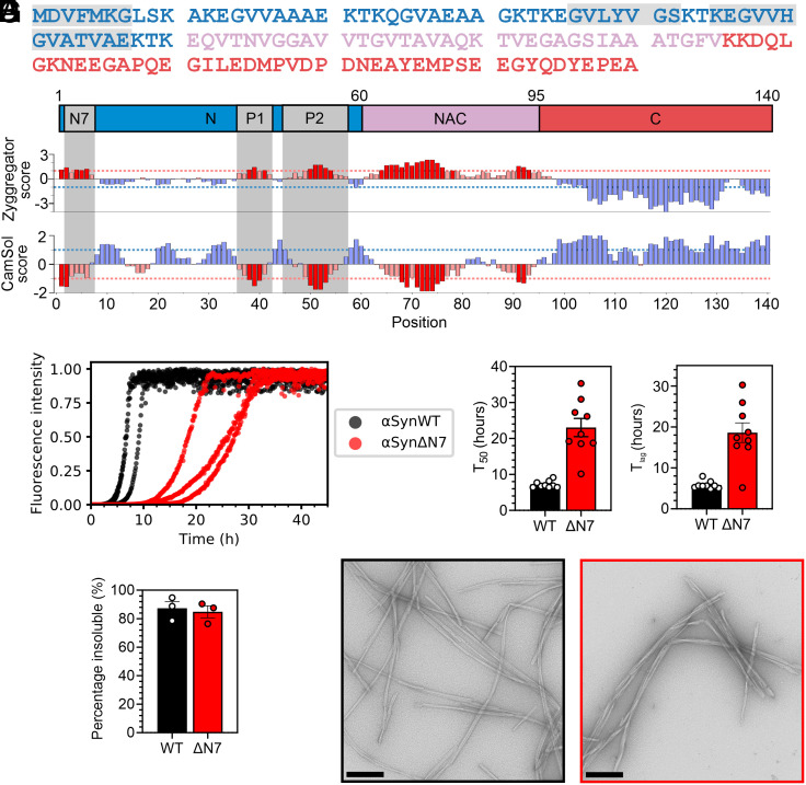
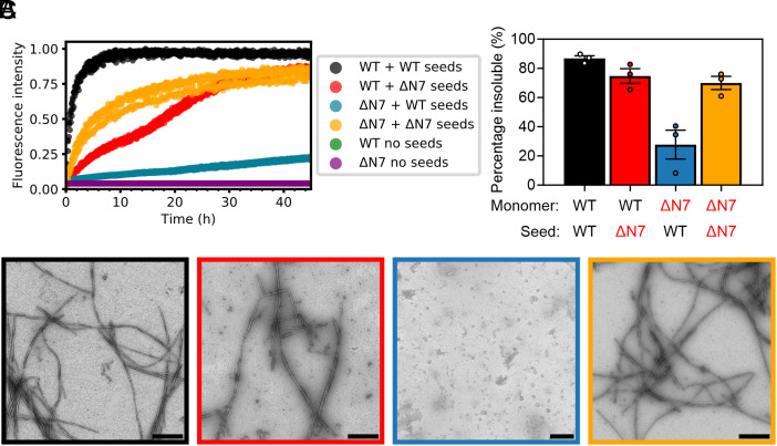
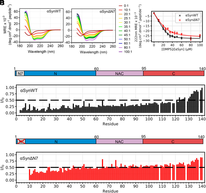
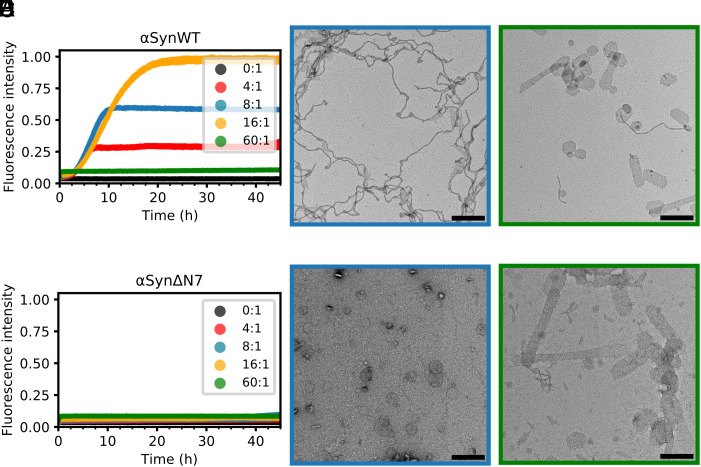
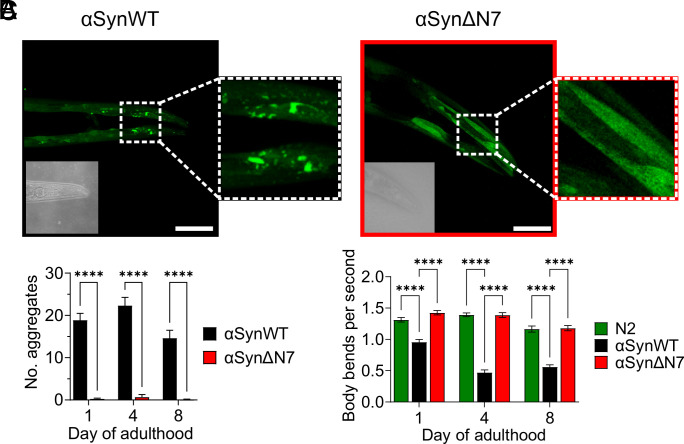
Amyloid formation by α-synuclein (αSyn) occurs in Parkinson's disease, multiple system atrophy, and dementia with Lewy bodies. Deciphering the residues that regulate αSyn amyloid fibril formation will not only provide mechanistic insight but may also reveal targets to prevent and treat disease. Previous investigations have identified several regions of αSyn to be important in the regulation of amyloid formation, including the non-amyloid-β component (NAC), P1 region (residues 36 to 42), and residues in the C-terminal domain. Recent studies have also indicated the importance of the N-terminal region of αSyn for both its physiological and pathological roles. Here, the role of residues 2 to 7 in the N-terminal region of αSyn is investigated in terms of their ability to regulate amyloid fibril formation in vitro and in vivo. Deletion of these residues (αSynΔN7) slows the rate of fibril formation in vitro and reduces the capacity of the protein to be recruited by wild-type (αSynWT) fibril seeds, despite cryo-EM showing a fibril structure consistent with those of full-length αSyn. Strikingly, fibril formation of αSynΔN7 is not induced by liposomes, despite the protein binding to liposomes with similar affinity to αSynWT. A Caenorhabditis elegans model also showed that αSynΔN7::YFP forms few puncta and lacks motility and lifespan defects typified by expression of αSynWT::YFP. Together, the results demonstrate the involvement of residues 2 to 7 of αSyn in amyloid formation, revealing a target for the design of amyloid inhibitors that may leave the functional role of the protein in membrane binding unperturbed.

Keywords: amyloid; liposome; membrane; synuclein.


Conflict of interest statement

Competing interests statement:The authors declare no competing interest.

Figures

Fig. 1.
Fig. 1.
Deletion of residues 2 to 7 of αSyn slows amyloid formation in vitro. (A) Amino acid sequence of αSynWT. Blue = N-terminal region; pink = NAC; and red = C-terminal region. (B) Zyggregator (24) and CamSol (25) profiles for αSyn. Red bars indicate predicted aggregation-prone and low-solubility regions, respectively. For A and B, the N7 (2DVFMKG7), P1 (36GVLYVGS42), and P2 (45KEGVVHGVATVAE57) regions are in gray. (C) Fibril formation kinetics of αSynWT (black) and αSynΔN7 (red). Data are normalized to maximum signal of each curve. (D) T50 and (E) Tlag values for nine replicates. (F) Quantification of the insoluble fraction at the end point of ThT assays. (G) Negative-stain electron micrographs of the ThT end points for αSynWT (black) and αSynΔN7 (red). (Scale bar, 250 nm.)
Fig. 2.
Fig. 2.
Residues 2 to 7 of αSyn are required to elongate αSynWT fibril seeds. (A) Seeded fibril growth for self- and cross-seeding of αSynWT and αSynΔN7 monomers with preformed fibril seeds [10% (v/v)]. Data are normalized to the maximum fluorescence of the dataset. Note that for “WT no seeds” (green) and “ΔN7 no seeds” (purple), there is no increase in ThT fluorescence signal, so the data cannot be seen readily behind each other. (B) Quantification of the insoluble fraction at the end point of ThT assays. (C) Negative-stain electron micrographs of the ThT end points, colored as in B. (Scale bar, 250 nm.)
Fig. 3.
Fig. 3.
Residues 2 to 7 of αSyn are not necessary for DMPS liposome binding. (A and B) Representative far-UV CD spectra of αSynWT and αSynΔN7 as a function of the lipid-to-protein ratio (LPR) (see key). (C) MRE at 222 nm as a function of LPR for αSynWT and αSynΔN7. Curves were fitted using equation 6 from ref. . Error bars are SEM. (D and E) Per-residue intensity ratios of 1H-15N HMQC NMR resonances for (D) αSynWT and (E) αSynΔN7. Spectra were collected at 30 °C at an LPR of 8:1. The dashed line indicates an I/I0 of 0.5.
Fig. 4.
Fig. 4.
Residues 2 to 7 of αSyn are critical for lipid-mediated fibrillation. (A) Fibril formation kinetics for αSynWT in the presence of DMPS liposomes. Key indicates [DMPS]:[αSyn] ratio. Data are normalized to the maximum fluorescence intensity of the dataset. (B and C) Negative-stain electron micrograph of the ThT assay end point for [DMPS]:[αSynWT] of 8:1 and 60:1, respectively. (Scale bar, 250 nm.) (DF), as (AC), but for αSynΔN7.
Fig. 5.
Fig. 5.
C. elegans expressing αSynΔN7::YFP exhibit fewer aggregates and motility defects than those expressing αSynWT::YFP. (A) Representative confocal images of C. elegans expressing αSynWT::YFP and αSynΔN7::YFP at day 8 of adulthood. (Scale bar, 50 μM.) Corresponding brightfield images are displayed in the Bottom Left of each image. (B) Quantification of puncta in the head region of C. elegans expressing αSynWT::YFP or αSynΔN7::YFP. (C) Quantification of the motility of N2, αSynWT::YFP, and αSynΔN7::YFP animals in terms of body bends per second. ****P < 0.0001.

References

    1. Koga S., Sekiya H., Kondru N., Ross O. A., Dickson D. W., Neuropathology and molecular diagnosis of Synucleinopathies. Mol. Neurodegener. 16, 83 (2021). - PMC - PubMed
    1. Krüger R., et al. , AlaSOPro mutation in the gene encoding α-synuclein in Parkinson’s disease. Nat. Genet. 18, 106–108 (1998). - PubMed
    1. Zarranz J. J., et al. , The new mutation, E46K, of α-synuclein causes parkinson and Lewy body dementia. Ann. Neurol. 55, 164–173 (2004). - PubMed
    1. Proukakis C., et al. , A novel α-synuclein missense mutation in Parkinson disease. Neurology 80, 1062–1064 (2013). - PMC - PubMed
    1. Kiely A. P., et al. , α-Synucleinopathy associated with G51D SNCA mutation: A link between Parkinson’s disease and multiple system atrophy? Acta Neuropathol. 125, 753–769 (2013). - PMC - PubMed

LinkOut - more resources