. 2024 Aug 20;121(34):e2315009121.
doi: 10.1073/pnas.2315009121. Epub 2024 Aug 12.

Insights into the interaction between UGGT, the gatekeeper of folding in the ER, and its partner, the selenoprotein SEP15

Affiliations

Insights into the interaction between UGGT, the gatekeeper of folding in the ER, and its partner, the selenoprotein SEP15

Robert V Williams et al. Proc Natl Acad Sci U S A. .

Abstract

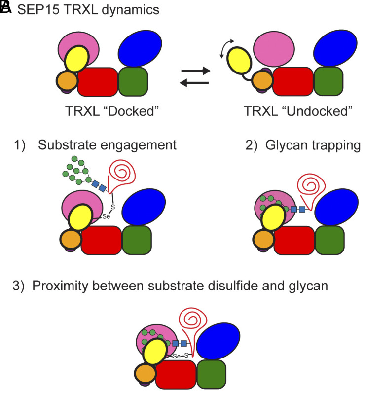
The enzyme UDP-glucose: glycoprotein glucosyltransferase (UGGT) is the gatekeeper of protein folding within the endoplasmic reticulum (ER). One-third of the human proteome traverses the ER where folding and maturation are facilitated by a complex protein homeostasis network. Both glycan modifications and disulfide bonds are of key importance in the maturation of these ER proteins. The actions of UGGT are intimately linked to the glycan code for folding and maturation of secretory proteins in the ER. UGGT selectively glucosylates the N-linked glycan of misfolded proteins so that they can reenter the lectin-folding chaperone cycle and be retained within the ER for further attempts at folding. An intriguing aspect of UGGT function is its interaction with its poorly understood cochaperone, the 15 kDa selenoprotein known as SELENOF or SEP15. This small protein contains a rare selenocysteine residue proposed to act as an oxidoreductase toward UGGT substrates. AlphaFold2 predictions of the UGGT1/SEP15 complex provide insight into this complex at a structural level. The predicted UGGT1/SEP15 interaction interface was validated by mutagenesis and coimmunoprecipitation experiments. These results serve as a springboard for models of the integrated action of UGGT1 and SEP15.

Keywords: SEP15; UGGT; endoplasmic reticulum; protein folding.


Conflict of interest statement

Competing interests statement:The authors declare no competing interest.

Figures

Fig. 1.
Fig. 1.
Glycoprotein maturation is overseen by a dedicated quality control system in the ER. 1) Oligosaccharyltransferase A (OST-A) scans a nascent polypeptide and glycosylates asparagine residues within N-X-S/T/C amino acid motifs. 2) The two terminal glucose residues of the glycan moiety are rapidly trimmed by glucosidases I and II (GlsI and GlsII). 3) The resulting monoglucosylated glycan serves as a binding site for the lectin chaperones, calnexin (CNX) and calreticulin (CRT), which promote protein folding. 4) Upon removal of the final glucose residue by GlsII the client is released. 5) If the released glycoprotein has not achieved its native fold, it is bound by UGGT, which senses the folding status of the glycoprotein and selectively reglucosylates the N-glycan. 6) The glycoprotein released from the lectin chaperones CNX/CRT and cochaperones, either in the first time through the cycle or after reentering the cycle if reglucosylated by UGGT, may achieve its native fold and proceed to exit the ER for export via ER exit sites (7). A significant fraction of the UGGT in the ER is in complex with the SEP15, but the function of this selenoprotein is unknown.
Fig. 2.
Fig. 2.
AlphaFold2 predicts a potential SEP15-binding domain in human UGGT1. (A) Cartoon of the human UGGT1 sequence. UGGT1 has a complex topology of eight domains: four thioredoxin-like domains (TRXL1 to 4), two β-sheet domains (βS1 and βS2), a glucosyltransferase domain (GT), and a domain absent in the fungal UGGTs (*). Note that TRXL4 and βS1 are composed of noncontiguous regions of the primary sequence. (B) AlphaFold2 prediction of the structure of human UGGT1. Domains are colored as in panel (A). The helixloophelix domain absent in previous crystal structures of UGGTs from thermophilic organisms lacking SEP15, which we propose to be the SEP15-binding region (SBR), is indicated by a dashed box. (C) A closer view of the SBR from (B), shown as a molecular lipophilicity potential surface created using ChimeraX (23). A hydrophobic patch (gold) in this region is a likely candidate for interacting with SEP15. (D) A multiple sequence alignment of eukaryotic UGGTs shows significant differences. The UGGTs from organisms that lack an SEP15 homolog have gaps in the region corresponding to the helixloophelix predicted in human UGGT1. Moreover, the region identified as a SEP15 binding region is shown by the purple box. (Note: For those organisms with multiple UGGT paralogs, only the UGGT1 sequence is shown). The α-helices observed in the predicted structure are indicated above the sequences.
Fig. 3.
Fig. 3.
AlphaFold2 prediction of SEP15 and the UGGT1/SEP15 complex. (A) SEP15 is composed of two domains: a cysteine-rich domain (CRD) and a thioredoxin-like domain (TRXL). The single selenocysteine (Sec, U) residue is found within the TRXL domain. The position of all Cys residues is indicated in black, and the Sec residue is indicated in bright green. The predicted disulfide pairings are shown. AlphaFold2 predicts a selenyl-sulfide pairing with the TRXL domain, but this reactive motif likely interconverts with a reduced form (indicated by an asterisk). (B) AlphaFold2 predicts a two-domain structure for SEP15. The CRD (gold) forms a bundle of three alpha helices, while the TRXL domain (yellow) adopts a mixed α/β structure. U65 is exposed to solvent. (C) The helices of the CRD are held together by three disulfide bonds: C10-C42, C21-C43, and C24-C39. (D) The predicted structure creates a hydrophobic patch on part of the CRD (gold). Hydrophilic residues are shown in cyan. (E) The AlphaFold-multimer prediction of the UGGT1/SEP15 complex shows SEP15 interacting with UGGT1 via both domains and contacting UGGT1 SBR and TRXL4. The reactive U65 residue is proximal to the catalytic pocket of the GT domain.
Fig. 4.
Fig. 4.
AlphaFold-predicted point mutants disrupt UGGT1/SEP15 interaction. (A) SEP15, WT UGGT1, and UGGT1 mutants were exogenously expressed for 24 h into HEK293 cells. SEP15 contains an HA tag, whereas UGGT1 and the point mutants possess a 3xFLAG. Both tags are located on the C terminus of the protein. Cells were transfected and the following day were lysed to isolate their soluble cellular fractions. The resulting lysate was split between a whole cell lysate (WCL) fraction to identify the total amount of protein and a FLAG immunoprecipitation (IP) to isolate either free or SEP15-bound UGGT1. The resulting WCL and FLAG IPs were resolved using a 9% and 14% SDS-PAGE gel to probe for UGGT1 and SEP15, respectively, by western blot. UGGT1 was detected using an endogenous polyclonal antibody raised toward UGGT1, while SEP15 was detected using an HA polyclonal antibody. (B) Quantification of the percentage of SEP15 bound to UGGT1. The percent SEP15 bound to UGGT1 was determined by quantifying and normalizing the amount of SEP15 that co-IPs with UGGT1 (lanes 8, 10, and 12) and dividing it by the normalized amount of total SEP15 (lanes 7, 9, and 11) and multiplying by 100. *** indicates P-value < 0.001.
Fig. 5.
Fig. 5.
Levels of UGGT glucosylation are altered in the absence of SELENOF. UGGT-modified substrates were isolated from SELENOF+/+/ALG6−/− and SELENOF−/−/ALG6−/− cells, and the changes in glucosylation were analyzed. Each point represents the change in glucosylation (log2 fold change) between the SELENOF−/− divided by the SELENOF+/+ cells in the ALG6−/− background and plotted against the −log10 P-value. The dotted line indicates a 50% increase or decrease in levels of UGGT-mediated glucosylation. The large gray circles denote substrates in the SELENOF−/− that had a significant change in glucosylation (P-value < 0.05), compared to the parental cell line, but did not have fold change resulting in a 50% increase or decrease. Red circles indicate substrates with a fold change resulting in a 50% increase or decrease in glucosylation. Data are a result of three independent biological replicates. See SI Appendix, Table S2 for details on the labeled substrates.
Fig. 6.
Fig. 6.
Hypotheses for integrated UGGT1 and SEP15 action toward glycoprotein substrates. (A) Within UGGT1/SEP15 complexes, SEP15 may exist in a conformational equilibrium between two states. In the “docked” state, SEP15 TRXL (yellow) binds to UGGT1 TRXL4 (red), while in the “undocked” state, SEP15 TRXL is unbound. The SEP15 CRD (gold) maintains the interaction with UGGT1 in both cases. This conformational equilibrium can be exploited in the mechanism of action on substrates. (B) Possible modes of action for SEP15 within a UGGT1/SEP15 complex are proposed. Diagram 1) illustrates a model in which SEP15 may assist UGGT1 in engaging substrates via formation of a mixed selenyl-sulfide. Diagram 2) shows a model in which the SEP15 TRXL may trap a substrate glycan within the catalytic site of the UGGT GT domain. And in diagram 3), action of SEP15 may be most important for substrates in which an N-glycan and a disulfide bond occur at particular distances from one another, either through space or through sequence, taking advantage of the proximity of the SEP15 Sec residue and the catalytic site of UGGT GT (pink). Additionally, the TRXL2 (blue) and TRXL3 (green) domains are shown.

References

    1. Kanapin A., et al. , Mouse proteome analysis. Genome Res. 13, 1335–1344 (2003). - PMC - PubMed
    1. Apweiler R., Hermjakob H., Sharon N., On the frequency of protein glycosylation, as deduced from analysis of the SWISS-PROT database. Biochim. et Biophys. Acta Gen. Subj. 1473, 4–8 (1999). - PubMed
    1. Hammond C., Braakman I., Helenius A., Role of N-linked oligosaccharide recognition, glucose trimming, and calnexin in glycoprotein folding and quality control. Proc. Natl. Acad. Sci. U.S.A. 91, 913–917 (1994). - PMC - PubMed
    1. Adams B. M., Oster M. E., Hebert D. N., Protein quality control in the endoplasmic reticulum. Protein J. 38, 317–329 (2019). - PMC - PubMed
    1. Zielinska D. F., Gnad F., Wiśniewski J. R., Mann M., Precision mapping of an in vivo N-glycoproteome reveals rigid topological and sequence constraints. Cell 141, 897–907 (2010). - PubMed

LinkOut - more resources