. 2024 Nov 26;8(22):5814-5825.
doi: 10.1182/bloodadvances.2024013175.

Platelet Ido1 expression is induced during Plasmodium yoelii infection, altering plasma tryptophan metabolites

Affiliations

Platelet Ido1 expression is induced during Plasmodium yoelii infection, altering plasma tryptophan metabolites

Sara K Blick-Nitko et al. Blood Adv. .

Abstract

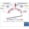
Platelets are immune responsive in many diseases as noted by changes in platelet messenger RNA in conditions such as sepsis, atherosclerosis, COVID-19, and many other inflammatory and infectious etiologies. The malaria causing Plasmodium parasite is a persistent public health threat and significant evidence shows that platelets participate in host responses to infection. Using a mouse model of nonlethal/uncomplicated malaria, non-lethal Plasmodium yoelii strain XNL (PyNL)-infected but not control mouse platelets expressed Ido1, a rate limiting enzyme in tryptophan metabolism that increases kynurenine at the expense of serotonin. Interferon-γ (IFN-γ) is a potent inducer of Ido1 and mice treated with recombinant IFN-γ had increased platelet Ido1 and IDO1 activity. PyNL-infected mice treated with anti-IFN-γ antibody had similar platelet Ido1 and metabolic profiles to that of uninfected controls. PyNL-infected mice become thrombocytopenic by day 7 after infection and transfusion of platelets from IFN-γ-treated wild-type mice but not Ido1-/- mice increased the plasma kynurenine-to-tryptophan ratio, indicating that platelets are a source of postinfection IDO1 activity. We generated platelet-specific Ido1 knockout mice to assess the contribution of platelet Ido1 during PyNL infection. Platelet-specific Ido1-/- mice had increased death and evidence of lung thrombi, which were not present in infected wild-type mice. Platelet Ido1 may be a significant contributor to plasma kynurenine in IFN-γ-driven immune processes and the loss of platelets may limit total Ido1, leading to immune and vascular dysfunction.


Conflict of interest statement

Conflict-of-interest disclosure: The authors declare no competing financial interests.

Figures
None
Graphical abstract
Figure 1.
Figure 1.
Plasmodium infection increased platelet Ido1. (A) Heat map of RNA-seq for IDO1 expression in platelets isolated from patients infected with P vivax and healthy controls. Individual bars represent samples derived from 7 controls and 7 patients with P vivax infection. (B) Percentage of SYBR+ parasitized RBCs measured at multiple time points using flow cytometry. (n = 5, ∗P < .05). (C) Platelet counts were obtained on multiple days after infection and expressed as fold change vs uninfected controls (n = 5). (D) RNA-seq of mouse platelets also revealed changes in Ido1 in a mouse infection model. (E) Platelet RNA was isolated from PyNL-infected mice and polymerase chain reaction performed for Ido1 expression at multiple time points. (∗P < 0,5, ∗∗P < .01, and ∗∗∗∗P < .0001).
Figure 2.
Figure 2.
Metabolic alterations within TRP/KYN pathway during P yoelii infection. Plasma was obtained from female and male control and infected mice at multiple time points. Normalized relative intensity measurements of metabolites were obtained from LC-MS/MS. (A) TRP (B) KYN (C) KTR as a measurement of Ido1 activity. (D) Serotonin levels measured in an ELISA and (E) normalized relative intensity of serotonin metabolite, 5-HIAA (∗P < .05, ∗∗P < .01, ∗∗∗P < .001).
Figure 3.
Figure 3.
IFN-γ increases platelet Ido1 expression. (A) IFN-γ treatment increased platelet Ido1 expression (n = 3). (B) Anti-IFN-γ treatment during PyNL infection inhibited platelet Ido1 expression at day 7 after infection. Normalized relative intensity levels on day 7 of (C) TRP, (D) KYN, and (E) KTR as an indicator of IDO1 activity. (F) Normalized relative intensity levels of 5-HIAA at day 7 after infection (∗P < .05).
Figure 4.
Figure 4.
Platelet transfusions alter plasma TRP metabolites in P yoelii infection. (A) Schematic diagram of platelet transfusion experimental set up (created with BioRender.com). (B) TRP and (C) KYN levels obtained from LC-MS/MS. (D) KTR as a measurement of IDO1 activity. (E) Serotonin quantification measured by ELISA, and (F) 5-HIAA levels obtained from LC-MS/MS (∗P < .05, ∗∗P < .01, ∗∗∗P < .001).
Figure 5.
Figure 5.
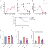
Platelet-specific Ido1−/− mice have altered plasma metabolites and decreased P yoelii infection survival. (A) Parasitemia was assessed by SYBR+ RBCs quantified by flow cytometry. No significant differences were found comparing both groups during PyNL infection. (B) Weight loss was tracked across time points for all groups and was similar except at day 10. (C) Platelet counts, shown as fold change to control, were similar between WT and psIdo1−/− mice during infection. (D) Kaplan Meier survival curve. PsIdo1−/− mice had decreased survival compared with WT control infected mice. (E-G) Normalized relative intensity on day 10 after infection for plasma (E) TRP, (F) KYN, and (G) KTR as a measurement of Ido1 activity. Data presented as mean ± standard error of the mean (SEM; ∗P < .05, ∗∗P < .01, ∗∗∗P < .001). Data were analyzed using unpaired 2-tailed Student t test.
Figure 6.
Figure 6.
Platelet-specific Ido1−/− mice had increased thrombi in their lungs after infection compared with WT infected mice. (A) Fibrinogen was stained and quantified on day 7 and 10 lung sections to detect thrombi. Images were taken at ×40 original magnification, scale bar represents 50 μm, and (B) immunohistochemistry quantification with ImageJ. (C) Lung sections were immunostained for CD42c on day 7 and 10 after infection to detect thrombi. Images were taken at ×40 original magnification, scale bar represents 25 μm. (D-E) Quantification of (D) thrombi <10 μm or (E) >10 μm averaged over 5 fields per mouse. Data presented as mean ± SEM (∗P < .05, ∗∗P < .01, ∗∗∗P < .001). Data were analyzed using unpaired 2-tailed Student t test.

References

    1. Machlus KR, Italiano JE. The incredible journey: from megakaryocyte development to platelet formation. J Cell Biol. 2013;201(6):785–796. - PMC - PubMed
    1. Nurden AT, Nurden P, Sanchez M, Andia I, Anitua E. Platelets and wound healing. Front Biosci. 2008;13:3532–3548. - PubMed
    1. Kapur R, Semple JW. Platelets as immune-sensing cells. Blood Adv. 2016;1(1):10–14. - PMC - PubMed
    1. Portier I, Campbell RA. Role of platelets in detection and regulation of infection. Arterioscler Thromb Vasc Biol. 2021;41(1):70–78. - PMC - PubMed
    1. Garraud O, Hamzeh-Cognasse H, Pozzetto B, Cavaillon JM, Cognasse F. Bench-to-bedside review: platelets and active immune functions - new clues for immunopathology? Crit Care. 2013;17(4):236. - PMC - PubMed

Publication types