. 2024 Sep:75:103306.
doi: 10.1016/j.redox.2024.103306. Epub 2024 Aug 8.

A reciprocal relationship between mitochondria and lipid peroxidation determines the chondrocyte intracellular redox environment

Affiliations

A reciprocal relationship between mitochondria and lipid peroxidation determines the chondrocyte intracellular redox environment

Madeline R Hines et al. Redox Biol. 2024 Sep.

Abstract

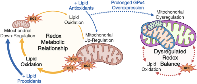
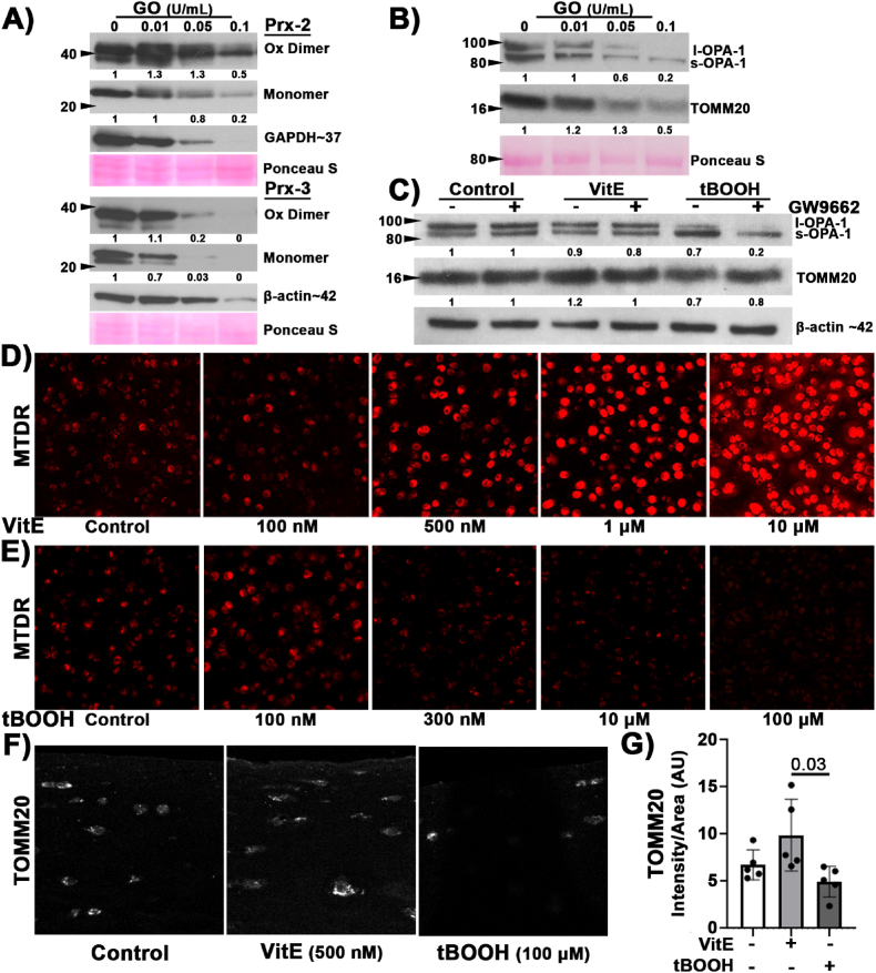
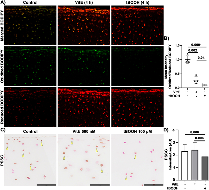
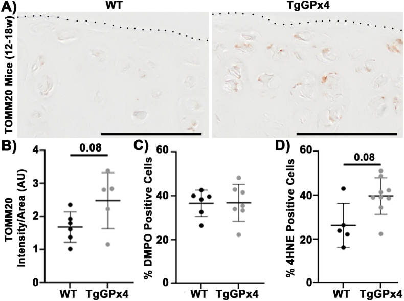
In orthopedic research, many studies have applied vitamin E as a protective antioxidant or used tert-butyl hydroperoxide to induce oxidative injury to chondrocytes. These studies often support the hypothesis that joint pathology causes oxidative stress and increased lipid peroxidation that might be prevented with lipid antioxidants to improve cell survival or function and joint health; however, lipid antioxidant supplementation was ineffective against osteoarthritis in clinical trials and animal data have been equivocal. Moreover, increased circulating vitamin E is associated with increased rates of osteoarthritis. This disconnect between benchtop and clinical results led us to hypothesize that oxidative stress-driven paradigms of chondrocyte redox function do not capture the metabolic and physiologic effects of lipid antioxidants and prooxidants on articular chondrocytes. We used ex vivo and in vivo cartilage models to investigate the effect of lipid antioxidants on healthy, primary, articular chondrocytes and applied immuno-spin trapping techniques to provide a broad indicator of high levels of oxidative stress independent of specific reactive oxygen species. Key findings demonstrate lipid antioxidants were pro-mitochondrial while lipid prooxidants decreased mitochondrial measures. In the absence of injury, radical formation was increased by lipid antioxidants; however, in the presence of injury, radical formation was decreased. In unstressed conditions, this relationship between chondrocyte mitochondria and redox regulation was reproduced in vivo with overexpression of glutathione peroxidase 4. In mice aged 18 months or more, overexpression of glutathione peroxidase 4 significantly decreased the presence of pro-mitochondrial peroxisome proliferation activated receptor gamma and deranged the relationship between mitochondria and the redox environment. This complex interaction suggests strategies targeting articular cartilage may benefit from adopting more nuanced paradigms of articular chondrocyte redox metabolism.

Keywords: Cartilage; Chondrocyte; Glutathione peroxidase 4; Immuno-spin trapping; Lipid peroxidation; Mitochondria.


Conflict of interest statement

Declaration of competing interest None.

Figures

Image 1
Graphical abstract
Fig. 1
Fig. 1
Lipid antioxidants increased mitochondrial markers while lipid prooxidants decreased mitochondrial markers. (A) Bovine cartilage was treated with increasing concentrations of GO to generate H2O2. Prx2 has a modest increase in oxidized dimer followed by a decrease in both the dimer and monomer with increasing concentrations of GO; whereas Prx3 had decreased dimer and monomer with increasing concentration of GO, n = 3. (B) Representative western blots with corresponding Ponceau s stain (red, loading control) illustrated increased GO concentration coincided with a change to OPA-1 and decreased TOMM20, n = 3. (C) Representative western blot of l-OPA-1 to s-OPA-1 isoform change after 4 h VitE (500 nM) or tBOOH (100 μM) treatment, n = 3. (D) Bovine osteochondral explants treated for 24 h with increasing concentrations of VitE. Mitochondria, visualized using MTDR, increased in a dose dependent manner. 20× objective. (E) Bovine osteochondral explants were treated with increasing concentrations of tBOOH. 24 h after treatment mitochondria were visualized with MTDR. As tBOOH increased, we observed a decrease in MTDR staining intensity. 20× objective. (F) Representative images of the superficial articular cartilage stained for TOMM20 (white) after 24 h of VitE or tBOOH. 40× objective. (G) TOMM20, staining intensity trended higher after 24 h treatment with VitE and lower 24 h after tBOOH. VitE and tBOOH groups were significantly different. (For interpretation of the references to colour in this figure legend, the reader is referred to the Web version of this article.)
Fig. 2
Fig. 2
Lipid Prooxidants Decrease Peroxyl Radical Formation and Protein Glutathionylation. Porcine osteochondral explants were incubated for 24 h with either 500 nM VitE or 100 μM tBOOH. (A) Representative images of BODIPY in articular cartilage. 20× objective. (B) The mean intensity ratio of oxidized:reduced shows that the control specimens had the most oxidized BODIPY. VitE decreased BODIPY and tBOOH had the least oxidized BODIPY, significantly less than both other groups. (C) Representative images of PSSG staining. Bar represents 50 μm. (D) The mean intensity of PSSG was unchanged with VitE treatment. PSSG intensity decreased after tBOOH treatment. Swine osteochondral explants were incubated for 4 h with either 500 nM VitE or 100 μM tBOOH, n = 4.
Fig. 3
Fig. 3
Lipid antioxidants increased basal IST but decreased IST after injury. Yellow arrows indicate positive staining for DMPO. (A) Representative images of DMPO staining in uninjured cartilage with DMPO and DMPO with VitE. (B) Representative images of DMPO staining 24 h after injury with DMPO and DMPO with VitE. (C) VitE increased DMPO staining in the absence of injury. Paired non-parametric t-test results shown. VitE shows lower DMPO staining after injury compared to the DMPO control. Impact increased DMPO staining in untreated tissue via unpaired non-parametric t-test. (D) Representative images of DMPO staining in samples treated with VitE or in combination of mitochondrial inhibitor Rot (2.5 μM). Scale bar is 50 μm. (E) Relative difference between % positive DMPO and treatment controls. VitE significantly increased the % positive DMPO cells. Addition of mitochondrial inhibitor Rot ablated this response to VitE, n = 6, unpaired t-test. (For interpretation of the references to colour in this figure legend, the reader is referred to the Web version of this article.)
Fig. 4
Fig. 4
TgGPx4 increased TOMM20 content and minimally altered the redox environment of healthy articular chondrocytes. (A) Representative images of TOMM20 from WT and TgGPx4 mice demonstrate TOMM20 was increased in TgGPx4 mice (12–18 w). Dotted line denotes the articular surface, scale bar 50 μm. (B) TOMM20 intensity trends higher in TgGPx4 compared to WT. (C) Mice (12–18 w) had no difference in %DMPO positive cells between the WT and TgGPx4. (D) 4HNE trended higher in with TgGPx4 compared to WT.
Fig. 5
Fig. 5
Lifelong overexpression of GPx4 led to suppressed PPAR-γ, mitochondrial dysregulation, and decoupling of redox homeostasis. (A) PPAR-γ was decreased in the TgGPx4 mice. Unpaired t-test. (B) TOMM20 intensity has a significantly wider spread in the TgGPx4 compared to WT, comparison of variance f = 0.028. (C) DMPO had higher variability in the TgGPx4 than WT, comparison of variance f = 0.19. (D) 4HNE positivity was more widespread in the TgGPx4 than the WT, comparison of variance f = 0.35.

References

    1. Brouillette M.J., et al. Strain-dependent oxidant release in articular cartilage originates from mitochondria. Biomech. Model. Mechanobiol. 2014;13(3):565–572. - PMC - PubMed
    1. Coleman M.C., et al. Injurious loading of articular cartilage compromises chondrocyte respiratory function. Arthritis Rheumatol. 2016;68(3):662–671. - PMC - PubMed
    1. Martin J.A., et al. N-acetylcysteine inhibits post-impact chondrocyte death in osteochondral explants. J Bone Joint Surg Am. 2009;91(8):1890–1897. - PMC - PubMed
    1. Goetz J.E., et al. Time-dependent loss of mitochondrial function precedes progressive histologic cartilage degeneration in a rabbit meniscal destabilization model. J. Orthop. Res. 2017;35(3):590–599. - PMC - PubMed
    1. Gomez-Contreras P.C., et al. Intersections between mitochondrial metabolism and redox biology mediate posttraumatic osteoarthritis. Curr. Rheumatol. Rep. 2021;23(5):32. - PubMed

LinkOut - more resources