. 2024 Aug 12;15(1):6891.
doi: 10.1038/s41467-024-51153-8.

HMGA1 orchestrates chromatin compartmentalization and sequesters genes into 3D networks coordinating senescence heterogeneity

Affiliations

HMGA1 orchestrates chromatin compartmentalization and sequesters genes into 3D networks coordinating senescence heterogeneity

Ioana Olan et al. Nat Commun. .

Abstract

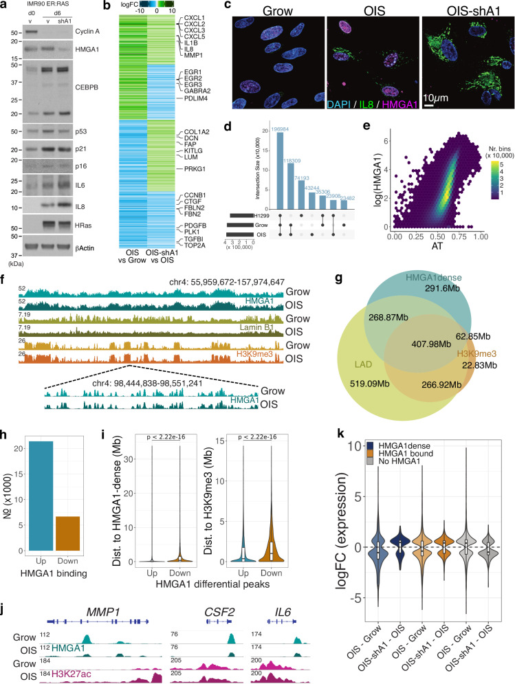
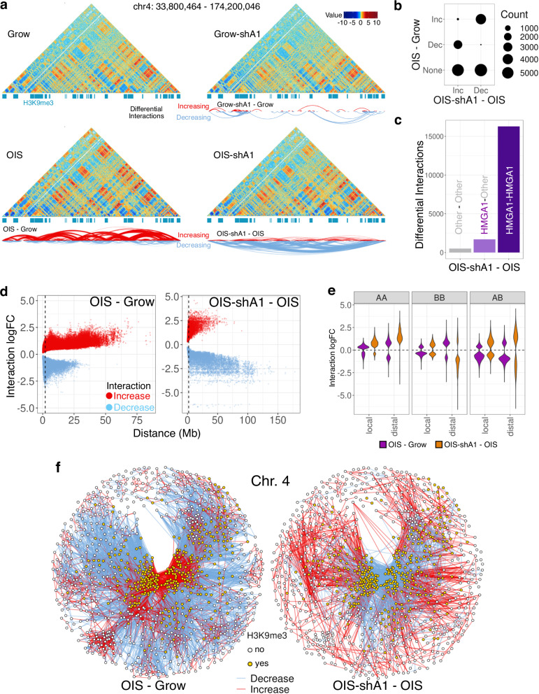
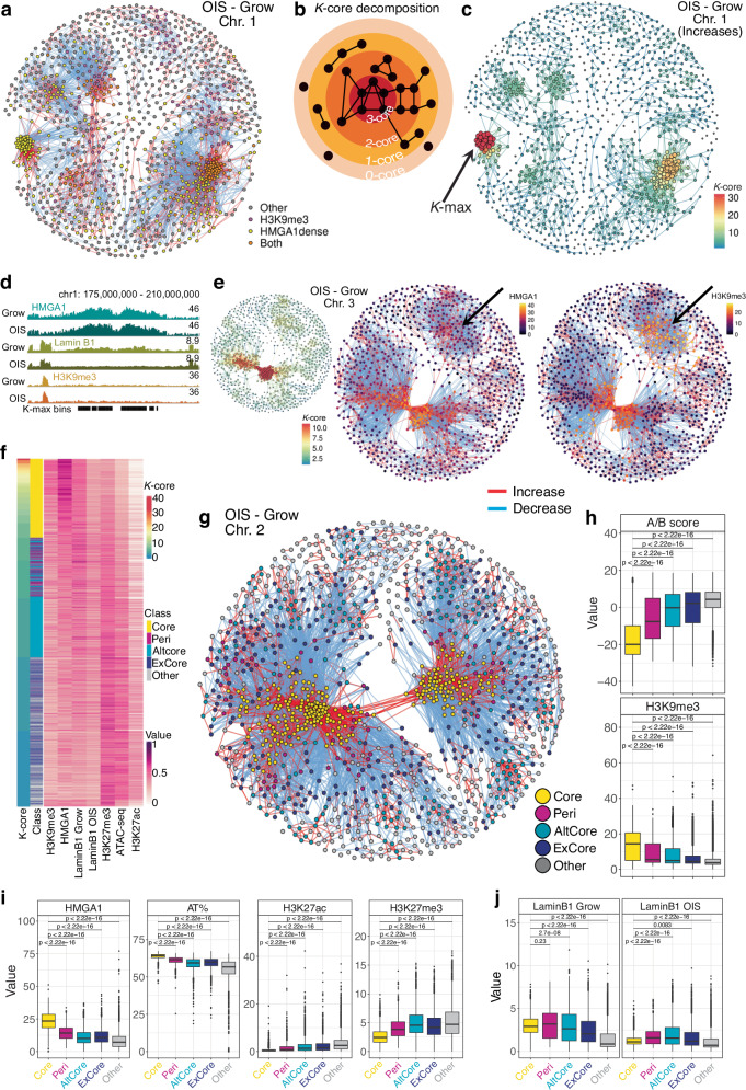
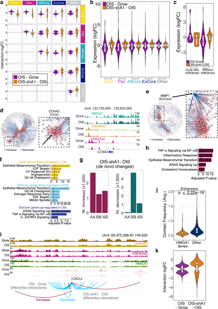
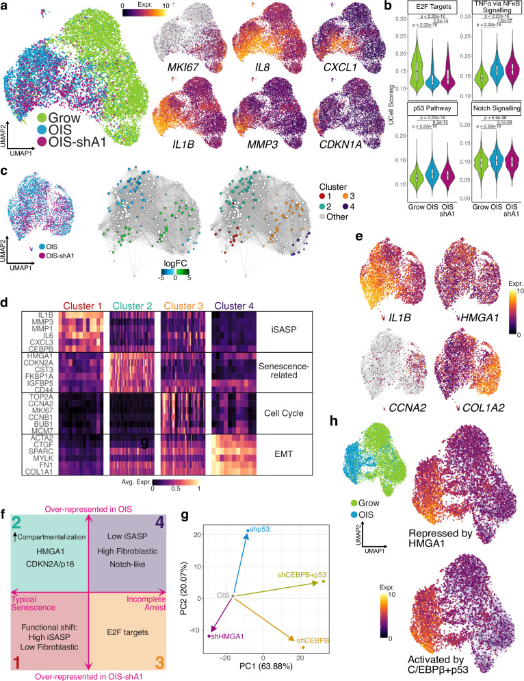
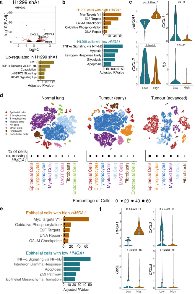
HMGA1 is an abundant non-histone chromatin protein that has been implicated in embryonic development, cancer, and cellular senescence, but its specific role remains elusive. Here, we combine functional genomics approaches with graph theory to investigate how HMGA1 genomic deposition controls high-order chromatin networks in an oncogene-induced senescence model. While the direct role of HMGA1 in gene activation has been described previously, we find little evidence to support this. Instead, we show that the heterogeneous linear distribution of HMGA1 drives a specific 3D chromatin organization. HMGA1-dense loci form highly interactive networks, similar to, but independent of, constitutive heterochromatic loci. This, coupled with the exclusion of HMGA1-poor chromatin regions, leads to coordinated gene regulation through the repositioning of genes. In the absence of HMGA1, the whole process is largely reversed, but many regulatory interactions also emerge, amplifying the inflammatory senescence-associated secretory phenotype. Such HMGA1-mediated fine-tuning of gene expression contributes to the heterogeneous nature of senescence at the single-cell level. A similar 'buffer' effect of HMGA1 on inflammatory signalling is also detected in lung cancer cells. Our study reveals a mechanism through which HMGA1 modulates chromatin compartmentalization and gene regulation in senescence and beyond.


Conflict of interest statement

A.J.P. is an employee of Altos Labs. P.F. and S.S. are co-founders and shareholders of Enhanc3D Genomics Ltd. All other authors declare no competing interests.

Figures

Fig. 1
Fig. 1. The HMGA1 effect on transcription and genome-wide binding distribution in proliferating and OIS cells.
a Protein-level changes of key senescence genes in proliferating IMR90 (Grow; day 0), OIS (day 6) with and without shHMGA1 (shA1)—three replicates per condition. Source data are provided as a Source Data file. b Gene expression log-fold changes (logFC) in OIS compared to Grow and OIS-shA1 compared to OIS of the genes differentially expressed in both comparisons, clustered by the direction of the change. c Immunofluorescence imaging of Grow, OIS and OIS-shA1 stained for DAPI (nuclear; blue), IL-8 (green), and HMGA1 (magenta) cells (n = 2 per condition, quantification—Supplementary Fig. 1f, Source data are provided as a Source Data file). d Overlap between HMGA1 consensus peaks in the IMR90 Grow and OIS and H1299 cells. e Correlation between AT% and average (log) HMGA1 signal in 200 kb bins. f Top: ChIP-seq normalised signal tracks of HMGA1, H3K9me3 and Lamin B1 in the Grow and OIS conditions (IGV browser); Bottom: close-up view of the HMGA1 peaks in a smaller genomic region. g Overlaps (genomic area covered in Mb) between HMGA1-dense regions, Lamin-associated domains (LADs), and H3K9me3 peaks in growing IMR90 cells. h Number of HMGA1 binding changes in OIS compared to Grow cells (Up—increased, Down—decreased). i Properties of the n = 16,420 peaks with increased (Up) and n = 4060 peaks with decreased (Down) HMGA1 binding from h: distance to the nearest HMGA1-dense region (left) and the nearest H3K9me3 peak (right). j ChIP-seq normalised signal tracks of HMGA1 and H3K27ac over representative genes with decreased HMGA1 on their gene body: MMP1, CSF2, IL6. k Distribution of log-fold changes in OIS compared to Grow and OIS-shA1 compared to OIS of the genes DE in each respective comparison classified as: 1) overlapping HMGA1-dense regions (n = 893 and 218 genes), 2) with any HMGA1 binding (n = 2580 and 518 including genes in HMGA1-dense regions), and 3) not bound by HMGA1 at all (n = 2756 and 445 genes), respectively. j, k Box plot centre line represents the median, the bounds correspond to the 0.25 and 0.75 quantiles, the whiskers represent the 0.1 and 0.9 quantiles. Significance testing was performed using two-sided Wilcoxon tests.
Fig. 2
Fig. 2. HMGA1 leads to global re-organisation of chromatin architecture in OIS.
a Hi-C interaction maps (resolution 200 kb) of Grow, Grow-shA1, OIS, and OIS-shA1 conditions, with H3K9me3 peaks marked (blue) and arcs representing increased (red) and decreased (blue) interactions. b Number of significantly increased (Inc) and decreased (Dec) interactions in OIS-shA1 compared to OIS, crosschecked against the changes in OIS compared to Grow. c The number of OIS-shA1 compared to OIS interaction changes where one or both regions involved is bound by HMGA1 (minimum 5 peaks per bin). d The distance between the regions involved in differential interactions against the interaction log-fold changes in OIS compared to Grow (left) and OIS-shA1 compared to OIS (right); the vertical dashed line marks the 2 Mb distance threshold denoting distal interactions. e Log-fold changes of the differential interactions between A-compartment regions (AA), B-compartment regions (BB) and between A- and B-compartment regions (AB), categorised by local (within 2 Mb) and distal (>2 Mb) interactions, in the OIS compared to Grow (purple) and OIS-shA1 compared to OIS (orange) comparisons. f Differential interaction networks of chromosome 4 between nodes representing 200 kb bins, coloured by H3K9me3 status, and edges representing increased (red) and decreased (blue) interactions; left: OIS compared to grow and right: OIS-shA1 compared to OIS; nodes are positioned according to the Fruchterman-Reingold layout calculated based on the edge weights representing the increased interaction log-fold changes.
Fig. 3
Fig. 3. Classification of genomic regions based on their differential interactions connectivity patterns.
a Differential interactions network of OIS compared to Grow on chromosome 1 with nodes coloured by their overlap with H3K9me 3 and HMGA1-dense regions. b Model representation of the k-core decomposition of an example network with k-max equal to 3. c The k-core decomposition of the network of increased interactions in OIS compared to Grow on chromosome 1, with the size of each node (200 kb bins) reflecting the degree (number of interactions) of the node and the k-core value as the node colour; edge colour reflects the minimum k-core of its nodes; arrow indicates the nodes with the maximum k-core value (k-max). d Normalised ChIP-seq signal tracks of HMGA1, Lamin B1, and H3K9me3 in Grow and OIS around the regions which form the degeneracy core (k-max) of chromosome 1 shown in c. e The k-core decomposition (left) of the chromosome 3 network (200 kb bins) of increased contacts in OIS compared to Grow, with the node colour corresponding to its k-core and the node size to its degree; the full network of differential interactions highlighting the HMGA1 (middle) and H3K9me3 (right) normalised ChIP-seq signal of each node; arrow indicates the cluster of regions with low k-core and HMGA1, but high H3K9me3 ChIP-seq signal. f Heatmap of the k-core values, classification, and epigenetic properties, represented by the ChIP-seq (or ATAC-seq) scaled normalised signal of H3K9me3, HMGA1, LaminB1 in Grow and OIS, H3K27me3, ATAC-seq (accessibility), and H3K27ac, of all the bins with k-core of at least 1. g The network of (all) differential interactions of OIS compared to Grow on chromosome 2 with nodes classified as: Core (yellow), Peri (peri-core, magenta), AltCore (alternative core, cyan), ExCore (excluded from cores, navy blue) and other (grey). h The A/B compartment score and H3K9me3 ChIP-seq signal of the genomic bins in each of the classes. i The distributions of the HMGA1 signal, the AT%, H3K27ac and H3K27me3 signal of the bins in each class. j Lamin B1 signal in Grow (left) and OIS (right) of all the bins in each of the classes. hj Compared regions: n = 2900 Core, n = 915 Peri, n = 2892 AltCore, n = 2368 ExCore, and 5235 Other. Box plot centre line represents the median, the bounds correspond to the 0.25 and 0.75 quantiles, the whiskers represent the 0.1 and 0.9 quantiles. P-values derived from two-sided Wilcoxon testing.
Fig. 4
Fig. 4. The effects of HMGA1 loss on the chromatin interactions network and gene expression.
a The log-fold changes of the differential interactions between the regions in each pair of classes in OIS compared to Grow (purple) and OIS-shA1 compared to OIS (orange). b The distributions of the gene expression log-fold changes in the OIS compared to Grow and OIS-shA1 compared to OIS comparisons of the genes within the five classes which are also DE in their respective comparisons (n = 253 and 130 genes in Core, n = 225 and 98 genes in Peri, n = 1015 and 398 genes in AltCore, n = 1088 and 418 genes in ExCore, and n = 4333 and 1411 genes in Other). c Gene expression log-fold changes in OIS compared to Grow and OIS-shA1 compared to OIS of the respective DE genes within ‘Core’ regions with (n = 9 and 6 genes) and without H3K9me3 (n = 208 and 101 genes). d The position of the Core CCNA2 gene (left) in the networks of differential interactions on chromosomes 4 in OIS compared to Grow and the ChIP-seq signal of HMGA1, H3K9me3 and H3K27ac at the CCNA2 locus (right). e The position of the ExCore gene MMP1 in the chromosome 11 network of differential interactions in OIS—Grow. f The top MSigDB Hallmarks gene sets enriched in the genes down-regulated (in OIS compared to Grow) and overlapping ‘Core’ and ‘AltCore’ regions, and the genes up-regulated (in OIS compared to Grow) and overlapping the ‘ExCore’ regions. g The increased (left) and decreased (right) de novo interactions in OIS-shA1 compared to OIS, according to the A- or B- compartment assignments of the interacting regions. h Gene set enrichment analysis against the MSigDB Hallmarks of the genes DE in OIS-shA1 compared to OIS and involved in the de novo increased interactions in the same comparison. P-values derived with EnrichR—Fischer’s exact test adjusted for multiple comparisons with Benjamini-Hochberg. i ChIP-seq normalised signal tracks of Grow and OIS Lamin B1, H3K9me3, HMGA1, and H3K27ac of the CXCL2 gene locus and the regions involved in increased (red) and decreased (blue) interactions in the OIS compared to Grow and OIS-shA1 compared to OIS, respectively. j The average contact frequencies in OIS of the enhancers within HMGA1-dense regions (n = 833 bins overlapping these enhancers) compared to other enhancers (n = 2610 bins overlapping these enhancers). k The interaction log-fold changes in OIS compared to Grow and OIS-shA1 compared to OIS of only the enhancers within HMGA1-dense regions (n = 833 bins overlapping these enhancers). b, c, j, k Box plot centre line represents the median, the bounds correspond to the 0.25 and 0.75 quantiles, the whiskers represent the 0.1 and 0.9 quantiles. P-values derived from two-sided Wilcoxon testing.
Fig. 5
Fig. 5. Senescence transcriptional programmes at single-cell level.
a Single-cell UMAP projection of Grow, OIS and OIS-shA1 cells (coloured by condition but each representing two replicates), highlighting key senescence genes: MKI67, IL8, CXCL1, IL1B, MMP3, and CDKN1A; expression values (log-transformed) were scaled to be between 0 and 10 for visualisation, with grey representing no expression detected. b Distribution of UCell single-cell scores for selected MSigDB Hallmarks gene sets in n = 6165 Grow, n = 2828 OIS, and n = 2073 OIS-shA1 cells; P-values derived from two-sided Wilcoxon testing. Box plot centre line represents the median, the bounds correspond to the 0.25 and 0.75 quantiles, the whiskers represent the 0.1 and 0.9 quantiles. c UMAP projection of the OIS and OIS-shA1 conditions only for Milo testing of cell neighbourhoods and clustering based on the log-fold changes between OIS and OIS-shA1. d Representative markers of the four Milo clusters of differential expression between OIS and OIS-shA1 cells at single-cell level, with expression values scaled between 0 and 1 and averaged over the cell neighbourhoods in each cluster from c. e Representative markers for clusters 1–4 coloured by scaled expression values on the UMAP projection of OIS and OIS-shA1 cells. f Schematic representation of the features of the four clusters of senescent cells identified using overrepresentation analysis, highlighting the clusters over-represented in OIS (2 and 4) and in OIS-shA1 (1 and 3), respectively. Although inflammatory SASP (iSASP) and p16 have been collectively considered senescence hallmarks, they represent distinct types of senescence at the single-cell level. Clusters 3 and 4 express cell-cycle genes and Cluster 4 resembles the previously described NOTCH-related ‘early phase senescence’ with augmented fibroblastic features,. g Dimensionality reduction (PCA) of the log-fold changes of OIS cells (bulk RNA-seq) in response to shHMGA1, shCEBPB, shp53 and double knock-down of p53 and CEBPB. h UMAP projection of the Grow and OIS cells, coloured by UCell scoring of the gene signatures of the genes activated in OIS and up-regulated by shA1 (repressed by HMGA1, top) and down-regulated by the double knock-down of p53 and CEBPB (activated by p53 + CEBPB, bottom).
Fig. 6
Fig. 6. The effect of HMGA1 on the transcriptome of lung adenocarcinoma.
a Expression changes in H1299 cells in response to shHMGA1, highlighting differentially expressed genes (top) and gene enrichment analysis against the MSigDB Hallmarks of the genes up-regulated by shHMGA1 in H1299 cells (bottom). Expression P-values derived from edgeR differential expression testing and adjusted for multiple testing using Benjamini-Hochberg correction. b Gene enrichment analysis of the markers of H1299 cells with high and low HMGA1 expression from a single-cell expression dataset of H1299 cells (see Methods). c Distribution of the expression at single-cell level of the representative markers of the same H1299 cells from b with low (n = 131) and high (n = 187) expression of HMGA1, respectively. d Cell populations of normal lung, early and advanced lung adenocarcinoma and the percentage of cells expressing HMGA1. e Top enrichment results (MSigDB Hallmarks) for the gene markers of epithelial cells in the advanced tumour with high (top) and low (bottom) HMGA1 expression. f Expression distribution of representative genes for the same cells from e, with high (n = 861) and low (n = 3084) HMGA1 expression. a, b, e Gene enrichment P values calculated with the EnrichR software using Fischer’s exact test and adjusted for multiple testing with Benjamini-Hochberg. c, f P values derived from two-sided Wilcoxon testing. Box plot centre line represents the median, the bounds correspond to the 0.25 and 0.75 quantiles, the whiskers represent the 0.1 and 0.9 quantiles.

References

    1. Olan, I. & Narita, M. Senescence: an identity crisis originating from deep within the nucleus. Annu. Rev. Cell Dev. Biol.38, 219–239 (2022). 10.1146/annurev-cellbio-120420-013537 - DOI - PubMed
    1. Gorgoulis, V. et al. Cellular senescence: defining a path forward. Cell179, 813–827 (2019). 10.1016/j.cell.2019.10.005 - DOI - PubMed
    1. Chandra, T. et al. Global reorganization of the nuclear landscape in senescent cells. Cell Rep.10, 471–483 (2015). 10.1016/j.celrep.2014.12.055 - DOI - PMC - PubMed
    1. Criscione, S. W. et al. Reorganization of chromosome architecture in replicative cellular senescence. Sci. Adv.2, e1500882 (2016). 10.1126/sciadv.1500882 - DOI - PMC - PubMed
    1. Zirkel, A. et al. HMGB2 loss upon senescence entry disrupts genomic organization and induces CTCF clustering across cell types. Mol. Cell70, 730–744.e6 (2018). 10.1016/j.molcel.2018.03.030 - DOI - PubMed