. 2024 Aug 12;15(1):6894.
doi: 10.1038/s41467-024-51046-w.

Variant-proof high affinity ACE2 antagonist limits SARS-CoV-2 replication in upper and lower airways

Affiliations

Variant-proof high affinity ACE2 antagonist limits SARS-CoV-2 replication in upper and lower airways

Matthew Gagne et al. Nat Commun. .

Abstract

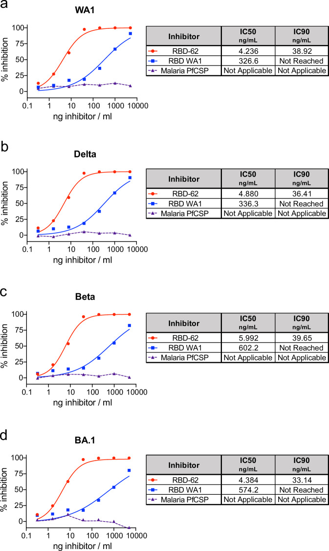
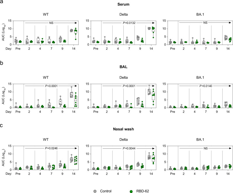
SARS-CoV-2 has the capacity to evolve mutations that escape vaccine- and infection-acquired immunity and antiviral drugs. A variant-agnostic therapeutic agent that protects against severe disease without putting selective pressure on the virus would thus be a valuable biomedical tool that would maintain its efficacy despite the ongoing emergence of new variants. Here, we challenge male rhesus macaques with SARS-CoV-2 Delta-the most pathogenic variant in a highly susceptible animal model. At the time of challenge, we also treat the macaques with aerosolized RBD-62, a protein developed through multiple rounds of in vitro evolution of SARS-CoV-2 RBD to acquire 1000-fold enhanced ACE2 binding affinity. RBD-62 treatment equivalently suppresses virus replication in both upper and lower airways, a phenomenon not previously observed with clinically approved vaccines. Importantly, RBD-62 does not block the development of virus-specific T- and B-cell responses and does not elicit anti-drug immunity. These data provide proof-of-concept that RBD-62 can prevent severe disease from a highly virulent variant.


Conflict of interest statement

The authors declare the following competing interests: M.S.S. serves on the scientific board of advisors for Moderna and Ocugen. D.C.D. is an inventor on US Patent Application No. 63/147,419 entitled “Antibodies Targeting the Spike Protein of Coronaviruses”. A.V.R., L.P., A.D., A.C., M.G.L. and H.A. are employees of Bioqual. The other authors declare no competing interests.

Figures

Fig. 1
Fig. 1. Inhibition of variant S–ACE2 binding.
SARS-CoV-2 S from WA1 (a), Delta (b), Beta (c) and BA.1 (d) were mixed with soluble ACE2 in combination with indicated concentrations of RBD-62, RBD from WA1 or an irrelevant malaria protein (PfCSP) to determine percentage binding inhibition relative to maximum binding without inclusion of inhibitor. Icons represent the average inhibition of duplicate technical replicates at each indicated dilution. IC50 and IC90 values (ng/mL) are indicated to the right of each graph and, along with the curves presented within the graphs, were calculated using the nonlinear regression analysis tool in Prism. The dotted lines indicate background inhibition observed for PfCSP. Source data are provided as a Source Data file.
Fig. 2
Fig. 2. Delta replication in upper and lower airways.
NHP (n = 8/group) were challenged with 2 × 105 TCID50 Delta and simultaneously treated with RBD-62 (green circles) or PBS (gray circles). a Subgenomic RNA encoding for N transcript was measured in the upper and lower airways at days 2, 4, 7, 9 and 14 post challenge. b Culturable virus was measured in the upper and lower airways at days 2 and 4 post challenge. Dotted lines indicate the assay limit of detection (LOD). Circles, boxes and horizontal lines represent individual animals, interquartile range and median, respectively, while minima and maxima are denoted at whisker termini. Statistical analyses were shown for comparison of groups at each timepoint and were performed using the Wilcoxon rank-sum test (two-sided) after Holm’s adjustment across timepoints. NS denotes that the indicated comparison was not significant, with P > 0.05. See also Supplementary Tables 2 and 3 for complete statistical analyses. Source data are provided as a Source Data file.
Fig. 3
Fig. 3. Anti-SARS-CoV-2 IgG binding titer kinetics.
NHP (n = 8/group) were challenged with 2 × 105 TCID50 Delta and simultaneously treated with RBD-62 (green circles) or PBS (gray circles). IgG binding titers were measured to wildtype, Delta and BA.1 RBD in a serum, b BAL and c nasal wash 1 month prior to challenge (pre-challenge) and on days 2, 4, 7, 9 and 14 post challenge. Serum was initially diluted 1:100 and then serially diluted 1:4. BAL and NW samples were initially diluted 1:5 and then serially diluted 1:5. Circles, boxes and horizontal lines represent individual animals, interquartile range and median, respectively, while minima and maxima are denoted at whisker termini. P values annotated on plots can be used to assess the statistical significance of a drug-specific treatment effect (difference between RBD-62 and control groups), based on two-sided generalized estimating equation (GEE) modeling, which included titers across all post challenge timepoints. No adjustments were made for multiple comparisons. NS denotes that the indicated comparison was not significant, with P > 0.05. See also Supplementary Table 4 for complete statistical analyses. Source data are provided as a Source Data file.
Fig. 4
Fig. 4. Kinetics of primary T cell responses following Delta challenge.
NHP (n = 8/group) were challenged with 2 × 105 TCID50 Delta and simultaneously treated with RBD-62 (green circles) or PBS (gray circles). ae Peripheral blood mononuclear cells (PBMC) or fh lymphocytes from BAL were collected prior to challenge (immediately preceding challenge for PBMC and 1 month pre-challenge for BAL) and on days 2, 4, 7, 9 and 14 post challenge. Cells were stimulated with WA1 S1 and S2 peptide pools and responses were measured by intracellular cytokine staining (ICS). a, f Percentage of memory CD4+ T cells expressing TH1 markers (IL-2, TNF or IFNγ). b, g Percentage of memory CD4+ T cells expressing TH2 markers (IL-4 or IL-13). c, h Percentage of memory CD8+ T cells expressing IL-2, TNF or IFNγ. d, e Percentage of TFH cells expressing CD40L or IL-21, respectively. Dotted lines set at 0%. Reported percentages may be negative due to background subtraction and may extend beyond the lower range of the y-axis. Circles, boxes and horizontal lines represent individual animals, interquartile range and median, respectively, while minima and maxima are denoted at whisker termini. Due to pre-specified minimum cell numbers per sample required for analysis, some timepoints include data from <8 NHP/group. Data are provided as a Source Data file.
Fig. 5
Fig. 5. Memory B-cell responses following Delta challenge and RBD-62 treatment.
NHP were challenged with 2 × 105 TCID50 Delta and simultaneously treated with RBD-62 or PBS. Memory B-cell specificity was determined at day 14 post challenge via binding to fluorochrome-labeled variant probe pairs as indicated in figure legends. Probe pairs include WA1 and Delta S-2P, Delta and BA.1 S-2P, and Delta S-2P and RBD-62. a Representative flow cytometry graphs for one animal in the control group (left) or treated group (right). Event frequencies denote the proportion of probe-binding cells within the total class-switched memory B-cell population. Cross-reactive memory B cells are represented by events in the top right quadrant whereas single-positive memory B cells reside in the top left or bottom right quadrants. b Pie charts indicating the geometric mean frequency of the entire S-specific memory B-cell compartment capable of binding to both members of a variant probe pair (dark gray) or a single variant within the pair (light gray or black) at day 14 post challenge. The control group is displayed on the left and the treated group is displayed on the right. n = 8 for treated group and n = 4 for control group. Source data are provided as a Source Data file.

Update of

References

    1. Planas, D. et al. Reduced sensitivity of SARS-CoV-2 variant Delta to antibody neutralization. Nature596, 276–280 (2021). 10.1038/s41586-021-03777-9 - DOI - PubMed
    1. Wang, R. et al. Analysis of SARS-CoV-2 variant mutations reveals neutralization escape mechanisms and the ability to use ACE2 receptors from additional species. Immunity54, 1611–1621 e1615 (2021). 10.1016/j.immuni.2021.06.003 - DOI - PMC - PubMed
    1. Edara, V. V. et al. mRNA-1273 and BNT162b2 mRNA vaccines have reduced neutralizing activity against the SARS-CoV-2 omicron variant. Cell Rep. Med.3, 100529 (2022). 10.1016/j.xcrm.2022.100529 - DOI - PMC - PubMed
    1. Hoffmann, M. et al. The Omicron variant is highly resistant against antibody-mediated neutralization: implications for control of the COVID-19 pandemic. Cell185, 447–456 e411 (2022). 10.1016/j.cell.2021.12.032 - DOI - PMC - PubMed
    1. Muik, A. et al. Neutralization of SARS-CoV-2 Omicron by BNT162b2 mRNA vaccine-elicited human sera. Science375, eabn7591 (2022). - PMC - PubMed

MeSH terms

Substances

Supplementary concepts