Review
. 2024 Dec;34(4):959-972.
doi: 10.1007/s00062-024-01447-7. Epub 2024 Aug 12.

Multiple Severe Intracranial Stenoses with Ischemic Stroke in Neuroborreliosis-associated Cerebral Vasculitis: Endovascular Treatment Strategies and Literature Review

Affiliations
Review

Multiple Severe Intracranial Stenoses with Ischemic Stroke in Neuroborreliosis-associated Cerebral Vasculitis: Endovascular Treatment Strategies and Literature Review

Kamran Hajiyev et al. Clin Neuroradiol. 2024 Dec.

Abstract

Introduction: Neuroborreliosis is the disseminated form of Lyme borreliosis and refers to the involvement of the central nervous system by Borrelia burgdorferi sensu lato spirochetes. Several reports suggest its emergence as a potential cause of cerebral vasculitis and stroke in children and young adults. The objective of this paper is to highlight endovascular treatment options within this context.

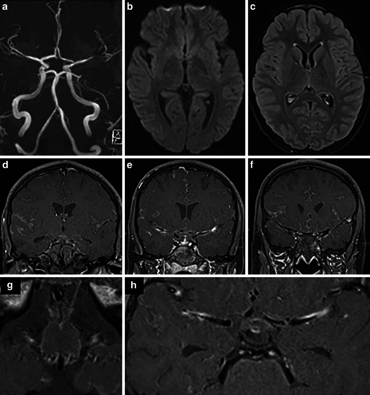
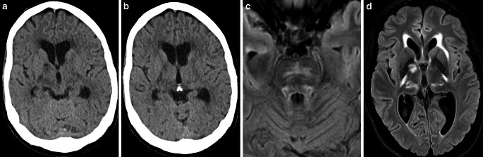
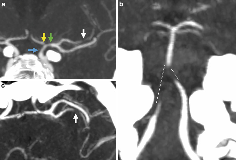
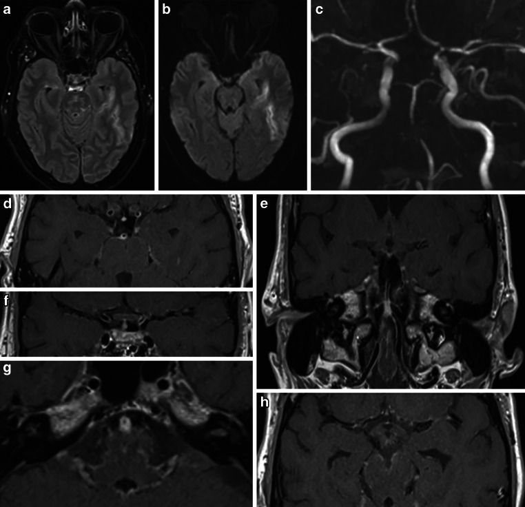
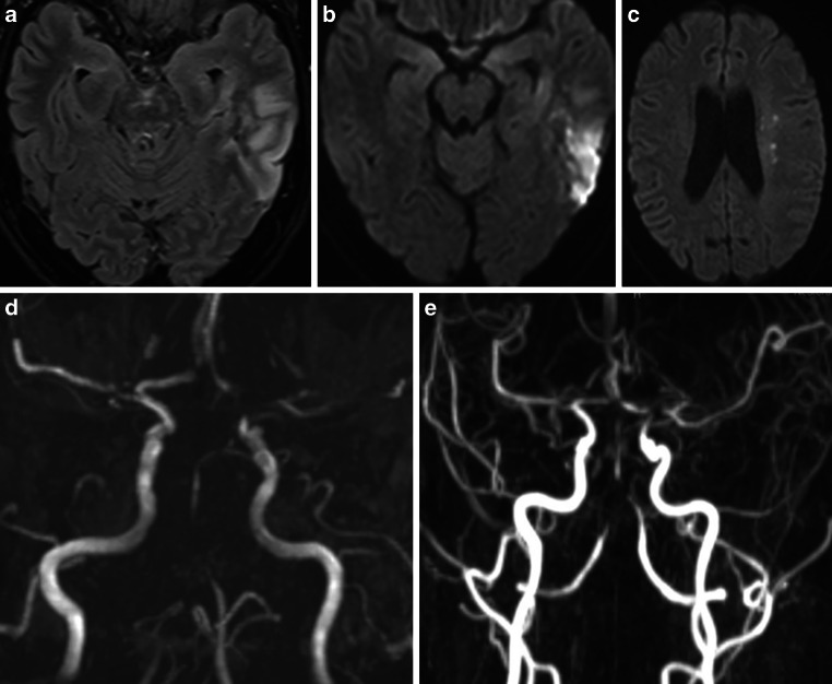
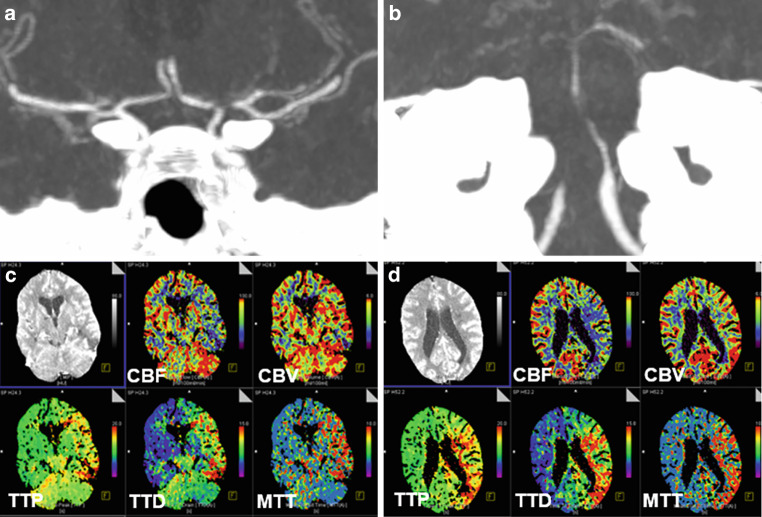
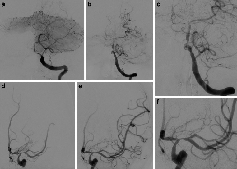
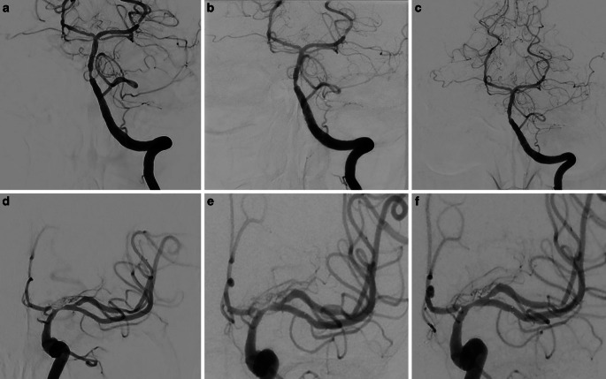
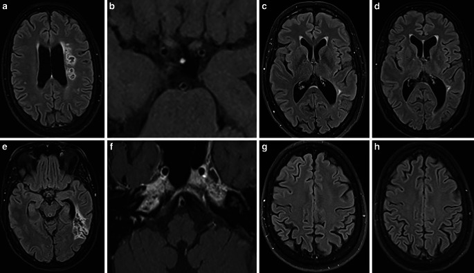
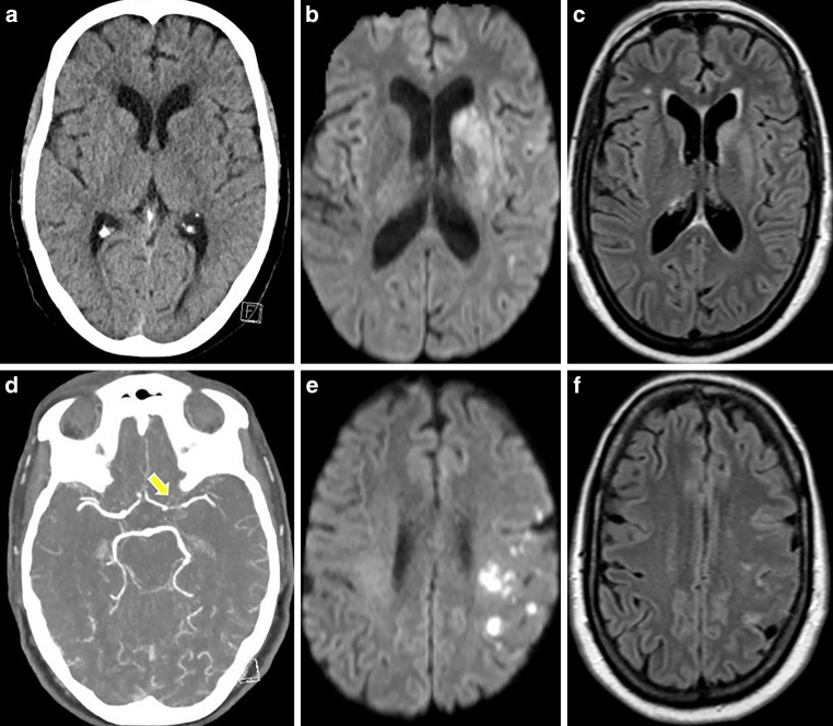
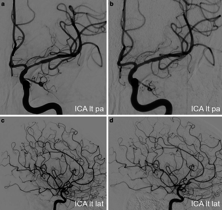
Methods: The medicinal and endovascular treatments of three patients-two adults and one child-with ischemic stroke resulting from neuroborreliosis-associated severe cerebral vasculitis were retrospectively assessed. Detailed descriptions of the clinical course, treatments, and follow-up data for each patient are provided. Additionally, a literature review focusing on endovascular treatment options within this topic was conducted.

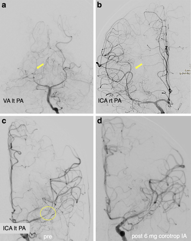
Results: Both endovascular and medicinal treatments resulted in excellent clinical outcomes in all three patients, with no observed periprocedural complications. Significant clinical improvement was noted during mid-term follow-up. Follow-up angiographies confirmed stent patency.

Conclusion: Endovascular interventions as a bailout strategy may enhance clinical outcomes in patients with vascular complications of neuroborreliosis, especially when medicinal therapy alone fails to achieve further improvement. In the setting of severe ischemic stroke with sub-occlusive large vessel stenosis or occlusion, the cause of which is often unknown, it should be considered to prioritize prompt endovascular treatment, even if neuroborreliosis is suspected on admission.

Keywords: Cerebral vasculitis; Endovascular treatment; Intracranial stenting; Ischemic stroke; Lyme neuroborreliosis; Vessel wall imaging.


Conflict of interest statement

Declarations Conflict of interest K. Hajiyev, M. Forsting, A. Cimpoca, A. Khanafer, H. Bäzner and H. Henkes declare that they have no competing interests. Ethical standards This study was conducted in accordance with the Declaration of Helsinki Written informed consent has been obtained from the patients to publish this paper. Further approval by the local ethics committee was waived.

Figures

Fig. 1
Fig. 1
MRI scan of a 13-year-old boy presenting with headache, right arm paresis, and facial palsy. MRI with diffusion-weighted imaging (DWI) (b = 1000 s/mm2, axial view; (a) and FLAIR (axial view; b) demonstrated a lesion with restricted diffusion of the left insular cortex (yellow arrow) without correlate on FLAIR images. Maximum intensity projection (MIP) images from TOF angiography (c) showed a left distal M1 stenosis (green arrow) with flow restriction
Fig. 2
Fig. 2
DSA of a 13-year-old boy with symptomatic stenosis of the left MCA. Angiograms of the left vertebral artery (VA) (PA view, a) and the right ICA (PA view, b) showed no focal stenosis but a generalized narrowing of several pial arteries (e.g., right posterior cerebral artery (PCA), yellow arrow, a; right MCA, yellow arrow, b). Angiograms of the left ICA showed a significant narrowing of the left distal M1 segment with involvement of the proximal M2 segments (PA view, yellow circle, c). A final DSA run of the left ICA after intra-arterial (IA) injection of 6 mg milrinone confirmed an increased diameter of the stenotic left M1/M2 junction and a general dilatation of the left anterior cerebral artery (ACA) and MCA (PA view, d)
Fig. 3
Fig. 3
MRI of a 13-year-old boy obtained 1 day after the clinical onset of cerebral vasculitis due to LNB. MIP images of TOF angiography (a) demonstrated better arterial flow in the left MCA branches. DWI (b = 1000 s/mm2) (axial view; b) and FLAIR images (axial view; c) showed a decrease in ischemic changes within the left insular cortex. Fat-saturated contrast-enhanced T1-weighted images (coronal views: def; axial views: gh) revealed diffuse leptomeningeal enhancement and arterial wall thickening with contrast enhancement in the both MCA and basilar artery
Fig. 4
Fig. 4
Three-month follow-up MRI after clinical onset and subsequent treatment of cerebral vasculitis due to LNB. FLAIR image (axial view; a) and T2-weighted image (axial view; b) showed no new cerebral lesions. MIP images from TOF angiography (c) demonstrated residual non-flow-limiting stenosis at the left MCA bifurcation. Fat-saturated contrast-enhanced T1-weighted images (coronal view, d; axial view, e) detected residual minimal vessel wall enhancement of the left distal M1 segment (yellow arrow, d) and no leptomeningeal enhancement
Fig. 5
Fig. 5
CT and MRI scans of a 57-year-old female presenting with confusion, speech disorder, bradykinesia, and mild right ptosis. Native CT images (axial views; ab) and MRI with FLAIR images (axial views; cd) demonstrated new lesions in the bilateral thalamus, bilateral internal capsule, left external capsule, and pons
Fig. 6
Fig. 6
CTA of the same patient showing stenoses of the left distal ICA (a, blue arrow), the left proximal A1 segment (a, yellow arrow), the left M1 segment (a, green arrow), the left M2 segment (ac, white arrows), both distal V4 segments, the proximal basilar artery (b, dotted lines)
Fig. 7
Fig. 7
MRI of a 57-year-old patient in the early acute phase of neuroborreliosis-induced vasculitis on day 1. FLAIR (axial view; a) and DWI image (axial view; b) demonstrating new ischemic area in the left temporal lobe. MIP of the TOF angiography (c) confirming previously seen intracranial stenoses (distal left ICA, left ACA/A1, left MCA—M1 and superior trunk, bilateral vertebral artery/V4, proximal basilar artery, bilateral PCA/P2). Fat-saturated post-contrast T1-weighted images (axial views—dfgh; coronal view—e) showing mural thickening and contrast enhancement in stenotic vessel segments, and basal leptomeningeal contrast enhancement
Fig. 8
Fig. 8
MRI of a patient with severe LNB-associated cerebral vasculitis on day 11 after clinical deterioration. FLAIR image (axial view; a) and DWI images (axial views; bc) showed progressed left temporal lobe infarction and new ischemic lesions in the deep white matter (watershed zone, hemodynamic infarcts). MIP of the TOF (d) and contrast-enhanced MR angiography (e) demonstrated worsening of the known intracranial stenoses
Fig. 9
Fig. 9
CTA and CTP obtained on day 14 after the onset of right hemiplegia and altered consciousness of a patient with LNB-associated severe cerebral vasculitis. CTA images (coronal views; ab) showing critical stenosis of the left ACA/A1, left MCA/M1/superior trunk, bilateral vertebral artery/V4, and the BA. The time-to-drain (TTD) and mean transit time (MTT), determined by CTP (cd), is prolonged in the entire posterior circulation and the left anterior circulation with concomitant cerebral blood volume (CBV)-cerebral blood flow (CBF) mismatch
Fig. 10
Fig. 10
Endovascular treatment of the severe intracranial stenoses in a patient with severe LNB-associated cerebral vasculitis. Diagnostic angiograms of the left VA (PA view; a) and the left ICA (PA view; d) demonstrating severe stenosis of the left VA-BA junction and the left proximal M1 segment. Angiograms after the stent implantation in the left VA-BA junction (Solitaire 3/20 mm; PA views – b, c) and in the left M1 segment (Solitaire 4/20 mm; PA views – e, f) showing the marked improvement of the antegrade arterial flow with residual stenosis
Fig. 11
Fig. 11
Follow-up DSA examinations after stenting for critical progressive intracranial stenoses due to LNB-related cerebral vasculitis. Angiograms of the left VA (PA views; abc) and the left ICA (PA views; def) after 3, 6 and 12 months demonstrating a positive remodeling effect of the stent, with no significant residual stenosis
Fig. 12
Fig. 12
Follow-up MRI scans after stent angioplasty for intracranial stenoses secondary to a LNB-associated cerebral vasculitis. FLAIR images (axial views; ae) and fat-saturated contrast-enhanced T1-weighted images (axial views; bf) after 3 months demonstrating unchanged chronic infarct areas in the left temporal lobe and in the left semioval center, and regressed mural thickening and contrast enhancement in the intracranial arteries. FLAIR images after 6 months (axial views; cg) and 12 months (axial views; dh) showed no new cerebral lesions
Fig. 13
Fig. 13
CT and MRI scans of a 56-year-old female presenting with right hemiparesis and a speech disorder. Native CT scan (axial view; a) showed a suspicious ischemic lesion in the left basal ganglia. DWI images (b = 1000 s/mm2, axial views; be) demonstrated acute ischemia in the left basal ganglia and the left pre- and post-central areas with correlation in FLAIR images (axial views; cf). CT angiography (axial view; d) revealed high-grade stenosis in the left proximal M1 segment (yellow arrow)
Fig. 14
Fig. 14
Endovascular treatment of the severe intracranial stenosis with non-occlusive thrombus in the proximal left M1 segment. Diagnostic angiograms of the left ICA (PA view, a; lateral view, d) demonstrating severe stenosis of the left M1 segment with adjacent thrombus (blue arrow), distal perfusion delay and a peripheral embolus in a precentral branch (yellow arrow). Angiograms after stent implantation (PA view, b; lateral view, e) showed complete coverage of the lesion with an improved distal perfusion. Native CT scan on day 1 after treatment (axial views; cf) demonstrated minimal SAH in the sulci of the left frontal and parietal lobes and no new cerebral infarction
Fig. 15
Fig. 15
MRI examination after stenting of the left MCA in a patient with severe left M1 stenosis due to LNB-associated vasculitis. DWI images (b = 1000 s/mm2, axial views; ad) and FLAIR images (axial views; be) demonstrated known ischemic lesions in the left basal ganglia and the left frontoparietal lobe, and residual sulcal SAH. Fat-saturated post-contrast T1-weighted images (coronal view—c; axial view—f) showing mural contrast enhancement in the left M1 segment and proximal A1 segment
Fig. 16
Fig. 16
Follow-up DSA examinations after stenting for severe left M1 stenosis due to LNB-related vasculitis. Angiograms of the left ICA after 6 (PA views; ac) and 12 months (PA views; bd) demonstrating a positive remodeling effect of the stent, with no significant residual stenosis

References

    1. Koedel U, Fingerle V, Pfister H‑W. Lyme neuroborreliosis—epidemiology, diagnosis and management. Nat Rev Neurol. 2015;11(8):446–56. 10.1038/nrneurol.2015.121. - PubMed
    1. Mygland Å, Ljøstad U, Fingerle V, Rupprecht T, Schmutzhard E, Steiner I. EFNS guidelines on the diagnosis and management of European lyme neuroborreliosis. Eur J Neurol. 2010;17(1):8. 10.1111/j.1468-1331.2009.02862.x. - PubMed
    1. Pachner AR, Steiner I. Lyme neuroborreliosis: infection, immunity, and inflammation. Lancet Neurol. 2007;6(6):544–52. 10.1016/S1474-4422(07)70128-X. - PubMed
    1. Rauer S, et al. ‘Guidelines for diagnosis and treatment in neurology—Lyme neuroborreliosis. GMS Ger Med Sci. 2020; 10.3205/000279. - PMC - PubMed
    1. Wittwer B, Pelletier S, Ducrocq X, Maillard L, Mione G, Richard S. Cerebrovascular events in lyme neuroborreliosis. J Stroke Cerebrovasc Dis. 2015;24(7):1671–8. 10.1016/j.jstrokecerebrovasdis.2015.03.056. - PubMed

MeSH terms

Substances

LinkOut - more resources