. 2025 Jul;43(7):1168-1176.
doi: 10.1038/s41587-024-02356-3. Epub 2024 Aug 12.

Removal of TREX1 activity enhances CRISPR-Cas9-mediated homologous recombination

Affiliations

Removal of TREX1 activity enhances CRISPR-Cas9-mediated homologous recombination

Mehmet E Karasu et al. Nat Biotechnol. 2025 Jul.

Abstract

CRISPR-Cas9-mediated homology-directed repair (HDR) can introduce desired mutations at targeted genomic sites, but achieving high efficiencies is a major hurdle in many cell types, including cells deficient in DNA repair activity. In this study, we used genome-wide screening in Fanconi anemia patient lymphoblastic cell lines to uncover suppressors of CRISPR-Cas9-mediated HDR. We found that a single exonuclease, TREX1, reduces HDR efficiency when the repair template is a single-stranded or linearized double-stranded DNA. TREX1 expression serves as a biomarker for CRISPR-Cas9-mediated HDR in that the high TREX1 expression present in many different cell types (such as U2OS, Jurkat, MDA-MB-231 and primary T cells as well as hematopoietic stem and progenitor cells) predicts poor HDR. Here we demonstrate rescue of HDR efficiency (ranging from two-fold to eight-fold improvement) either by TREX1 knockout or by the use of single-stranded DNA templates chemically protected from TREX1 activity. Our data explain why some cell types are easier to edit than others and indicate routes for increasing CRISPR-Cas9-mediated HDR in TREX1-expressing contexts.


Conflict of interest statement

Competing interests: J.E.C. is a co-founder and board member of Spotlight Therapeutics, a scientific advisory board (SAB) member of Mission Therapeutics, an SAB member of Relation Therapeutics, an SAB member of Hornet Bio, an SAB member for the Joint AstraZeneca–CRUK Functional Genomics Centre and a consultant for Cimeio Therapeutics. The laboratory of J.E.C. has funded collaborations with Allogene and Cimeio. The other authors declare no competing interests.

Figures

Fig. 1
Fig. 1. Identification of TREX1 as a restrictive factor of CRISPR–Cas9-mediated HDR.
a, FA patient-derived LCLs are compromised in a BFP-to-GFP reporter assay for CRISPR–Cas9-mediated HDR. The efficiency of HDR was measured by flow cytometry 5 d after Cas9 targeting (n = 5 for FA LCLs and n = 3 for healthy donor, biologically independent experiments, P = 1.94 × 10−7 and 6.0 × 10−6). b, Genome-wide CRISPRi/CRISPRn screening identified TREX1 as the sole gene whose knockdown strongly rescues HDR in FANCA−/− LCLs. Gene-level effects and statistics were calculated using drugZ and ranked by the normZ score. The size of each point reflects the FDR (Supplementary Table 2). c,d, CRISPRi knockdown of TREX1 significantly increases CRISPR–Cas9-mediated HDR in FANCA (c) and FANCD2 (d) deficient LCLs. Both cell backgrounds were stably transduced with up to three different sgRNAs targeting TREX1, and a BFP-to-GFP assay was used to measure HDR efficiency. In Fig. 1a,c,d, each dot represents an individual biological replicate, and bars represent the mean (n = 4 for sgNT in c and sgTREX1-3 in d, n = 3 for the rest of the samples in c and d, biologically independent experiments, P = 0.031, 0.0065 and 0.0088 for c and P = 0.0234, 0.0096 for d, respectively). All P values were calculated using an unpaired and two-sided t-test, *P < 0.05, **P < 0.01, ***P < 0.001. NT, non-targeting. Source data are provided as a Source Data file. Source data
Fig. 2
Fig. 2. TREX1 interacts with ssODN HDR templates and is inhibited by phosphorothioate protection.
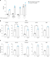
a, RPE-1 hTERT TREX1–/–cells perform high levels of HDR, and TREX1 cDNA complementation abrogates HDR as measured by the BFP-to-GFP assay. Dots (n = 8 for RPE1 wild-type and RPE-1 TREX1–/–, n = 5 for RPE-1 hTERT TREX1–/– + wild-type TREX1 cDNA) represent individual biological replicate measurements; bars represent the mean values; and error bars represent the standard deviation. b,TREX1 co-immunoprecipitates with 5′-biotin-labeled ssODN template. HDR donors were delivered by electroporation to RPE-1 hTERT wild-type and RPE-1 hTERT TREX1–/– cells. After 20 min, cells were collected, and lysates were prepared for immunoprecipitation with streptavidin beads. Blots were probed with anti-TREX1, anti-RPA32 and anti-β actin (n = 1). c, Incorporation of four phosphorothioate (PT) bonds on the 5′ and 3′ or only 3′ ends of an ssODN rescues HDR efficiency in FANCA–/– LCLs, FANCD2–/– LCLs and RPE1 cells. 5′ end protection behaves as unprotected (un) ssODN. HDR was measured using the BFP-to-GFP assay. Each dot represents an individual biological replicate (n = 2 for FANCA–/– LCLs and FANCD2–/– LCLs and n = 4 for RPE-1 hTERT cells), and bars represent the mean. Source data are provided as a Source Data file. IB, immunoblotting; IP, immunoprecipitation; wt, wild-type. Source data
Fig. 3
Fig. 3. Protected ssODN templates increase HDR in TREX1-expressing cell contexts at multiple loci.
a, Cell types expressing TREX1 exhibit compromised HDR, but this is rescued by using phosphorothioate-protected ssODNs. A cell type with normally low levels of TREX1 is already efficient at CRISPR–Cas9 HDR, and this is not further improved by a protected ssODN. HDR was measured by the BFP-to-GFP assay in Jurkat, MDA-MB-231, U2OS and K562 cell lines. Black dots represent measurements with unprotected (un) ssODN template, and blue dots represent measurements with 5′ and 3′ protected (PT) ssODN templates. Each dot represents individual biological replicate measurements (n = 2, biologically independent experiments). b,c, CRISPR–Cas9-mediated HDR efficiency is rescued at multiple endogenous loci in TREX1-expressing RPE-1 cells (b) (n = 3, except HBB site n = 2, biologically independent experiments) (P = 0.0005 for CXCR4, 0.0262 for ABCA3, 0.0029 for FANCD2 and 0.0028 for UROS, respectively) and U2OS cells (c) (n = 2, biologically independent experiments) by phosphorothioate ssODN protection. Editing sites and HDR mutations are shown in Supplementary Fig. 9. Black dots indicate use of unprotected (un) ssODN templates, and blue dots represent 5′ and 3′ PT ssODN templates. Each dot represents an individual biological replicate, and bars represent the mean. All P values were calculated using an unpaired and two-sided t-test, *P < 0.05, **P < 0.01, ***P < 0.001. Source data are provided as a Source Data file. Source data
Fig. 4
Fig. 4. TREX1 suppresses HDR in TREX1-expressing primary cells.
a, Primary cells, such as activated T cells and HSPCs, express TREX1. Protein extracts from the indicated primary cells and cell culture lines were analyzed using western blotting. Blots were probed with anti-TREX1 and anti-HSP60 antibodies (n = 2). bd, CRISPR–Cas9-mediated HDR was improved in activated T cells and HSPCs using protected ssODN templates. HDR efficiency was measured in three different loci in activated T cells (n = 10, P = 0.0006 for CXCR4, n = 6, P = 6.6 × 10−6 for FANCD2 and n = 6, P = 9.3 × 10−5 for UROS loci—n indicates the number of biologically independent experiments) (b), two different loci in HSPCs (n = 2, biologically independent experiments) (c) and a single locus in iPSCs (n = 4 biologically independent experiments, P = 0.0865) (d). Black dots represent measurements with unprotected (un) ssODN templates, and blue dots represent measurements with 5′ and 3′ protected (PT) ssODN templates. e, Protein extracts of activated T-cells from the indicated samples were analyzed using western blotting. Blots were probed with anti-TREX1 and anti-HSP60 antibodies (n = 2). f, Knockout of TREX1 in activated T-cells increases CRISPR–Cas9-mediated HDR from unprotected ssODN templates (n = 5, P = 2.8 × 10−5, P = 0.3815 for CXCR4 and P = 6.1 × 10−8, P = 0.1103 for FANCD2, n = 2 for UROS). Black dots represent measurements with unprotected (un) ssODN templates, and blue dots represent measurements with 5′ and 3′ protected (PT) ssODN templates. Each dot represents an individual biological replicate, and bars represent the mean. All P values were calculated using an unpaired and two-sided t-test:  < 0.05, **P < 0.01, ***P < 0.001. Source data are provided as a Source Data file. NT, non-targeting; IB, immunoblotting; KO, knockout; NS, not significant. Source data
Fig. 5
Fig. 5. TREX1 has differential activity on various types of HDR donors.
a, CRISPRi suppression or pooled CRISPR knockout increases HDR efficiency from linear dsDNA templates in multiple cell backgrounds. GFP was introduced into RAB11A and FBL loci in HeLa cells (n = 8, P = 1.1 × 10−6 for RAB11A and n = 4, P = 2.4 × 10−7 for FBL, respectively), activated T cells (from Fig. 4e, with RAB11A only) and Jurkat cells (FBL only) (n = 2). Each dot represents an individual biological replicate measurement. b, Overexpression of TREX1 in K562 cells reduces the efficiency of linear dsDNA-templated HDR of mCherry into the LMNB1 locus (n = 3, P = 0.0002). Each dot represents an individual biological replicate measurement. c, Western blot shows the overexpression of the TREX1-3×FLAG construct in K562 cells. Blots were probed with anti-TREX1 and anti-HSP60 antibodies (n = 1). d, TREX1 overexpression significantly decreases CRISPR–Cas9-mediated HDR in a BFP-to-GFP assay when using an ssODN template (n = 3, P = 5.4 × 10−5). Each dot represents an individual biological replicate measurement. e, TREX1 overexpression only moderately represses rAAV-templated HDR in a BFP-to-GFP assay. rAAV donor templates carrying promoterless GFP were used in K562 wild-type and TREX1-overexpressed cells. After CRISPR–Cas9 targeting with the gRNA, rAAV was incubated with the cells overnight (n = 2) or for 2 d (n = 3, P = 0.013), and HDR was measured by the presence of GFP-positive cells in FACS 5 d after nucleofection. Each dot represents an individual biological replicate measurement. f, Overall model of TREX1’s role in repressing CRISPR–Cas9-mediated HDR. In TREX1-low cells, ssODN templates are abundant throughout the cell and available in the nucleus for efficient CRISPR–Cas9-mediated HDR. In cases where TREX1 expression is high, ssODN templates are degraded 3′ to 5′ by TREX1 at the endoplasmic reticulum. This reduces ssODN availability in the nucleus and decreases HDR efficiency. Phosphorothioate protection prevents TREX1 digestion of the ssODN, maintaining a high template concentration throughout the cell and enabling efficient CRISPR–Cas9-mediated HDR. Each dot represents an individual biological replicate, and bars represent the mean. All P values were calculated using an unpaired and two-sided t-test: *P < 0.05, **P < 0.01, ***P < 0.001. Source data are provided as a Source Data file. NT, non-targeting; IB, immunoblotting; o/n, overnight. Source data

References

    1. Rouet, P., Smih, F. & Jasin, M. Expression of a site-specific endonuclease stimulates homologous recombination in mammalian cells. Proc. Natl Acad. Sci. USA91, 6064–6068 (1994). - PMC - PubMed
    1. Smih, F., Rouet, P., Romanienko, P. J. & Jasin, M. Double-strand breaks at the target locus stimulate gene targeting in embryonic stem cells. Nucleic Acids Res.23, 5012–5019 (1995). - PMC - PubMed
    1. Choulika, A., Perrin, A., Dujon, B. & Nicolas, J. F. Induction of homologous recombination in mammalian chromosomes by using the I-SceI system of Saccharomyces cerevisiae. Mol. Cell. Biol.15, 1968–1973 (1995). - PMC - PubMed
    1. Lin, S., Staahl, B. T., Alla, R. K. & Doudna, J. A. Enhanced homology-directed human genome engineering by controlled timing of CRISPR/Cas9 delivery. eLife3, e04766 (2014). - PMC - PubMed
    1. Miyaoka, Y. et al. Systematic quantification of HDR and NHEJ reveals effects of locus, nuclease, and cell type on genome-editing. Sci. Rep.6, 23549 (2016). - PMC - PubMed

MeSH terms

Substances

LinkOut - more resources