

Influence of circadian rhythm on effects induced by mechanical strain in periodontal ligament cells
- PMID: 39134880
- PMCID: PMC12540528
- DOI: 10.1007/s00056-024-00542-1
Influence of circadian rhythm on effects induced by mechanical strain in periodontal ligament cells
Abstract
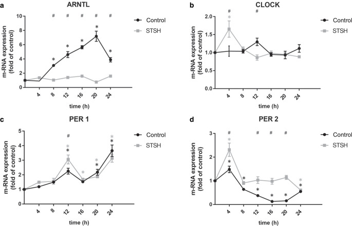
Purpose: The aim of this study was to investigate the influence of mechanical strain on clock gene function in periodontal ligament (PDL) cells. Furthermore, we wanted to analyze whether effects induced by mechanical stress vary in relation to the circadian rhythm.
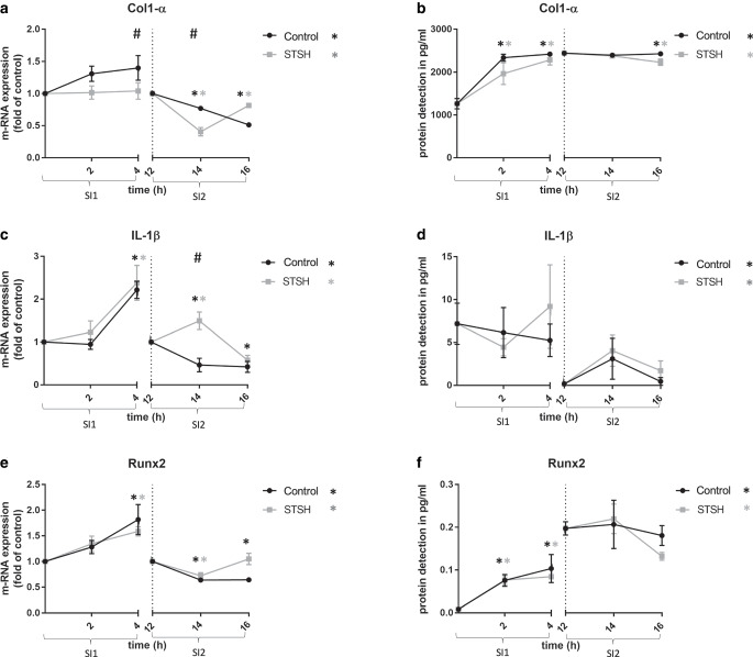
Methods: Human PDL fibroblasts were synchronized in their circadian rhythm with dexamethasone and stretched over 24 h. Unstretched cells served as controls. Gene expression of the core clock genes were analyzed at 4 h intervals by quantitative real-time polymerase chain reaction (qRT-PCR). Time points 0 h (group SI1) and 12 h (group SI2) after synchronization served as starting points of a 4 h force application period. Collagen-1α (COL-1α/Col-1α), interleukin-1β (IL1-β), and runt-related transcription factor 2 (RUNX2/Runx2) were assessed by qRT-PCR and enzyme-linked immunosorbent assay (ELISA) after 2 and 4 h. Statistical analysis comprised one-way analysis of variance (ANOVA) and post hoc tests.
Results: After synchronization, the typical pattern for clock genes was visible in control cells over the 24 h period. This pattern was significantly altered by mechanical strain. Under tensile stress, ARNTL gene expression was reduced, while Per1 and 2 gene expression were upregulated. In addition, mechanical stress had a differential effect on the expression of Col-1α and IL1‑β depending on its initiation within the circadian rhythm (group SI1 vs group SI2). For RUNX2, no significant differences in the two groups were observed.
Conclusion: Our results suggest that mechanical stress affects the molecular peripheral oscillator of PDL cells. Vice versa, the circadian rhythm also seems to partially influence the effects that mechanical stress exerts on PDL cells.
Zusammenfassung: ZIEL: Ziel dieser Arbeit war es, den Einfluss von mechanischer Belastung, wie sie auch im Rahmen einer kieferorthopädischen Zahnbewegung auf das Parodontalligament (PDL) appliziert wird, auf Funktionen von Clock-Genen zu untersuchen. Zusätzlich sollte analysiert werden, ob sich eine mechanische Belastung in Abhängigkeit der zirkadianen Rhythmik unterschiedlich auf wichtige Proteine der PDL-Zellen auswirkt.
Material und methoden: Die periphere zirkadiane Rhythmik humaner PDL-Zellen wurde mittels Dexamethason synchronisiert und einer statischen Dehnung von 20% über bis zu 24 h ausgesetzt. Parallel wurde eine Kontrollgruppe ohne Dehnung angesetzt. In Zeitintervallen von 4 h wurde die Genexpression der Clock-Gene mittels qRT-PCR („quantitative real-time polymerase chain reaction“) bestimmt. Weiter wurden die Zellen einem Dehnungsintervall von 4 h zu den Zeitpunkten 0 h (Gruppe SI1) und 12 h (Gruppe SI2) nach Synchronisation ausgesetzt. Die Expression der Gene Collagen-1α (Col-1α), Interleukin-1β (IL1-β) und Runt-verwandter Transkriptionsfaktor 2 (RUNX2) wurde jeweils nach 2 und 4 h mittels qRT-PCR und ELISA („enzyme-linked immunosorbent assay“) quantifiziert. Die statistische Analyse erfolgte über eine einseitige Varianzanalyse (ANOVA) und Post-hoc-Tests.
Ergebnisse: In den Kontrollzellen war nach Synchronisation das für die Clock-Gene typische Muster im Verlauf der 24 h erkennbar. Dieses wurde durch die mechanische Belastung signifikant verändert. Unter Zugbelastung wurde eine Verringerung der ARNTL-Genexpression verzeichnet, während die Per1- und Per2-Genexpression hochreguliert wurden. Die mechanische Belastung hatte abhängig von der Initiierung nach Synchronisation (Gruppe SI1 vs. Gruppe SI2) einen unterschiedlichen Einfluss auf die Expression von Col-1α und IL1‑β. Für Runx2 wurde in beiden Gruppen kein Unterschied beobachtet.
Schlussfolgerung: Die Ergebnisse legen nahe, dass mechanische Belastung den molekularen peripheren Oszillator der PDL-Zellen beeinflusst. Umgekehrt scheint auch die zirkadiane Rhythmik die Auswirkungen von mechanischem Stress auf PDL-Zellen teilweise zu beeinflussen.
Keywords: Circadian rhythm; Clock genes; Mechanical stimulation; PDL-cells; Tooth movement.
© 2024. The Author(s).
Conflict of interest statement
Conflict of interest: L.I. Peters, J. Marciniak, E. Kutschera, C. Luiz, E. Calvano Küchler, C. Kirschneck, A. Jäger and S. Beisel-Memmert declare that they have no competing interests.
Figures
References
-
- Albrecht U (2012) Timing to perfection: the biology of central and peripheral circadian clocks. Neuron 74:246–260 - PubMed
-
- Balsalobre A, Brown SA, Marcacci L, Tronche F, Kellendonk C, Reichardt HM et al (2000) Resetting of circadian time in peripheral tissues by glucocorticoid signaling. Science 289:2344–2347 - PubMed
MeSH terms
LinkOut - more resources
Full Text Sources
