

A novel variant in the SPTB gene underlying hereditary spherocytosis and a literature review of previous variants
- PMID: 39135028
- PMCID: PMC11318180
- DOI: 10.1186/s12920-024-01973-w
A novel variant in the SPTB gene underlying hereditary spherocytosis and a literature review of previous variants
Abstract
Background: Hereditary spherocytosis (HS, MIM#612641) is one of the most common hereditary hemolytic disorders. This study aimed to confirm a novel variant's pathogenicity and reveal a patient's genetic etiology.
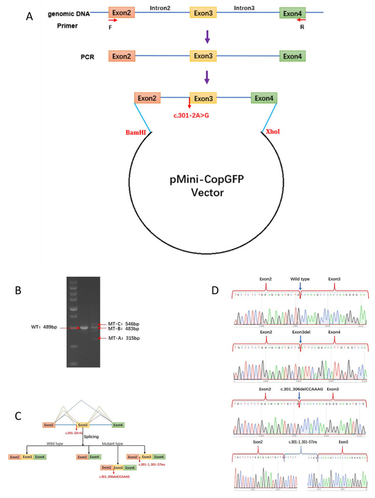
Methods: The clinical data of a patient with HS who underwent genetic sequencing at the Children's Hospital of Chongqing Medical University were reviewed retrospectively. In silico prediction and in vitro minigene splicing reporter system were then conducted on the detected variant to analyze its intramolecular impact. A summary of the literature related to HS due to SPTB gene variants was also presented.
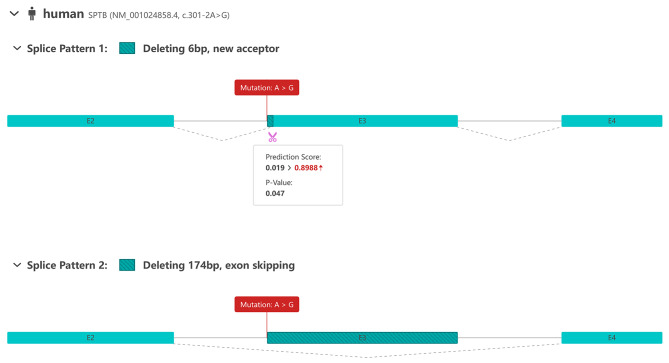
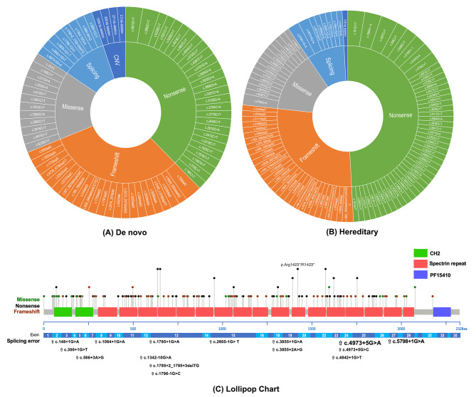
Results: A novel variant (c.301-2 A > G) in the SPTB gene (NM_001024858.4) was identified in the proband. Using Sanger sequencing, we conclusively confirmed that the inheritance of the variant could not be traced to the biological parents. The in vitro minigene assay revealed three different transcripts derived from the c.301-2 A > G variant: r.301_474del, r.301_306delCCAAAG, and r.301-1_301-57ins. Through a literature review, patients with HS who had been genotypically validated were summarized and the SPTB gene variant profile was mapped.
Conclusion: We identified a splicing variant of the SPTB gene, thus confirming its aberrant translation. The novel variant was the probable genetic etiology of the proband with HS. Our findings expanded the variant spectrum of the SPTB gene, thus improving the understanding of the associated hereditary hemolytic disorders from a clinical and molecular perspective and contributing to the foundation of genetic counseling and diagnosis.
Keywords: SPTB gene; Hereditary spherocytosis; Minigene; Novel variant.
© 2024. The Author(s).
Conflict of interest statement
The authors declare no competing interests.
Figures

References
-
- Gallagher PG. Difficulty in diagnosis of Hereditary Spherocytosis in the Neonate. Pediatrics, 2021. 148(3). - PubMed
Publication types
MeSH terms
Substances
Grants and funding
- xyxthjb-2023-006/Public Welfare Project for Rare Blood Disorders in the field of Hematology in China
- xyxthjb-2023-006/Public Welfare Project for Rare Blood Disorders in the field of Hematology in China
- xyxthjb-2023-006/Public Welfare Project for Rare Blood Disorders in the field of Hematology in China
- xyxthjb-2023-006/Public Welfare Project for Rare Blood Disorders in the field of Hematology in China
- xyxthjb-2023-006/Public Welfare Project for Rare Blood Disorders in the field of Hematology in China
- xyxthjb-2023-006/Public Welfare Project for Rare Blood Disorders in the field of Hematology in China
- xyxthjb-2023-006/Public Welfare Project for Rare Blood Disorders in the field of Hematology in China
- xyxthjb-2023-006/Public Welfare Project for Rare Blood Disorders in the field of Hematology in China
- xyxthjb-2023-006/Public Welfare Project for Rare Blood Disorders in the field of Hematology in China
LinkOut - more resources
Full Text Sources
