. 2024 Aug 12;24(1):287.
doi: 10.1186/s12935-024-03453-8.

Upregulation of MELK promotes chemoresistance and induces macrophage M2 polarization via CSF-1/JAK2/STAT3 pathway in gastric cancer

Affiliations

Upregulation of MELK promotes chemoresistance and induces macrophage M2 polarization via CSF-1/JAK2/STAT3 pathway in gastric cancer

Pengfei Su et al. Cancer Cell Int. .

Abstract

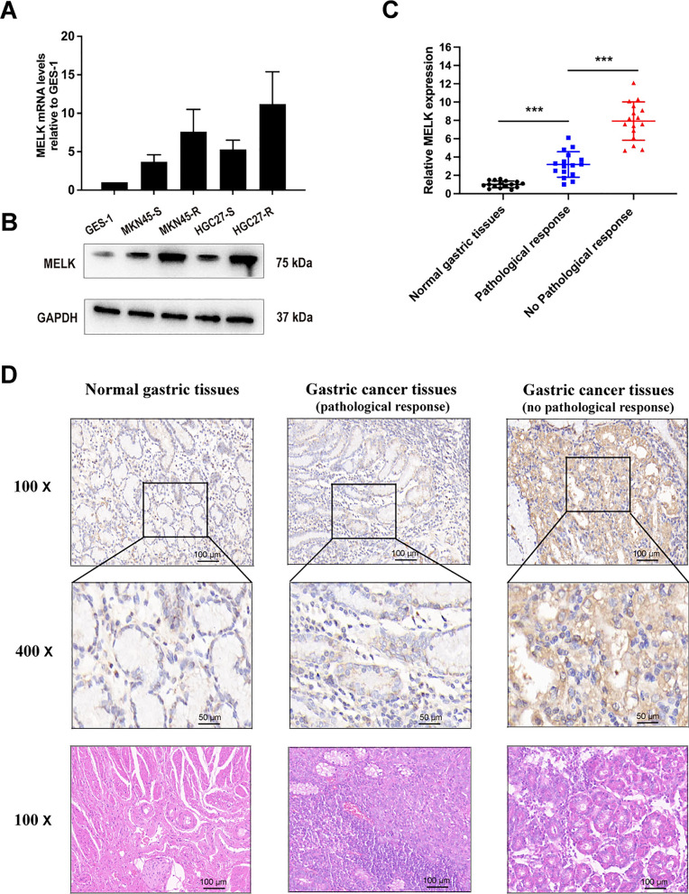
Background: Gastric cancer (GC) stands out as one of the most prevalent malignancies affecting the digestive system, characterized by a substantial incidence rate and mortality. Maternal embryonic leucine zipper kinase (MELK) has been implicated in the advancement of various cancer types and the modulation of the tumor microenvironment. This study aims to delve into the involvement of MELK in chemoresistance and the tumor microenvironment of GC.

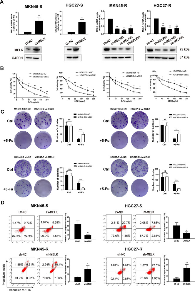
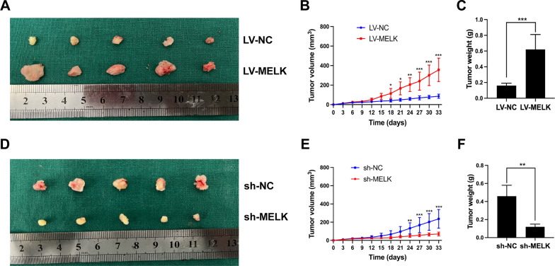
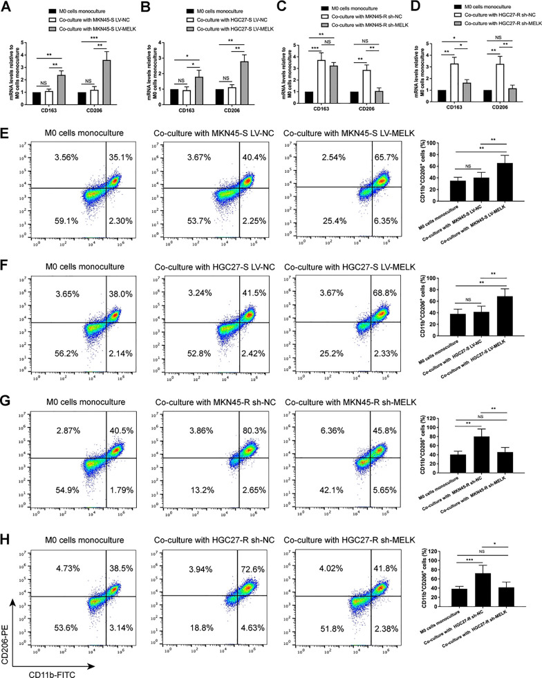
Methods: The MELK expression was detected using quantitative real-time polymerase chain reaction (qRT-PCR), western blotting and immunohistochemistry. Lentiviral transfection was employed to establish stable cell lines with either overexpressed or silenced MELK. The impact of MELK on the chemoresistance of GC cells and the polarization of macrophages was investigated through in vitro and in vivo functional assays. Additionally, the correlation between MELK and the cytokines colony-stimulating factor 1 (CSF-1), as well as stromal macrophages, was analysed. The prognostic significance of MELK, CSF-1, and CD206 expression levels in clinical samples was further investigated.

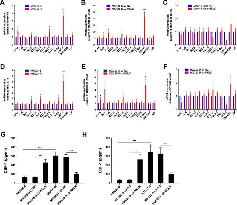
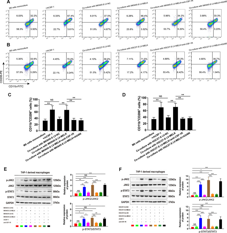
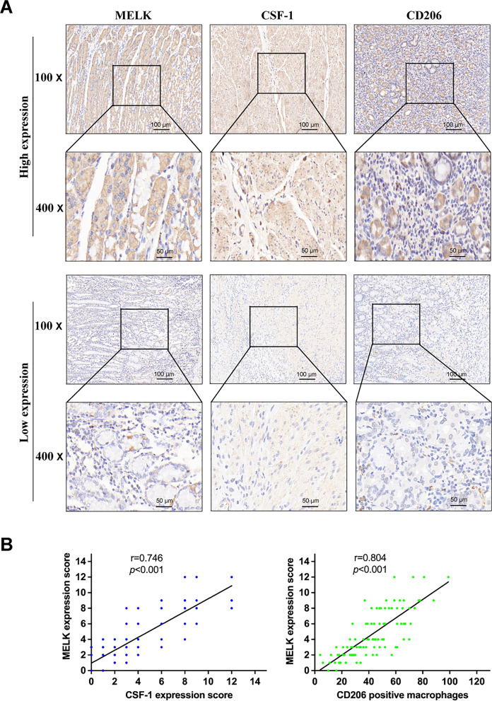
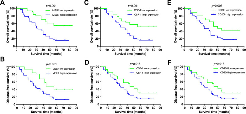
Results: MELK was found to be highly expressed in chemoresistant GC cells and tissues. Furthermore, both in vitro and in vivo assays indicated that MELK overexpression conferred chemoresistance in GC cells. Additionally, MELK overexpression was observed to induce M2 macrophage polarization via the CSF-1/JAK2/STAT3 pathway, thereby contributing to chemoresistance within the tumor microenvironment. The expression of MELK in GC tissues from neoadjuvant chemotherapy patients correlated positively with CSF-1 and CD206. Moreover, patients with higher expression levels of MELK, CSF-1, or CD206 exhibited significantly shorter OS and DFS rates.

Conclusions: Our investigation underscores the critical role of MELK in promoting chemoresistance and inducing M2 macrophage polarization in GC. It proposes novel targets and methods for the treatment of GC, as well as prognostic factors for neoadjuvant chemotherapy.

Keywords: Chemoresistance; Gastric cancer; MELK; Polarization; Prognosis; Tumor-associated macrophages.


Conflict of interest statement

The authors declare no competing interests.

Figures

Fig. 1
Fig. 1
MELK was highly expressed in chemoresistant GC cells and tissues. A, B MELK expression in gastric epithelial cells, parental and chemoresistant GC cells were evaluated by qRT-PCR and western blot assay. C, D MELK expression in normal gastric tissues and GC tissues was evaluated by qRT-PCR and IHC. Data were presented as mean ± SD of at least three independent experiments. ***p < 0.001
Fig. 2
Fig. 2
Effect of MELK on 5-Fu-resistance of GC cells in vitro. A MELK expressions in GC cells with MELK overexpression or knockdown were detected and validated by qRT-PCR and western blot. B GC cells with MELK overexpression or knockdown were incubated with different concentrations of 5-Fu (0, 5, 10, 20, 40, 80, 120, 160, 200 μg/ml) for 24 h, and then cell viability was detected by CCK-8 assay. C GC cells treated with or without 5-Fu for two weeks, the numbers of colonies were counted. D Apoptotic flow cytometry assay was performed to verify that MELK inhibited 5-Fu-induced apoptosis. Data were presented as mean ± SD of three independent experiments. *p < 0.05, **p < 0.01, ***p < 0.001
Fig. 3
Fig. 3
Effect of MELK on 5-Fu-resistance of MKN45 cells in vivo. AC The comparison of tumor volume and weight in nude mice inoculated with MKN45-S cells overexpressing MELK and negative control cells. DF The comparison of tumor volume and weight in nude mice inoculated with MKN45-R cells silencing MELK and negative control cells. *p < 0.05, **p < 0.01, ***p < 0.001
Fig. 4
Fig. 4
Co-culturing with GC cells overexpressing MELK induced macrophages polarization to M2 phenotype. AD CD163 and CD206 in macrophages were detected by qRT-PCR in mRNA levels, and the results were normalized by GAPDH mRNA levels. EH The percentages of CD11b+ CD206+ cells (M2 macrophages) were measured through flow cytometry analysis. Data were presented as mean ± SD of three independent experiments. NS not significantly, *p < 0.05, **p < 0.01, ***p < 0.001
Fig. 5
Fig. 5
CSF-1 deriving from GC cells was associated with the expression of MELK. AF qRT-PCR detected the mRNA levels of thirteen cytokines in GC cells, the change trend of CSF-1 was consistent with previous results and the difference was statistically significant. G, H ELISA detected CSF-1 in culture supernatants of GC cells. Data were presented as mean ± SD of three independent experiments. *p < 0.05, **p < 0.01, ***p < 0.001
Fig. 6
Fig. 6
MELK induced macrophages polarization towards M2 phenotype via CSF-1/JAK2/STAT3 pathway. AD Flow cytometry detected the percentages of CD11b+CD206+ cells in macrophages incubating with CSF-1, co-culturing with parental GC cells with MELK overexpression in the presence of anti-CSF-1R, AG490 or not. E, F Macrophages were incubated with CSF-1, co-cultured with parental GC cells overexpressing MELK and their negative control cells, and co-cultured with chemoresistant GC cells silencing MELK and their negative control cells in the presence of anti-CSF-1R or not, then the JAK2/STAT3 pathway proteins in these macrophages were detected by western blot. Data were presented as mean ± SD of three independent experiments. NS not significantly, *p < 0.05, **p < 0.01, ***p < 0.001
Fig. 7
Fig. 7
The correlation of MELK with CSF-1 and CD206. A Representative images of immunohistochemical staining for MELK, CSF-1 and CD206 in human GC tissues. B The correlation of MELK with CSF-1 and CD206 in GC tissue was analyzed through Spearman rank-order correlation
Fig. 8
Fig. 8
Correlation of MELK, CSF-1 and CD206 expression with the prognosis of GC patients. A, B Overall survival (OS) and disease-free survival (DFS) analysis based on MELK in our cohort. C, D OS and DFS analysis based on CSF-1. (E and F) OS and DFS analysis based on CD206 (high expression vs. low expression)

Similar articles

Cited by

References

    1. Sung H, Ferlay J, Siegel RL, et al. Global Cancer Statistics 2020: GLOBOCAN estimates of incidence and mortality worldwide for 36 cancers in 185 countries. CA Cancer J Clin. 2021;71(3):209–49. 10.3322/caac.21660 - DOI - PubMed
    1. Biagioni A, Skalamera I, Peri S, et al. Update on gastric cancer treatments and gene therapies. Cancer Metastasis Rev. 2019;38(3):537–48. 10.1007/s10555-019-09803-7 - DOI - PubMed
    1. Tang BF, Yan RC, Wang SW, et al. Maternal embryonic leucine zipper kinase in tumor cells and tumor microenvironment: an emerging player and promising therapeutic opportunity. Cancer Lett. 2023;560: 216126. 10.1016/j.canlet.2023.216126 - DOI - PubMed
    1. Beullens M, Vancauwenbergh S, Morrice N, et al. Substrate specifificity and activity regulation of protein kinase MELK. J Biol Chem. 2005;280(48):40003–11. 10.1074/jbc.M507274200 - DOI - PubMed
    1. Badouel C, Chartrain I, Blot J, et al. Maternal embryonic leucine zipper kinase is stabilized in mitosis by phosphorylation and is partially degraded upon mitotic exit. Exp Cell Res. 2010;316(13):2166–73. 10.1016/j.yexcr.2010.04.019 - DOI - PubMed

LinkOut - more resources