. 2024 Aug 12;22(1):477.
doi: 10.1186/s12951-024-02753-5.

Neutrophil membrane-derived nanoparticles protect traumatic brain injury via inhibiting calcium overload and scavenging ROS

Affiliations

Neutrophil membrane-derived nanoparticles protect traumatic brain injury via inhibiting calcium overload and scavenging ROS

Hongqing Li et al. J Nanobiotechnology. .

Abstract

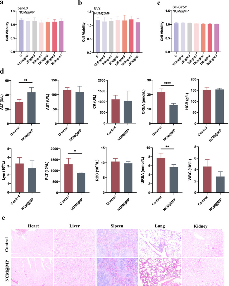
The secondary injury is more serious after traumatic brain injury (TBI) compared with primary injury. Release of excessive reactive oxygen species (ROS) and Ca2+ influx at the damaged site trigger the secondary injury. Herein, a neutrophil-like cell membrane-functionalized nanoparticle was developed to prevent ROS-associated secondary injury. NCM@MP was composed of three parts: (1) Differentiated neutrophil-like cell membrane (NCM) was synthesized, with inflammation-responsive ability to achieve effective targeting and to increase the retention time of Mn3O4 and nimodipine (MP) in deep injury brain tissue via C-X-C chemokine receptor type 4, integrin beta 1 and macrophage antigen-1. (2) Nimodipine was used to inhibit Ca2+ influx, eliminating the ROS at source. (3) Mn3O4 further eradicated the existing ROS. In addition, NCM@MP also exhibited desirable properties for T1 enhanced imaging and low toxicity which may serve as promising multifunctional nanoplatforms for precise therapies. In our study, NCM@MP obviously alleviated oxidative stress response, reduced neuroinflammation, protected blood-brain barrier integrity, relieved brain edema, promoted the regeneration of neurons, and improved the cognition of TBI mice. This study provides a promising TBI management to relieve the secondary spread of damage.

Keywords: Mn3O4; Neutrophil membrane; Nimodipine; Reactive oxygen species; Traumatic brain injury.


Conflict of interest statement

The authors declare that they have no competing interests.

Figures

Fig. 1
Fig. 1
a TEM images of Mn3O4. b The lattice of Mn3O4 shown in TEM. c HAADF-STEM-EDX mapping of Mn3O4. d Dynamic light scattering (DLS) measurements of Mn3O4. e X-ray Diffraction (XRD) pattern of Mn3O4. f Mn 2p1/2 and Mn 2p3/2 XPS spectra of Mn3O4. g Representative western blot images of Integrin beta 1, MAC-1 and CXCR4, respectively. h TEM image of NCM@MP. i Dynamic light scattering (DLS) of NCM@MP. jk The insets were the corresponding T1WI and T2WI of NCM@MP solution, respectively. lm r1 and r2 relaxivity of NCM@MP at the different concentrations of Mn. Data were mean ± SD (n = 3 per group). *p < 0.05, **p < 0.01, ***p < 0.001, ****p < 0.0001
Fig. 2
Fig. 2
a ·O2−, b DPPH, and c·OH scavenging rate by MP and NCM@MP, respectively. d Fluorescence values of ROS level in BV2 microglia with different treatments. Indicators for oxidative stress, including e SOD, f MDA activity under different and neuroinflammation indicators g IL-1β, h TNF-α levels in LPS-triggered BV2 cells under different treatment conditions showed significant anti-oxidative stress response and anti-inflammatory of NCM@MP. i Calcium overloading degree tested by Fluo-4/AM Labeling which showed NCM@MP alleviated intracellular calcium levels. jl Cell apoptosis representative images of H2O2-triggered SH-SY5Y cells treated by NCM@Mn3O4 and NCM@MP, which revealed neuroprotective function of NCM@MP in vitro. Data were mean ± SD (n = 3 per group). *p < 0.05, **p < 0.01, ***p < 0.001, ****p < 0.0001
Fig. 3
Fig. 3
ab Targeting ability of NCM@MP in vitro evaluated by ICP-MS analysis. cd In vivo T1WI and relaxation time of TBI lesion pre- and post-injection of NCM@MP at different time points. e The BBB-crossing ability of NCM@MP evaluated by T1 relaxation time. f T1 relaxation time were used to evaluate the ability of NCM@MP to target TBI. Data were mean ± SD (n = 5 per group). *p < 0.05, **p < 0.01, ***p < 0.001, ****p < 0.0001
Fig. 4
Fig. 4
ab Injury area percentage was decreased by NCM@MP treatment on the 1st day after TBI using TTC staining. cd Representative Ktrans map derived from DCE-MR imaging and quantitative analysis of the brain at 1-day post-treatment. ef Representative T2WI and quantitative analysis of brain edema at 1-day post-treatment. gh Representative T2WI and quantitative analysis of lesion volumes at 4 weeks post-injection. i Hematoxylin–eosin staining at 28-day post-treatment. These results showed NCM@MP improved BBB permeability and edema in TBI acute stage, and increased lesion defect recovery in chronic stage. jk Swimming trajectories of mice and the time to find the platform in the training phase in four groups in the testing phase of water maze test. lm Latency to first entry and platform entries on the test day. Results illustrated that the NCM@MP promoting the recovery of neuronal cognition and the spatial learning and memory abilities of TBI mice. Data were mean ± SD (n = 5 per group). *p < 0.05, **p < 0.01, ***p < 0.001, ****p < 0.0001
Fig. 5
Fig. 5
ab ELISA analysis of inflammatory factors IL-1β and TNF-α levels of the mice on day 1 post TBI with NCM@Mn3O4 or NCM@MP treatment confirmed the abilities on regulating inflammatory factors. cd The brain SOD and MDA levels of the mice on day 1 after different treatment confirmed the anti-oxidative abilities of the NCM@MP on TBI mice. e Representative immunohistochemical images and quantification of GFAP in astrocytes and lBA1 in microglia from the injured tissues in TBI models at day 3 post-injection, which showed NCM@MP reduced glial cell activation in vivo. The scale bar denotes 50 μm. f Representative immunohistochemical images and quantification of SOX and NeuN in regenerated tissues at day 28 post-injection, which showed NCM@MP promoted neuronal regeneration. The scale bar denotes 20 μm for SOX and 50 μm for NeuN. g Representative western blot images and quantification of signaling pathway. Data were mean ± SD (n = 3 per group). *p < 0.05, **p < 0.01, ***p < 0.001, ****p < 0.0001
Fig. 6
Fig. 6
ac The cell viability of bEnd.3, BV2 and SH-SY5Y treated with a variety of concentrations of NCM@MP and there were no significant vitality changes. de Serum biochemical indexes and HE staining of the main organs of the mice at 14 days post-treatment. These results showed great biocompatibility of NCM@MP. Data were mean ± SD (n = 5 per group). *p < 0.05, **p < 0.01, ***p < 0.001, ****p < 0.0001

Similar articles

Cited by

References

    1. Kalra S, Malik R, Singh G, Bhatia S, Al-Harrasi A, Mohan S, Albratty M, Albarrati A, Tambuwala MM. Pathogenesis and management of traumatic brain injury (TBI): role of neuroinflammation and anti-inflammatory drugs. Inflammopharmacology. 2022;30:1153–66. 10.1007/s10787-022-01017-8 - DOI - PMC - PubMed
    1. Werner C, Engelhard K. Pathophysiology of traumatic brain injury. Br J Anaesth. 2007;99:4–9. 10.1093/bja/aem131 - DOI - PubMed
    1. Tehse J, Taghibiglou C. The overlooked aspect of excitotoxicity: glutamate-independent excitotoxicity in traumatic brain injuries. Eur J Neurosci. 2019;49:1157–70. 10.1111/ejn.14307 - DOI - PubMed
    1. Feno S, Butera G, Reane DV, Rizzuto R, Raffaello A. Crosstalk between calcium and ROS in pathophysiological conditions. Oxid Med Cell Longev. 2019. 10.1155/2019/9324018. 10.1155/2019/9324018 - DOI - PMC - PubMed
    1. Han Z, Han Y, Huang X, Ma H, Zhang X, Song J, Dong J, Li S, Yu R, Liu H. A novel targeted nanoparticle for traumatic brain injury treatment: combined effect of ROS depletion and calcium overload inhibition. Adv Healthc Mater. 2022;11:e2102256. 10.1002/adhm.202102256 - DOI - PubMed

LinkOut - more resources