. 2024 Aug 12;22(1):476.
doi: 10.1186/s12951-024-02743-7.

Age-dependent changes in phagocytic activity: in vivo response of mouse pulmonary antigen presenting cells to direct lung delivery of charged PEGDA nanoparticles

Affiliations

Age-dependent changes in phagocytic activity: in vivo response of mouse pulmonary antigen presenting cells to direct lung delivery of charged PEGDA nanoparticles

Emma R Sudduth et al. J Nanobiotechnology. .

Erratum in

Abstract

Background: Current needle-based vaccination for respiratory viruses is ineffective at producing sufficient, long-lasting local immunity in the elderly. Direct pulmonary delivery to the resident local pulmonary immune cells can create long-term mucosal responses. However, criteria for drug vehicle design rules that can overcome age-specific changes in immune cell functions have yet to be established.

Results: Here, in vivo charge-based nanoparticle (NP) uptake was compared in mice of two age groups (2- and 16-months) within the four notable pulmonary antigen presenting cell (APC) populations: alveolar macrophages (AM), interstitial macrophages (IM), CD103+ dendritic cells (DCs), and CD11b+ DCs. Both macrophage populations exhibited preferential uptake of anionic nanoparticles but showed inverse rates of phagocytosis between the AM and IM populations across age. DC populations demonstrated preferential uptake of cationic nanoparticles, which remarkably did not significantly change in the aged group. Further characterization of cell phenotypes post-NP internalization demonstrated unique surface marker expression and activation levels for each APC population, showcasing heightened DC inflammatory response to NP delivery in the aged group.

Conclusion: The age of mice demonstrated significant preferences in the charge-based NP uptake in APCs that differed greatly between macrophages and DCs. Carefully balance of the targeting and activation of specific types of pulmonary APCs will be critical to produce efficient, age-based vaccines for the growing elderly population.

Keywords: Aging; Antigen presenting cells; Nanoparticles; Phagocytosis; Pulmonary delivery; Pulmonary immunity.


Conflict of interest statement

The authors declare no competing interests.

Figures

Fig. 1
Fig. 1
Advanced age demonstrates shift in APC population phenotype. Whole murine lung samples were digested into a single cell suspension for multicolor flow cytometry analysis of four pulmonary antigen presenting cells (APCs) of interest (alveolar macrophages (macs.; AMs), interstitial macrophages (IMs), CD103+ dendritic cell (DCs), and CD11b+ DCs across two ages (2- and 16-months). (A) Study overview schematic including cells and age models of interest. (B) Primary gating for cells, singlets, live cells, and CD45+ cells. C-D.) Representative flow gating scheme for 2-months (C) and 16-months (D)
Fig. 2
Fig. 2
16-month lung presents unique cellular immunophenotype. Cellular comparison was performed of the murine lung for two age groups of mice (2- and 16-months). (A) Fold change in surface marker MFI of 16-month isolated cell types compared to average expression of the 2-month group. Boxes with Xs indicate markers that were not expressed in those cell types. (B) Multicolor flow cytometry analysis of overall CD45+ cell counts and CD45 median fluorescence intensity (MFI). C-F.) Flow cytometry isolated individual cell counts for alveolar macrophages (AM; C), interstitial macrophages (IM; D), CD103+ dendritic cells (DCs; E), and CD11b+ DCs (F) in the two ages. Displayed numerical results represents mean ± SD (n = 5) from untreated group results. Indicated significance is calculated via unpaired Student’s t-tests [p < 0.05 (*), 0.01 (**), 0.001 (***), < 0.001 (****)]
Fig. 3
Fig. 3
PEGDA Nanoparticle Formulation and Dosing Timeline. Poly(ethylene-glycol) diacrylate (PEGDA) hydrogel nanoparticles (NPs) at 50 wt% solid were synthesized using a reverse emulsion photopolymerization technique for uptake studies. (A) Composition of PEGDA pre-particle composition (left) and specific co-monomer chemical structures (right) of PEGDA NPs. (B) Surface presentation of synthesized NPs (pink = (+) NP, purple = (-) NP) (C) Dynamic light scattering data for NPs. Results represent mean ± SD, n = 2 independent batch replicates. Dh = hydrodynamic size, PDI = polydispersity index (D) Timeline of dosage experiments with NPs leading to collection of bronchoalveolar lavage fluid (BALF), serum, and whole lung tissue for analysis
Fig. 4
Fig. 4
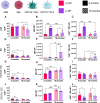
Nanoparticle uptake shifts with charge, age, and type of cell. Two ages of mice (2- and 16- months) were dosed via orotracheal instillation with 100 µg of positive [(+)NP] or negative [(-)NP] PEGDA NPs. After 72 h, flow cytometry determined NP uptake in four APCs in the lung including alveolar macrophages (AMs, A-C), interstitial macrophages (IMs, D-F), CD103+ dendritic cells (DCs, G-I), and CD11b+ DCs (J-L). Parameters shown include percent of cell population with NP uptake (%NP; A, D, G, J), median fluorescence intensity of the NP channel (MFI; B, E, H, K), and the total fluorescence (%NP*MFI; C, F, I, L). Data represents mean ± SD (n = 5). Indicated significance is calculated via two-way ANOVA [Tukey Test, p < 0.05 (*), 0.01 (**), 0.001 (***), < 0.001 (****)]
Fig. 5
Fig. 5
Aged phagocytotic cells in BALF show reduced NP uptake. After 72 h orotracheal instillation of positive (+) or negative (-) PEGDA NPs, cells isolated from BALF from 2- and 16-month mice. Representative stained images to identify nuclei (DAPI; blue), actin (Phalloidin; grey), and NP uptake (pink) were taken for 2-month (+)NP (A), 2-month (-)NP (B), 16-month (+)NP (C), and 16-month (-)NP (D). Images were taken using Biotek Cytation 5 Multimode Imager in which exposure was modified to visualize NPs in various samples. 2-month (-) NP samples (B) were at least 2x shorter in integration time than all other conditions. Scale bar represents 50 μm
Fig. 6
Fig. 6
Immunofluorescence histology highlights CD11c+cellular uptake of NPs. Frozen whole lung murine tissue was sectioned at 10 μm thickness after 72 h dosage of either positive (+; A, C,E, G) or negative (-; B, D,F, H) nanoparticles (NPs; pink) via orotracheal instillation. The tissue was stained with primary anti-hamster CD11c, secondary IgG Alexa Fluor 488 (green), and DAPI (nuclei; blue). Representative images (n = 1) represent 2-month specimens (A, B, E, F) and 16-month specimens (C, D, G, H). White arrows indicate overlap between CD11c+, NP+ cells, while pink arrows represent non-overlapping “non-localized” NPs. Images acquired using Biotek Cytation 5 Multimode Imager and exposure was modified for viewing of nanoparticles and CD11c stains in each image. Top and bottom row scale bars represent 100 μm and 25 μm, respectively
Fig. 7
Fig. 7
Nanoparticle uptake in aged cells changes activation profile. A.) Multicolor flow cytometry results from partial lung tissue digests after 72-hr pulmonary delivery of PEGDA NPs. Alveolar macrophages (AMs; CD45+, CD11c+, SiglecF+) and dendritic cells (DCs; CD45+, CD11c+, MHCIIhigh) were isolated from partial lung digest. AMs (B-D) and DCs (E-G) expression of CD80 (B, E), CD86 (C, F), and MHCII (D, G) were compared across NP treatments using fold change from age-appropriate UT. Data represents mean ± SD (n = 5). Indicated significance is calculated via two-way ANOVA [Tukey Test, p < 0.05 (*), 0.01 (**), 0.001 (***), < 0.001 (****)]

Similar articles

Cited by

  • Nanoparticle-Based Pulmonary Immune Engineering.
    Trautmann-Rodriguez M, Fromen CA. Trautmann-Rodriguez M, et al. Annu Rev Chem Biomol Eng. 2025 Jun;16(1):249-270. doi: 10.1146/annurev-chembioeng-082223-105117. Epub 2025 Mar 12. Annu Rev Chem Biomol Eng. 2025. PMID: 40073112 Free PMC article. Review.

References

    1. Häder A, Köse-Vogel N, Schulz L, Mlynska L, Hornung F, Hagel S, Teichgräber U, Lang SM, Pletz MW, Saux CJL, et al. Respiratory infections in the aging lung: implications for diagnosis, therapy, and Prevention. Aging Dis. 2023;14:1091–104. - PMC - PubMed
    1. Schneider JL, Rowe JH, Garcia-de-Alba C, Kim CF, Sharpe AH, Haigis MC. The aging lung: physiology, disease, and immunity. Cell. 2021;184:1990–2019. - PMC - PubMed
    1. Boe DM, Boule LA, Kovacs EJ. Innate immune responses in the ageing lung. Clin Exp Immunol. 2017;187:16–25. - PMC - PubMed
    1. Skloot GS. The effects of Aging on Lung structure and function. Clin Geriatr Med. 2017;33:447–57. - PubMed
    1. Amanna IJ. Balancing the efficacy and safety of vaccines in the Elderly. Open Longev Sci. 2012;6:64–72. - PMC - PubMed

Substances

LinkOut - more resources