. 2024 Aug 12;21(1):186.
doi: 10.1186/s12985-024-02460-5.

SARS-CoV-2 nucleocapsid protein promotes self-deacetylation by inducing HDAC6 to facilitate viral replication

Affiliations

SARS-CoV-2 nucleocapsid protein promotes self-deacetylation by inducing HDAC6 to facilitate viral replication

Arpita Mukherjee et al. Virol J. .

Abstract

Background: The global outbreak of COVID-19 caused by the SARS-CoV-2 has led to millions of deaths. This unanticipated emergency has prompted virologists across the globe to delve deeper into the intricate dynamicity of the host-virus interface with an aim to identify antiviral targets and elucidate host and viral determinants of severe disease.

Aim: The present study was undertaken to analyse the role of histone deacetylase 6 (HDAC6) in regulating SARS-CoV-2 infection.

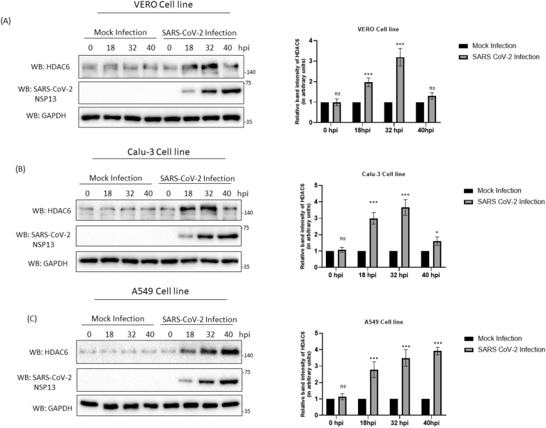
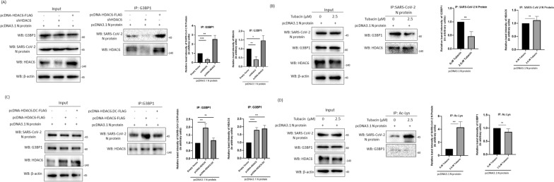
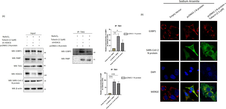
Results: Gradual increase in HDAC6 expression was observed in different SARS-CoV-2-permissive cell lines following SARS-CoV-2 infection. The SARS-CoV-2 nucleocapsid protein (N protein) was identified as the primary viral factor responsible for upregulating HDAC6 expression. Downregulation of HDAC6 using shRNA or a specific inhibitor tubacin resulted in reduced viral replication suggesting proviral role of its deacetylase activity. Further investigations uncovered the interaction of HDAC6 with stress granule protein G3BP1 and N protein during infection. HDAC6-mediated deacetylation of SARS-CoV-2 N protein was found to be crucial for its association with G3BP1.

Conclusion: This study provides valuable insights into the molecular mechanisms underlying the disruption of cytoplasmic stress granules during SARS-CoV-2 infection and highlights the significance of HDAC6 in the process.

Keywords: Deacetylation; G3BP1; HDAC6; SARS-CoV-2; Stress granules (SGs).


Conflict of interest statement

The authors declare no competing interests.

Figures

Fig. 1
Fig. 1
SARS-CoV-2 infection leads to increased expression of HDAC6. A VERO E6 cells (at a MOI of 1), B Calu-3 cells (at a MOI of 1) and C A549 cell (at a MOI of 2) infected with SARS-CoV-2 or kept mock-infected were harvested at indicated time post-infection (18, 32, and 40 hpi) and cellular lysates were prepared. Lysates were run on SDS-PAGE followed by western blot analysis using anti-HDAC6 antibody. GAPDH and NSP13 were used as internal loading control and SARS-CoV-2 infection marker respectively
Fig. 2
Fig. 2
Inhibition of HDAC6 resulted in reduction of SARS CoV-2 replication. A A549 cells were either transfected with shHDAC6 or pcDNA-HDAC6-FLAG followed by SARS-CoV-2 infection (at a MOI = 1). Total RNA was isolated after 24 h of infection followed by qRT-PCR using SARS-CoV-2 orf1a, hdac6 and gapdh-specific primers. Data represent means ± SD of three independent experiments. ***p ≤ .001, unpaired t-test and **p ≤ .01, unpaired t-test. B Protein was extracted from SARS-CoV-2 infected cells at 24 hpi (MOI = 1) ectopically expressing shHDAC6 or pcDNA-HDAC6-FLAG. S, NSP13 and HDAC6 expression levels were analysed via western blotting. C SARS-CoV-2 infected (24 hpi) A549 cells were treated with increasing doses of tubacin (0–2.5 μM). Cellular extracts were subjected to immunoblot to analyse levels of S and NSP13. GAPDH was used as internal loading control. D Total RNA was isolated from tubacin treated SARS-CoV-2 infected cells (MOI = 1) at 24 hpi followed by qRT-PCR using SARS-CoV-2 orf1a and gapdh specific primers. Data represent means ± SD of three independent experiments. ***p ≤ .001, unpaired t-test and **p ≤ .01, unpaired t-test. E A549 cells were either transfected with empty vector or pcDNA-HDAC6-FLAG or pcDNA-HDAC6.DC-FLAG followed by SARS-CoV-2 infection (at a MOI = 0.5). Total RNA was isolated after 24 h of infection followed by qRT-PCR using SARS-CoV-2 orf1a, hdac6 and gapdh-specific primers. Data represent means ± SD of three independent experiments. ***p ≤ 0.001, unpaired t-test, *p ≤ 0.1, unpaired t-test and ns = not significant, unpaired t-test. F A549 cells were infected with SARS-CoV-2 (MOI = 1) either in presence or absence of tubacin (1–2.5 μM) for 24, 48 and 72 h followed by plaque assay. Viral titers were measured as plaque forming units [log (pfu/ml)]. Data represent means ± SD of three independent experiments. **p ≤ .01,multiple t-tests,***p ≤ .001, multiple t-tests and ns = not significant, multiple t-tests. G A549 cells were either transfected with empty vector or shHDAC6 or pcDNA-HDAC6-FLAG or pcDNA-HDAC6.DC-FLAG followed by SARS-CoV-2 infection (at a MOI = 0.5) for 24, 48 and 72 h followed by plaque assay. Viral titers were measured as plaque forming units [log (pfu/ml)]. Data represent means ± SD of three independent experiments. ***p ≤ .001, multiple t-tests, *p ≤ 0.05,  multiple t-tests and ns = not significant, multiple t-tests
Fig. 3
Fig. 3
Increased interaction between G3BP1 and HDAC6 during SARS-CoV-2 infection. A Cellular lysates from A549 cells either infected with SARS-CoV-2 at a MOI of 2 (18–32 hpi) or kept mock-infected were run on SDS-PAGE and protein level expression of acetylated α-tubulin, HDAC6 and NSP13 protein was analysed via western blotting. B SARS-CoV-2-infected (18–32 hpi) or mock-infected lysates were pulled down using HDAC6-specific antibody followed by western blot analysis of the immunoprecipitates by anti-G3BP1, anti-HSP90 and anti-NSP13 antibodies. Inputs were probed with anti-HSP90, anti-G3BP1, anti-HDAC6, anti-NSP13 and β-actin antibodies confirming protein expression. (C) Reciprocal co-immunoprecipitation analysis was carried out in SARS-CoV-2-infected (18–32 hpi) or mock-infected lysates; anti-G3BP1 was used for pull down and immunoblot analysis was performed with HDAC6-specific antibody. Inputs were probed with anti-G3BP1, anti-HDAC6 and β-actin antibodies
Fig. 4
Fig. 4
SARS-CoV-2 N Protein interacts with HDAC6. A Cellular lysates from SARS-CoV-2-infected or mock-infected A549 cells were co-immunoprecipitated with anti-HDAC6 antibody. Western blot analysis was performed to check the expression of N protein and SARS-CoV-2 Spike protein within the HDAC6 immunoprecipitates. Inputs were probed with anti-HDAC6, anti-N and anti-Spike antibodies confirming protein expression. B A549 cells were either transfected with pcDNA3.1-N plasmid or empty vector followed by immunoprecipitation with anti-HDAC6 antibody. Immunoblot analysis was performed with anti-N protein and anti-HDAC6 antibody. C Reciprocal co-immunoprecipitation assay from A549 cells either transfected with pcDNA3.1-N or empty vector using anti-N protein antibody was performed. Immunoprecipitates were analysed for the expression of HDAC6 and N protein. Inputs were probed with anti-HDAC6, anti-N and anti-β-actin antibodies confirming protein expression
Fig. 5
Fig. 5
SARS-CoV-2 N Protein induces expression of HDAC6. A Cellular lysates were prepared from A549 cells either transfected with increasing concentration of pcDNA3.1-N (1–3 μg) or transfected with empty pcDNA3.1 vector. Expression of HDAC6, N protein and GAPDH was analysed via western blotting. B Co-immunoprecipitation analysis was performed in A549 cells either pcDNA3.1-N protein-transfected or empty vector-transfected A549 cells using anti-G3BP1 antibody and immunoblot analysis was performed using HDAC6 and N protein-specific antibodies. C Total RNA was isolated from A549 cells either transfected with empty vector or pcDNA3.1-N protein followed by qRT-PCR using hdac6 and gapdh-specific primers. ns = not significant, unpaired t-test. D A549 cells were either transfected with empty vector or pcDNA3.1 N protein followed by CHX treatment at 18 h post-transfection. Cells were harvested at indicated time post-CHX treatment (0, 6, 12, 18) and HDAC6, SARS-CoV-2 N protein and GAPDH expressions were analysed via immunoblotting. E A549 cells were either transfected with pcDNA3.1-N protein or empty vector followed by immunoprecipitation with anti-K48-ubiquitin antibody. Immunoblot analysis was performed with anti-HDAC6 antibody. F SARS-CoV-2-infected (24–32 hpi) or mock-infected lysates were pulled down using with anti-K48-ubiquitin antibody. Immunoblot analysis was performed with anti-HDAC6 antibody
Fig. 6
Fig. 6
HDAC6 mediates interaction between G3BP1 and SARS-CoV-2 N protein. A A549 cells were transfected with pcDNA3.1-N and co-transfected with either pcDNA-HDAC6-FLAG or shHDAC6 were analysed by co-immunoprecipitation by using anti-G3BP1 antibody. Immunoprecipitates were subjected to western blot analysis using HDAC6 and SARS-CoV-2 N protein-specific antibody. B A549 cells transfected with pcDNA3.1-N plasmid either in presence or absence of tubacin (2.5 μM), followed by co-immunoprecipitation of cellular lysates with anti-SARS-CoV-2 N protein antibody. Immunoblot analysis was performed to check the expression of G3BP1 and HDAC6. C A549 cells were transfected with pcDNA3.1-N and co-transfected with either pcDNA-HDAC6-FLAG or pcDNA-HDAC6.DC-FLAG. Cellular lysates were subjected to co-immunoprecipitation using anti-G3BP1 antibody. Western blot analysis was performed using SARS-CoV-2 N protein and HDAC6-specific antibodies. Input lanes were probed with anti-G3BP1, anti-HDAC6 and anti-SARS-CoV-2 N antibodies. D A549 cells transfected with pcDNA3.1-N and either treated with tubacin (2.5 μM) or treated with DMSO. Cell lysates were subjected to immunoprecipitation using anti-Ac-Lys antibody. Western blot was performed using G3BP1 and HDAC6-specific antibodies
Fig. 7
Fig. 7
Induction of HDAC6 expression during SARS-CoV-2 infection facilities disruption of Stress Granules. A A549 cells were transfected with pcDNA3.1-N protein and/or co-transfected with shHDAC6 followed by either tubacin (2.5 µM) or DMSO treatment. Cells were treated with NaAsO2 1 h before harvesting. Co-immunoprecipitation was performed from the cellular lysates in using anti-TIA-1 antibody. Western blot was performed using PABP, N protein, G3BP1, HDAC6 and TIA-1-specific antibodies. B A549 cells were either transfected with pcDNA3.1-N protein or empty vector and either treated with tubacin (2.5 µM) or left untreated. 1 h before fixation cells were treated with NaAsO2 followed by permeabilization and staining with anti-G3BP1 and anti-N protein primary antibody. Secondary staining was performed with Rhodamine-conjugated anti-mouse (for G3BP1) and DyLight™ 488-conjugated anti-rabbit (for N protein) secondary antibodies. DAPI was used for mounting. Imaging was done with a confocal microscope (63× oil immersion) and scale bar was set at 5 μm
Fig. 8
Fig. 8
Schematic representation showing crosstalk between HDAC6 and SARS-CoV-2 infection. SARS-CoV-2 N protein interacts with and induces the expression of HDAC6. Subsequently, HDAC6 promotes the deacetylation of the N protein, which facilitates its association with G3BP1, leading to the disruption of cellular stress granules

Similar articles

Cited by

References

    1. Agrawal P, Sambaturu N, Olgun G, Hannenhalli S. A path-based analysis of infected cell line and COVID-19 patient transcriptome reveals novel potential targets and drugs against SARS-CoV-2. Front Immunol. 2022;13:918817. 10.3389/fimmu.2022.918817 - DOI - PMC - PubMed
    1. Ashour HM, Elkhatib WF, Rahman MM, Elshabrawy HA. Insights into the recent 2019 novel coronavirus (SARS-CoV-2) in light of past human coronavirus outbreaks. Pathogens. 2020;9(3):186. 10.3390/pathogens9030186 - DOI - PMC - PubMed
    1. Baer A, Kehn-Hall K. Viral concentration determination through plaque assays: using traditional and novel overlay systems. J Vis Exp. 2014;93:e52065. 10.3791/52065.PMID:25407402;PMCID:PMC4255882.10.3791/52065.PMID:25407402;PMCID:PMC4255882 - DOI - PMC - PubMed
    1. Banerjee I, Miyake Y, Nobs SP, Schneider C, Horvath P, Kopf M, Matthias P, Helenius A, Yamauchi Y. Influenza A virus uses the aggresome processing machinery for host cell entry. Science. 2014;346(6208):473–7. 10.1126/science.1257037 - DOI - PubMed
    1. Bayati A, Kumar R, Francis V, McPherson PS. SARS-CoV-2 infects cells after viral entry via clathrin-mediated endocytosis. J Biol Chem. 2021;296:100306. 10.1016/j.jbc.2021.100306 - DOI - PMC - PubMed

Publication types

MeSH terms

LinkOut - more resources