Review
. 2024 Aug 13;12(1):81.
doi: 10.1186/s40364-024-00624-7.

The role of macrophage plasticity in neurodegenerative diseases

Affiliations
Review

The role of macrophage plasticity in neurodegenerative diseases

Hongyue Ma et al. Biomark Res. .

Abstract

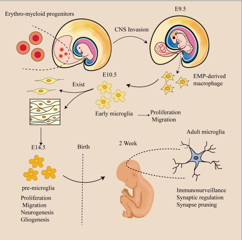
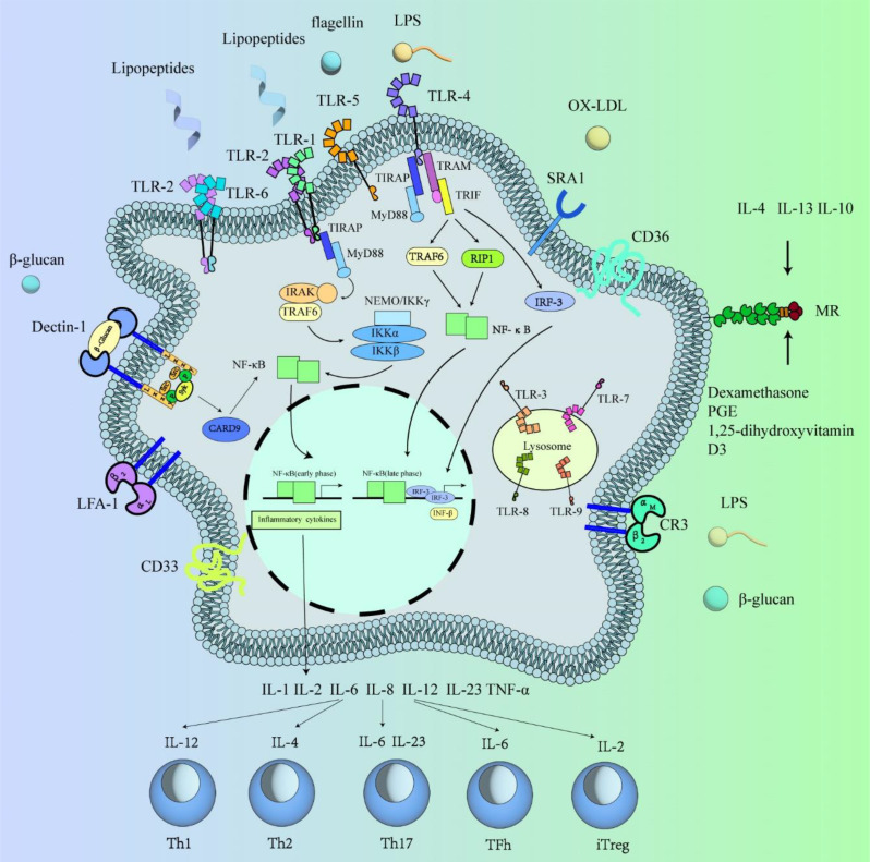
Tissue-resident macrophages and recruited macrophages play pivotal roles in innate immunity and the maintenance of brain homeostasis. Investigating the involvement of these macrophage populations in eliciting pathological changes associated with neurodegenerative diseases has been a focal point of research. Dysregulated states of macrophages can compromise clearance mechanisms for pathological proteins such as amyloid-β (Aβ) in Alzheimer's disease (AD) and TDP-43 in Amyotrophic lateral sclerosis (ALS). Additionally, recent evidence suggests that abnormalities in the peripheral clearance of pathological proteins are implicated in the pathogenesis and progression of neurodegenerative diseases. Furthermore, numerous genome-wide association studies have linked genetic risk factors, which alter the functionality of various immune cells, to the accumulation of pathological proteins. This review aims to unravel the intricacies of macrophage biology in both homeostatic conditions and neurodegenerative disorders. To this end, we initially provide an overview of the modifications in receptor and gene expression observed in diverse macrophage subsets throughout development. Subsequently, we outlined the roles of resident macrophages and recruited macrophages in neurodegenerative diseases and the progress of targeted therapy. Finally, we describe the latest advances in macrophage imaging methods and measurement of inflammation, which may provide information and related treatment strategies that hold promise for informing the design of future investigations and therapeutic interventions.

Keywords: Gene expression; Imaging methodologies; Immune cells; Macrophages; Neurodegenerative disease.


Conflict of interest statement

The authors declare no competing interests.

Figures

Fig. 1
Fig. 1
Microglial development. Microglia emerge from yolk sac precursors, and PreMacs are able to colonize the whole embryo and differentiate into tissue-specific resident macrophages during organ development. They colocate in the developing brain around E9.5. At approximately E14.5, a transition from early to premicroglia occurs, with premicroglia progressively acquiring adult properties after birth and different developmental phenotypes
Fig. 2
Fig. 2
Macrophage receptors and their secreted factors. Polysaccharide-activated macrophages first bind to cell surface polysaccharide receptors, activate intracellular signaling pathways, mediate the release of inflammatory factors such as TNF-α, IL-1β, and IL-6, and subsequently enhance their immunomodulatory ability. The main polysaccharide receptors identified include TLRs, MR, Dectin-1, SR, and CR3. Inflammatory factors can activate different T-cell subsets
Fig. 3
Fig. 3
The structures, surface markers, cytokine secretion and biological functions of the M1 and M2 macrophage subpopulations are summarized. Macrophages differentiate into two heterogeneous and opposing macrophage subtypes: classically activated (“M1-type”) macrophages stimulated by LPS and IFN-γ and alternatively activated (“M2-type”) macrophages stimulated by IL-4 and IL-13. M2 macrophages can dedifferentiate into four subtypes: 2a, 2b, 2c and 2d. With the exception of M2b macrophages, the other three cell types are activated by the secretion of the appropriate chemokines and anti-inflammatory factors
Fig. 4
Fig. 4
Macrophage ontogeny and specification. Arrows indicate developmental relationships between cells. Macrophage-specific transcription factors and tissue signals are marked in red and blue, respectively. Tissue-resident macrophages are tissue-specific subpopulations that arise during organogenesis. They establish and maintain stable spatial and functional relationships with specialized tissue cells. For example, microglia coexist with neurons in the brain, osteoclasts with osteoblasts in bone, and adipose-associated macrophages with white adipocytes in adipose tissue. They sense and integrate local and systemic information to provide growth factors, nutrient recycling and waste removal to specialized tissue cells essential for tissue growth, homeostasis and repair

Similar articles

Cited by

References

    1. Metschnikoff E. Lecture on phagocytosis and immunity. BMJ. 1891;1:213–7.10.1136/bmj.1.1570.213 - DOI - PMC - PubMed
    1. Wang Z, Wu Z, Wang H, Feng R, Wang G, Li M, et al. An immune cell atlas reveals the dynamics of human macrophage specification during prenatal development. Cell. 2023;186:4454–e7119. 10.1016/j.cell.2023.08.019 - DOI - PubMed
    1. Utz SG, See P, Mildenberger W, Thion MS, Silvin A, Lutz M et al. Early fate defines microglia and non-parenchymal brain macrophage development. Cell. 2020;181:557 – 73.e18. - PubMed
    1. Chakarov S, Lim HY, Tan L, Lim SY, See P, Lum J, et al. Two distinct interstitial macrophage populations coexist across tissues in specific subtissular niches. Science. 2019;363:eaau0964. 10.1126/science.aau0964 - DOI - PubMed
    1. Dick SA, Wong A, Hamidzada H, Nejat S, Nechanitzky R, Vohra S, et al. Three tissue resident macrophage subsets coexist across organs with conserved origins and life cycles. Sci Immunol. 2022;7:eabf7777. 10.1126/sciimmunol.abf7777 - DOI - PubMed

LinkOut - more resources