. 2024 Jul 29:15:1447757.
doi: 10.3389/fphar.2024.1447757. eCollection 2024.

Melatonin improves cognitive dysfunction and decreases gliosis in the streptozotocin-induced rat model of sporadic Alzheimer's disease

Affiliations

Melatonin improves cognitive dysfunction and decreases gliosis in the streptozotocin-induced rat model of sporadic Alzheimer's disease

Zsolt Gáll et al. Front Pharmacol. .

Abstract

Introduction: Alzheimer's disease (AD) and other forms of dementia have a devastating effect on the community and healthcare system, as neurodegenerative diseases are causing disability and dependency in older population. Pharmacological treatment options are limited to symptomatic alleviation of cholinergic deficit and accelerated clearance of β-amyloid aggregates, but accessible disease-modifying interventions are needed especially in the early phase of AD. Melatonin was previously demonstrated to improve cognitive function in clinical setting and experimental studies also.

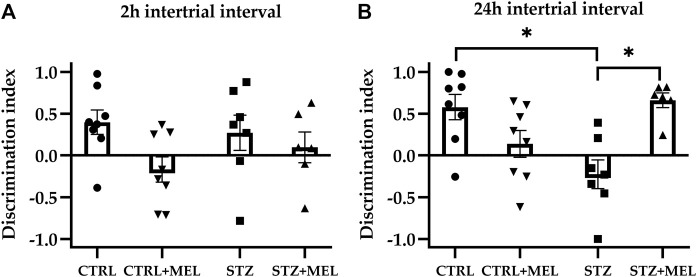
Methods: In this study, the influence of melatonin supplementation was studied on behavioral parameters and morphological aspects of the hippocampus and amygdala of rats. Streptozotocin (STZ) was injected intracerebroventricularly to induce AD-like symptoms in male adult Wistar rats (n = 18) which were compared to age-matched, sham-operated animals (n = 16). Melatonin was administered once daily in a dose of 20 mg/kg body weight by oral route. Behavioral analysis included open-field, novel object recognition, and radial-arm maze tests. TNF-α and MMP-9 levels were determined from blood samples to assess the anti-inflammatory and neuroprotective effects of melatonin. Immunohistological staining of brain sections was performed using anti-NeuN, anti-IBA-1, and anti-GFAP primary antibodies to evaluate the cellular reorganization of hippocampus.

Results and discussion: The results show that after 40 days of treatment, melatonin improved the cognitive performance of STZ-induced rats and reduced the activation of microglia in both CA1 and CA3 regions of the hippocampus. STZ-injected animals had higher levels of GFAP-labeled astrocytes in the CA1 region, but melatonin treatment reduced this to that of the control group. In conclusion, melatonin may be a potential therapeutic option for treating AD-like cognitive decline and neuroinflammation.

Keywords: Alzheimer’s disease; animal model; cognitive dysfunction; melatonin; neuroinflammation.


Conflict of interest statement

The authors declare that the research was conducted in the absence of any commercial or financial relationships that could be construed as a potential conflict of interest.

Figures

FIGURE 1
FIGURE 1
Timeline of the study design. Legend: OF, open field test; NOR, novel object recognition test; RAM, radial arm maze test; ICV, intracerebroventricular. Solid arrows represent behavioral tests, while striped arrows represent surgical interventions.
FIGURE 2
FIGURE 2
Open field test performance of melatonin treated rats underwent ICV-STZ injection. Quantified parameters: distance moved (A), freezing time (B), number of entries in the center zone (C), and the number of rearings (D). Legend: *p < 0.05, **p < 0.01, ***p < 0.001, CTRL–control group icv injected with buffer and treated with vehicle (n = 8), CTRL + MEL–group icv injected with buffer and treated with melatonin (n = 8), STZ–group icv injected with streptozotocin and treated with vehicle (n = 7), STZ + MEL–group icv injected with streptozotocin and treated with melatonin (n = 6).
FIGURE 3
FIGURE 3
The discrimination index derived from novel object recognition task as a measure of recognition memory. Two intertrial intervals were tested: (A) 2-hrs and (B) 24-hrs. Data is expressed as mean ± SEM. Legend: *p < 0.05, CTRL–control group icv injected with buffer and treated with vehicle (n = 8), CTRL + MEL–group icv injected with buffer and treated with melatonin (n = 8), STZ–group icv injected with streptozotocin and treated with vehicle (n = 7), STZ + MEL–group icv injected with streptozotocin and treated with melatonin (n = 6).
FIGURE 4
FIGURE 4
Radial arm maze test performance of melatonin treated rats underwent ICV-STZ injec-tion. Quantified parameters: target visits (A), total errors (B), working (C), and reference memory errors (D). Legend: *p < 0.05, CTRL–control group icv injected with buffer and treated with ve-hicle (n = 8), CTRL + MEL–group icv injected with buffer and treated with melatonin (n = 8), STZ–group icv injected with streptozotocin and treated with vehicle (n = 7), STZ + MEL–group icv in-jected with streptozotocin and treated with melatonin (n = 6).
FIGURE 5
FIGURE 5
Serum levels of (A) MMP-9 and (B) TNF-α after melatonin treatment in ICV-STZ model of Alzheimer’s disease. Legend: *p < 0.05, CTRL–control group icv injected with buffer and treated with vehicle (n = 7), CTRL + MEL–group icv injected with buffer and treated with melatonin (n = 8), STZ–group icv injected with streptozotocin and treated with vehicle (n = 5), STZ + MEL–group icv injected with streptozotocin and treated with melatonin (n = 6).
FIGURE 6
FIGURE 6
Effects of chronic melatonin (20 mg/kg body weight) treatment on the hippocampal re-organization induced by ICV-STZ in rats. Representative confocal microscopy images of the cornu Ammonis 3 (CA3) region (A) in the control group injected ICV with citrate buffer; (B) in the control group treated with melatonin; (C) in the ICV-STZ injected group; (D) in the ICV-STZ injected and melatonin treated group. The percentage area of neuron specific nuclear protein (NeuN) immunostaining (expressed as percentage of the area covered by positive signal compared to the total area of a region) revealed no effect of the melatonin treatment on STZ-induced cell loss in the (E) CA1 region and (F) CA3 region of the hippocampus. The magnification was ×20. The scale bar indicates 50 μm. Legend: ***p < 0.001, CTRL–control group icv injected with buffer and treated with vehicle (n = 10 hippocampi from n = 5 animals), CTRL + MEL–group icv injected with buffer and treated with melatonin (n = 16 hippocampi from n = 8 animals), STZ–group icv injected with streptozotocin and treated with vehicle (n = 20 hippocampi from n = 7 animals), STZ + MEL–group icv injected with streptozotocin and treated with melatonin (n = 12 hippocampi from n = 6 animals). The red rectangle indicates the region where significant modifications were observed.
FIGURE 7
FIGURE 7
Effects of chronic melatonin (20 mg/kg body weight) treatment on the GFAP-positive astrocytes in the ICV-STZ sporadic model of Alzheimer’s disease in rats. Representative confocal microscopy images of the cornu Ammonis 3 (CA3) region (A) in the control group injected ICV with citrate buffer; (B) in the control group treated with melatonin; (C) in the ICV-STZ injected group; (D) in the ICV-STZ injected and melatonin treated group. The magnification was ×20. The scale bar indicates 50 μm. The astrocyte density (expressed as cell/mm2) revealed no effect of the melatonin treatment in the CA1 region (E) but it did reduce astrocyte density in the CA3 region (F) of the hippocampus. The astrocyte dimension (expressed as mean cell surface in μm2) showed significant decrease in both melatonin treated groups in the CA1 region (G), however, the modifications did not reach the limit of significance in the CA3 region (H). Data in column graphs are expressed as mean ± SEM (n = 5–8).; Legend: *p < 0.05, CTRL–control group icv injected with buffer and treated with vehicle (n = 10 hippocampi from n = 5 animals), CTRL + MEL–group icv injected with buffer and treated with melatonin (n = 16 hippocampi from n = 8 animals), STZ–group icv injected with streptozotocin and treated with vehicle (n = 20 hippocampi from n = 7 animals), STZ + MEL–group icv injected with streptozotocin and treated with melatonin (n = 12 hippocampi from n = 6 animals). Red arrowheads indicate the regions where significant differences were observed.
FIGURE 8
FIGURE 8
Effects of chronic melatonin (20 mg/kg body weight) treatment on the IBA-1-positive microglia in the ICV-STZ sporadic model of Alzheimer’s disease in rats. Representative confocal microscopy images of the cornu Ammonis 3 (CA3) region (A) in the control group injected ICV with citrate buffer; (B) in the control group treated with melatonin; (C) in the ICV-STZ injected group; (D) in the ICV-STZ injected and melatonin treated group. The magnification was ×20. The scale bar indicates 50 μm. The microglia density (expressed as cell/mm2) showed significant increase due to ICV-STZ injection in both CA1 and CA3 regions and melatonin treatment reduced this increase in the CA1 region (E), however it did not reach significance limit in the CA3 region (F) of the hippocampus. Data in column graphs are expressed as mean ± SEM (n = 5–8).; Legend: *p < 0.05, **p < 0.01, CTRL–control group icv injected with buffer and treated with vehicle (n = 10 hippocampi from n = 5 animals), CTRL + MEL–group icv injected with buffer and treated with melatonin (n = 16 hippocampi from n = 8 animals), STZ–group icv injected with streptozotocin and treated with vehicle (n = 20 hippocampi from n = 7 animals), STZ + MEL–group icv injected with streptozotocin and treated with melatonin (n = 12 hippocampi from n = 6 animals). Green arrowheads indicate the regions where significant differences were observed.

References

    1. Andrade M. K., Souza L. C., Azevedo E. M., Bail E. L., Zanata S. M., Andreatini R., et al. (2023). Melatonin reduces β-amyloid accumulation and improves short-term memory in streptozotocin-induced sporadic Alzheimer’s disease model. IBRO Neurosci. Rep. 14, 264–272. 10.1016/j.ibneur.2023.01.005 - DOI - PMC - PubMed
    1. Antunes M., Biala G. (2012). The novel object recognition memory: neurobiology, test procedure, and its modifications. Cogn. Process 13 (2), 93–110. 10.1007/s10339-011-0430-z - DOI - PMC - PubMed
    1. Berg S., Kutra D., Kroeger T., Straehle C. N., Kausler B. X., Haubold C., et al. (2019). ilastik: interactive machine learning for (bio)image analysis. Nat. Methods 16 (12), 1226–1232. 10.1038/s41592-019-0582-9 - DOI - PubMed
    1. Bubenik G. A., Konturek S. J. (2011). Melatonin and aging: prospects for human treatment. J. Physiol. Pharmacol. 62 (1), 13–19. - PubMed
    1. Cardinali D P., Furio A M., Brusco L I. (2010). Clinical aspects of melatonin intervention in Alzheimers disease progression. Curr. Neuropharmacol. 8 (3), 218–227. 10.2174/157015910792246209 - DOI - PMC - PubMed

LinkOut - more resources