. 2024 Jun:13:29-89.
doi: 10.3114/fuse.2024.13.03. Epub 2024 Apr 11.

Species diversity in Pseudocercospora

Affiliations

Species diversity in Pseudocercospora

J Z Groenewald et al. Fungal Syst Evol. 2024 Jun.

Abstract

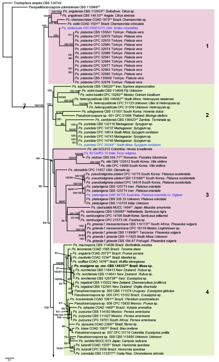
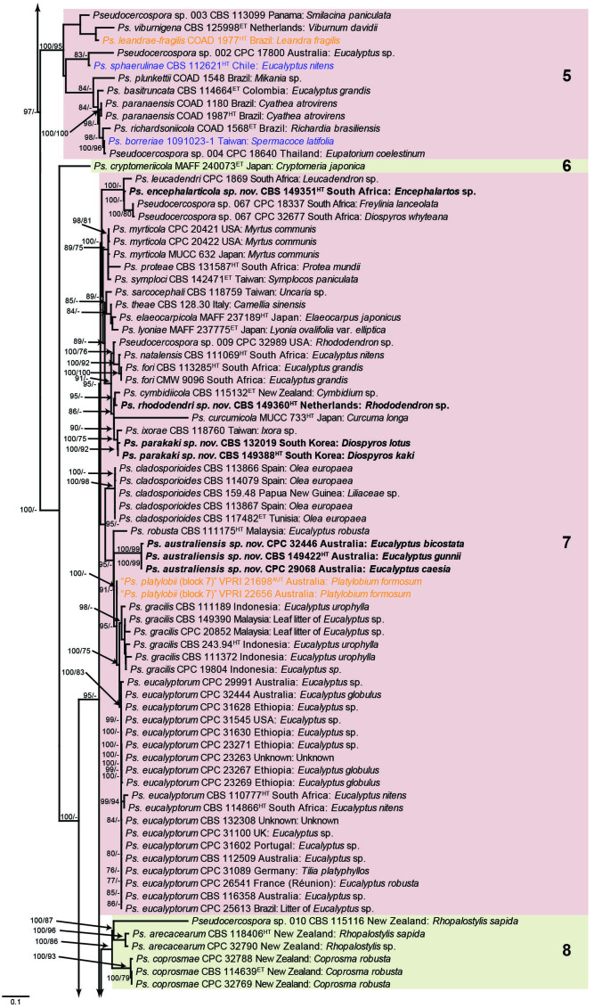
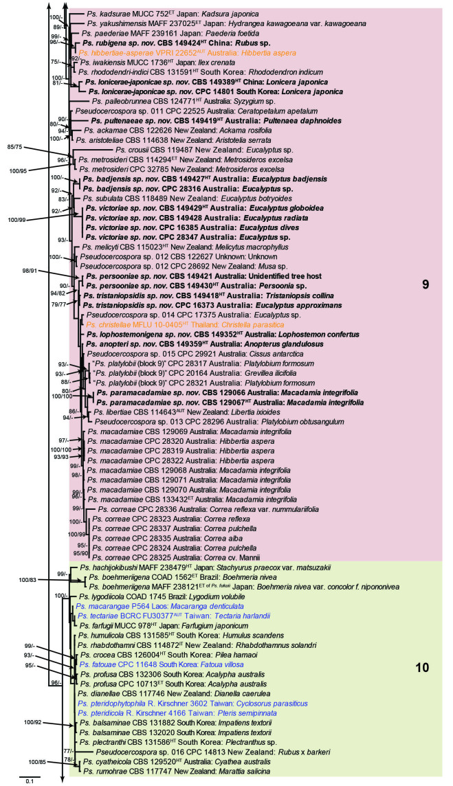
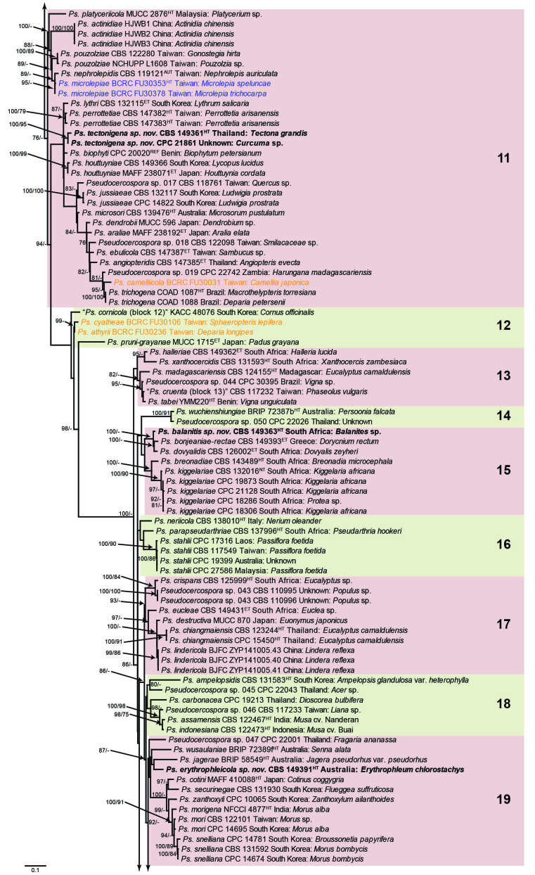
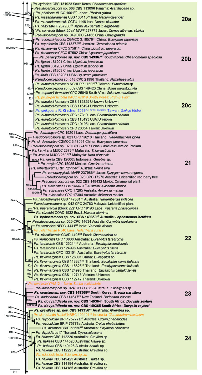
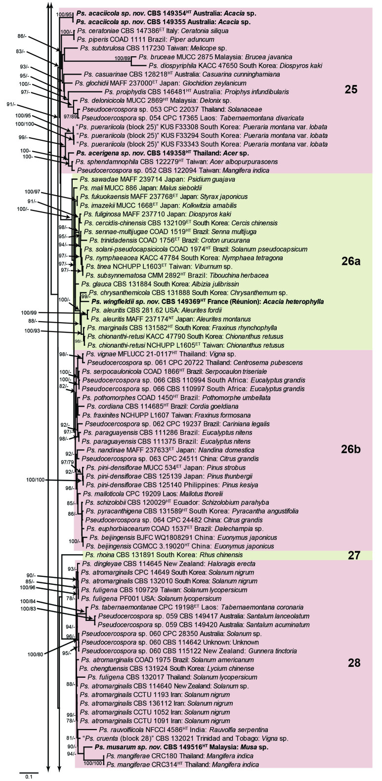
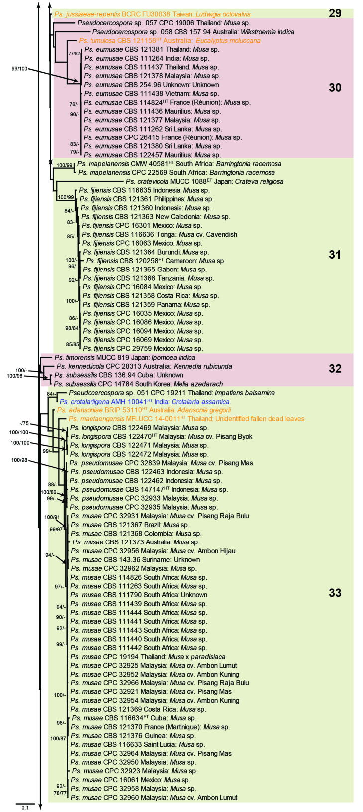
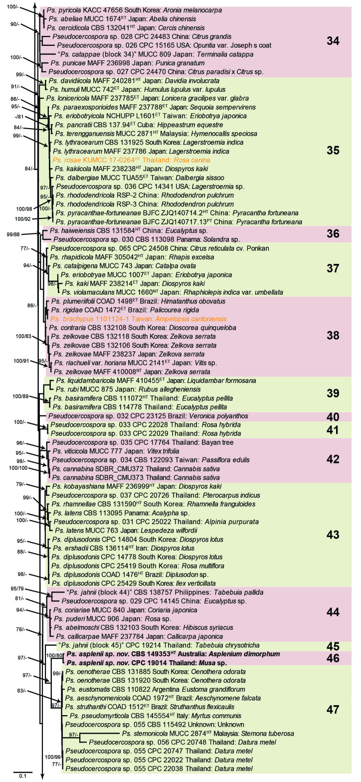
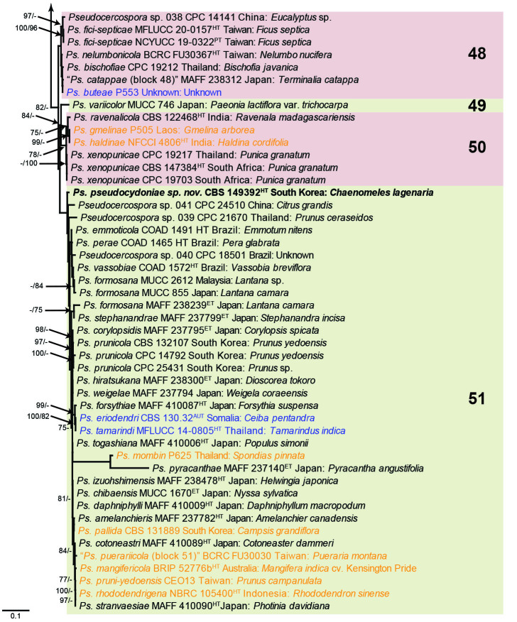
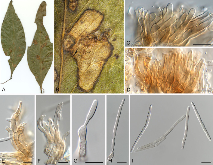
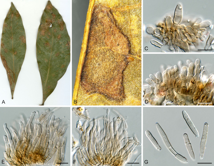
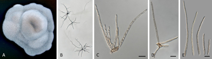
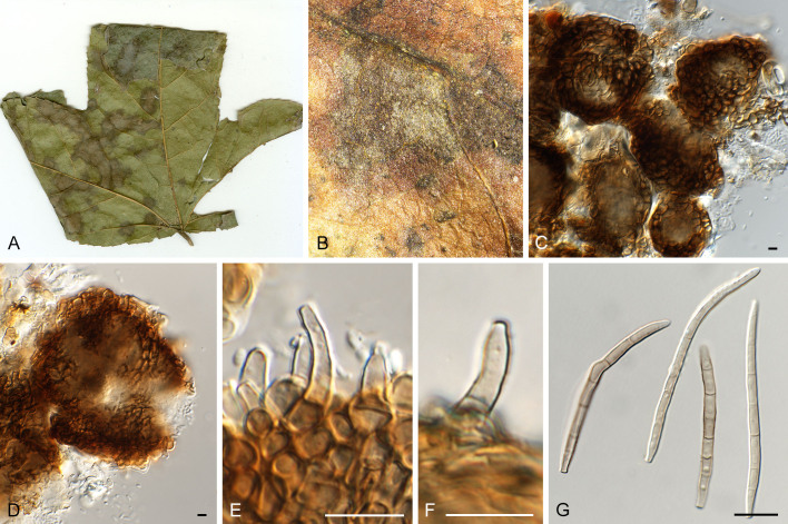
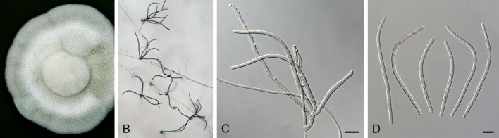
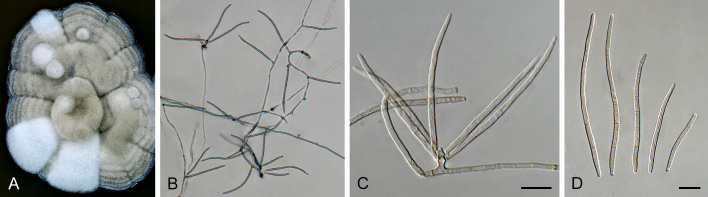
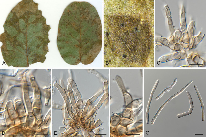
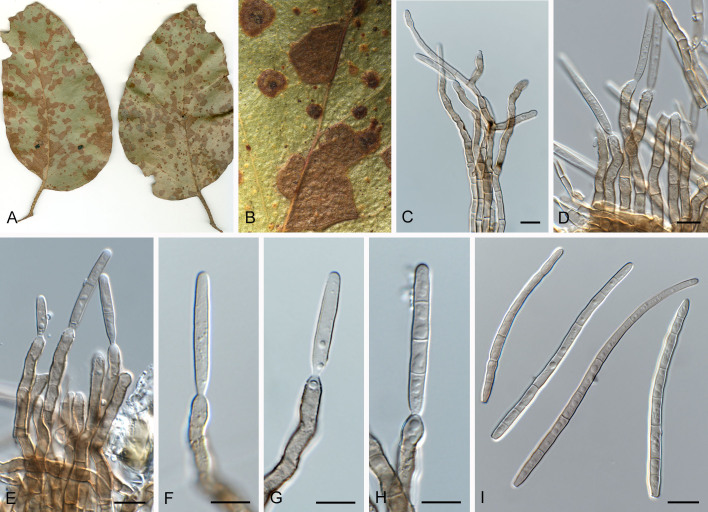
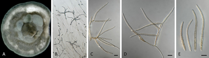
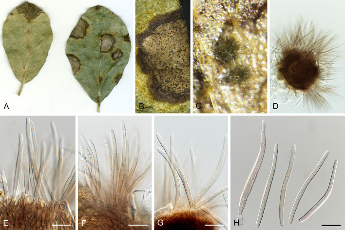
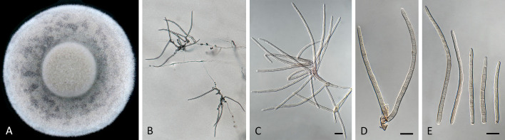
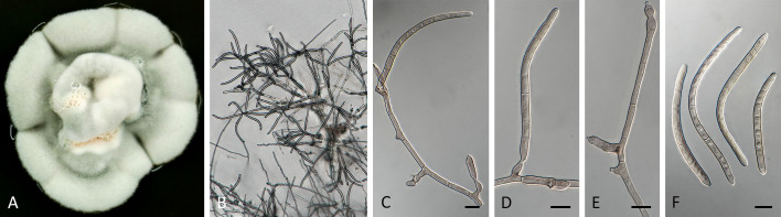
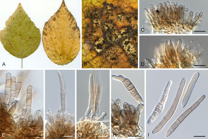
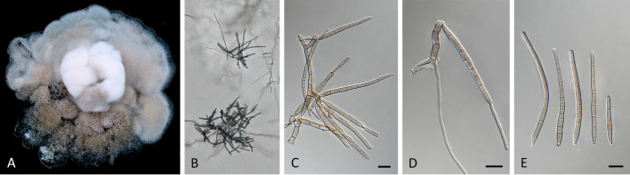
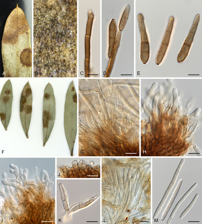
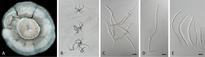
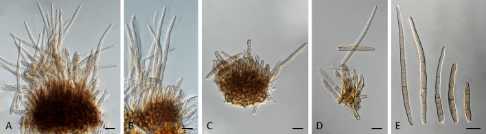
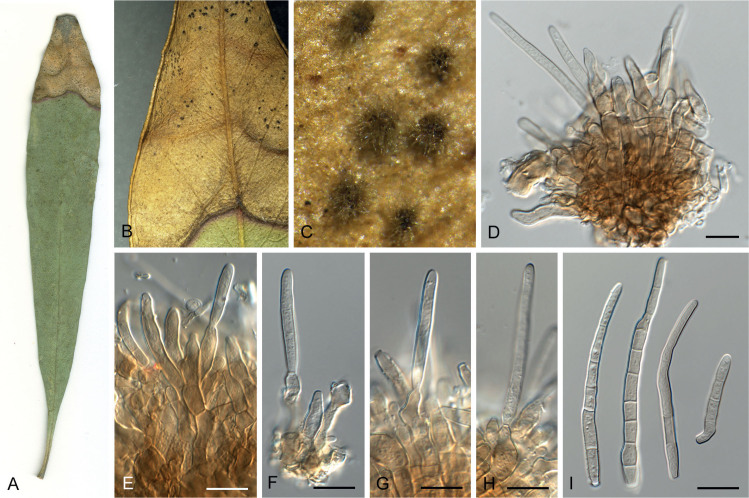
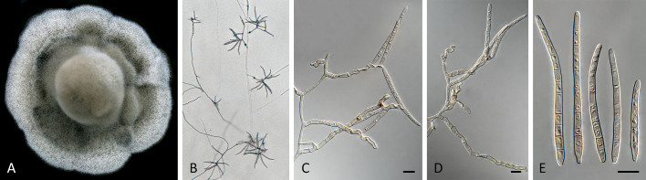
Species of Pseudocercospora are commonly associated with leaf and fruit spots on diverse plant hosts in sub-tropical and tropical regions. Pseudocercospora spp. have mycosphaerella-like sexual morphs, but represent a distinct genus in Mycosphaerellaceae (Mycosphaerellales, Dothideomycetes). The present study adds a further 29 novel species of Pseudocercospora from 413 host species representing 297 host genera occurring in 60 countries and designates four epitypes and one lectotype for established names. This study recognises 329 species names, with an additional 69 phylogenetic lineages remaining unnamed due to difficulty in being able to unambiguously apply existing names to those lineages. To help elucidate the taxonomy of these species, a phylogenetic tree was generated from multi-locus DNA sequence data of the internal transcribed spacers and intervening 5.8S nuclear nrRNA gene (ITS), partial actin (actA), and partial translation elongation factor 1-alpha (tef1), as well as the partial DNA-directed RNA polymerase II second largest subunit (rpb2) gene sequences. Novel species described in this study include those from various countries as follows: Australia, Ps. acaciicola from leaf spots on Acacia sp., Ps. anopter from leaf spots on Anopterus glandulosus, Ps. asplenii from leaf spots on Asplenium dimorphum, Ps. australiensis from leaf spots on Eucalyptus gunnii, Ps. badjensis from leaf spots on Eucalyptus badjensis, Ps. erythrophloeicola from leaf spots on Erythrophleum chlorostachys, Ps. grevilleae from leaf spots on Grevillea sp., Ps. lophostemonigena from leaf spots on Lophostemon confertus, Ps. lophostemonis from leaf spots on Lophostemon lactifluus, Ps. paramacadamiae from leaf spots on Macadamia integrifolia, Ps. persooniae from leaf spots on Persoonia sp., Ps. pultenaeae from leaf spots on Pultenaea daphnoides, Ps. tristaniopsidis from leaf spots on Tristaniopsis collina, Ps. victoriae from leaf spots on Eucalyptus globoidea. Brazil, Ps. musigena from leaf spots on Musa sp. China, Ps. lonicerae-japonicae from leaf spots on Lonicera japonica, Ps. rubigena leaf spots on Rubus sp. France (Réunion), Ps. wingfieldii from leaf spots on Acacia heterophylla. Malaysia, Ps. musarum from leaf spots on Musa sp. Netherlands, Ps. rhododendri from leaf spots on Rhododendron sp. South Africa, Ps. balanitis from leaf spots on Balanites sp., Ps. dovyalidicola from leaf spots on Dovyalis zeyheri, Ps. encephalarticola from leaf spots on Encephalartos sp. South Korea, Ps. grewiana from leaf spots on Grewia biloba, Ps. parakaki from leaf spots on Diospyros kaki, Ps. pseudocydoniae from leaf spots on Chaenomeles lagenaria, Ps. paracydoniae from leaf spots on Chaenomeles speciosa. Thailand, Ps. acerigena from leaf spots on Acer sp., Ps. tectonigena from leaf spots on Tectona grandis. Epitypes are designated for Cercospora bonjeaneae-rectae, Cercospora halleriae, Ps. eucleae, and an epitype as well as a lectotype for Ps. macadamiae. Results obtained in the present study contribute to a better understanding of the host specificity and distribution in Pseudocercospora spp., many of which represent important pathogens of food or fibre crops, or organisms of quarantine concern. Citation: Groenewald JZ, Chen YY, Zhang Y, Roux J, Shin H-D, Shivas RG, Summerell BA, Braun U, Alfenas AC, Ujat AH, Nakashima C, Crous PW (2024). Species diversity in Pseudocercospora. Fungal Systematics and Evolution 13: 29-89. doi: 10.3114/fuse.2024.13.03.

Keywords: Multi-gene phylogeny Mycosphaerellaceae new taxa; plant pathogen taxonomy.


Conflict of interest statement

The authors declare that there is no conflict of interest.

Figures

Fig. 1
Fig. 1
Consensus phylogram (50 % majority rule) obtained from the maximum likelihood analysis with IQ-TREE v. 2.1.3 of the concatenated nucleotide alignment. Bootstrap support values (> 74 %) from 1 000 non-parametric bootstrap replicates are shown at the nodes, followed by RAxML 1 000 replicate bootstrap support values (> 74 %). Culture collection or voucher numbers are followed by the country and host information where available. Sequences derived from material with a type status are indicated with a superscript HT (from (ex-)type), IT (from (ex-)isotype), PT (from (ex-)paratype), NT (from (ex-)neotype, AUT (from authentic), REF (from reference) and ET (from (ex-)epitype). Strains in dark blue font represent strains with only an ITS sequence in the dataset but with no conflict in their positions between the IQ-TREE and RAxML analyses, whereas an orange font indicates such a strain with a conflict in its position between the two analyses. Numbered coloured blocks are provided to facilitate referencing to the position of a species in the phylogenetic tree. Taxa for which it was not possible to assign the correct name due to lack of type material are indicated together with their block number between parentheses in the phylogenetic tree, Suppl. Tables S1 and S3 and the Taxonomy section. Novel species described in this study are highlighted with bold font. The tree was rooted to Trochophora simplex with Parapallidocercospora colombiensis as internal distant genus. The scale bar indicates the expected number of changes per site.
Fig. 1
Fig. 1
Consensus phylogram (50 % majority rule) obtained from the maximum likelihood analysis with IQ-TREE v. 2.1.3 of the concatenated nucleotide alignment. Bootstrap support values (> 74 %) from 1 000 non-parametric bootstrap replicates are shown at the nodes, followed by RAxML 1 000 replicate bootstrap support values (> 74 %). Culture collection or voucher numbers are followed by the country and host information where available. Sequences derived from material with a type status are indicated with a superscript HT (from (ex-)type), IT (from (ex-)isotype), PT (from (ex-)paratype), NT (from (ex-)neotype, AUT (from authentic), REF (from reference) and ET (from (ex-)epitype). Strains in dark blue font represent strains with only an ITS sequence in the dataset but with no conflict in their positions between the IQ-TREE and RAxML analyses, whereas an orange font indicates such a strain with a conflict in its position between the two analyses. Numbered coloured blocks are provided to facilitate referencing to the position of a species in the phylogenetic tree. Taxa for which it was not possible to assign the correct name due to lack of type material are indicated together with their block number between parentheses in the phylogenetic tree, Suppl. Tables S1 and S3 and the Taxonomy section. Novel species described in this study are highlighted with bold font. The tree was rooted to Trochophora simplex with Parapallidocercospora colombiensis as internal distant genus. The scale bar indicates the expected number of changes per site.
Fig. 2
Fig. 2
Pseudocercospora acaciicola (CPC 17039, ex-type culture). A. Leaf spots on upper and lower leaf surface. B. Close-up of lesion. C–H. Fascicles with conidiophores and conidiogenous cells giving rise to conidia. I. Conidia. Scale bars = 10 μm.
Fig. 3
Fig. 3
Pseudocercospora acaciicola (CPC 17121). A. Leaf spots on upper and lower leaf surface. B. Close-up of lesion. C–F. Fascicles with conidiophores and conidiogenous cells giving rise to conidia. G. Conidia. Scale bars = 10 μm.
Fig. 4
Fig. 4
Pseudocercospora acaciicola (CPC 17121). A. Colony on MEA. B. Sporulation on SNA. C, D. Conidiogenous cells giving rise to conidia. E. Conidia. Scale bars = 10 μm.
Fig. 5
Fig. 5
Pseudocercospora acerigena (CPC 18629, ex-type culture). A. Leaf spots on lower leaf surface. B. Close-up of lesion. C, D. Spermatogonia. E, F. Conidiophores and conidiogenous cells giving rise to conidia. G. Conidia. Scale bars = 10 μm.
Fig. 6
Fig. 6
Pseudocercospora anopteri (CPC 20152, ex-type culture). A. Colony on MEA. B. Sporulation on SNA. C. Conidiogenous cells giving rise to conidia. D. Conidia. Scale bars = 10 μm.
Fig. 7
Fig. 7
Pseudocercospora asplenii (CPC 17011, ex-type culture). A. Colony on MEA. B. Sporulation on SNA. C. Conidiogenous cells giving rise to conidia. D. Conidia. Scale bars = 10 μm.
Fig. 8
Fig. 8
Pseudocercospora australiensis (CPC 29218, ex-type culture). A. Leaf spots on lower and upper leaf surface. B. Close-up of lesion. C–F. Fascicles with conidiophores and conidiogenous cells giving rise to conidia. G. Conidia. Scale bars = 10 μm.
Fig. 9
Fig. 9
Pseudocercospora badjensis (CPC 32376, ex-type culture). A. Leaf spots on upper and lower leaf surface. B. Close-up of lesion. C–H. Fascicles with conidiophores and conidiogenous cells giving rise to conidia. I. Conidia. Scale bars = 10 μm.
Fig. 10
Fig. 10
Pseudocercospora badjensis (CPC 32376, ex-type culture). A. Colony on MEA. B. Sporulation on SNA. C, D. Conidiogenous cells giving rise to conidia. E. Conidia. Scale bars = 10 μm.
Fig. 11
Fig. 11
Pseudocercospora balanitis (CPC 25271, ex-type culture). A. Leaf spots on lower and upper leaf surface. B. Close-up of lesion. C, D. Fascicles of conidiophores. E–G. Fascicles with conidiophores and conidiogenous cells giving rise to conidia. H. Conidia. Scale bars = 10 μm.
Fig. 12
Fig. 12
Pseudocercospora balanitis (CPC 25271, ex-type culture). A. Colony on MEA. B. Sporulation on SNA. C, D. Conidiogenous cells giving rise to conidia. E. Conidia. Scale bars = 10 μm.
Fig. 13
Fig. 13
Pseudocercospora bonjeaneae-rectae (CPC 31698, ex-epitype culture). A–C. Fascicles with conidiogenous cells giving rise to conidia. D. Conidia. Scale bars = 10 μm.
Fig. 14
Fig. 14
Pseudocercospora dovyalidicola (CPC 25273, ex-type culture). A. Leaf spots on upper and lower leaf surface. B, C. Close-up of lesions. D–I. Fascicles with conidiophores and conidiogenous cells giving rise to conidia. J. Conidia. Scale bars = 10 μm.
Fig. 15
Fig. 15
Pseudocercospora dovyalidicola (CPC 25275). A. Colony on MEA. B. Sporulation on SNA. C, D. Conidiogenous cells giving rise to conidia. E. Conidia. Scale bars = 10 μm.
Fig. 16
Fig. 16
Pseudocercospora encephalarticola (CPC 15278, ex-type culture). A. Colony on MEA. B. Sporulation on SNA. C, D. Conidiogenous cells giving rise to conidia. E. Conidia. Scale bars = 10 μm.
Fig. 17
Fig. 17
Pseudocercospora erythrophleicola (CPC 17241, ex-type culture). A–C. Fascicles with conidiogenous cells giving rise to conidia. D. Conidia. Scale bars = 10 μm.
Fig. 18
Fig. 18
Pseudocercospora eucleae (CPC 32792, ex-epitype culture). A. Leaf spots on lower and upper leaf surface. B. Close-up of lesions. C–F. Fascicles with conidiophores and conidiogenous cells giving rise to conidia. G. Conidia. Scale bars = 10 μm.
Fig. 19
Fig. 19
Pseudocercospora gracilis (CPC 20850). A. Leaf spots on lower and upper leaf surface. B. Close-up of lesion with ascomata. C–E. Asci and ascospores. Scale bars = 10 μm.
Fig. 20
Fig. 20
Pseudocercospora grevilleae (CPC 17585, ex-type culture). A. Leaf spots on lower and upper leaf surface. B. Close-up of lesion. C, D. Fascicles with conidiophores and conidiogenous cells giving rise to conidia. E, F. Conidia. Scale bars = 10 μm.
Fig. 21
Fig. 21
Pseudocercospora grevilleae (CPC 17585, ex-type culture). A. Colony on MEA. B. Sporulation on SNA. C–E. Conidiogenous cells giving rise to conidia. F. Conidia. Scale bars = 10 μm.
Fig. 22
Fig. 22
Pseudocercospora grewiana (CPC 25564, ex-type culture). A. Leaf spots on upper and lower leaf surface. B. Close-up of lesion. C–H. Fascicles with conidiophores and conidiogenous cells giving rise to conidia. I. Conidia. Scale bars = 10 μm.
Fig. 23
Fig. 23
Pseudocercospora grewiana (CPC 25564, ex-type culture). A. Colony on MEA. B. Sporulation on SNA. C, D. Conidiogenous cells giving rise to conidia. E. Conidia. Scale bars = 10 μm.
Fig. 24
Fig. 24
A–E. Verrucisporota proteacearum. A. Leaf spots on lower leaf surface. B. Close-up of lesion and sporulation. C, D. Conidiophores and conidiogenous cells giving rise to conidia. E. Conidia. F–M. Pseudocercospora hakeae (CPC 32190). F. Leaf spots on lower and upper leaf surface. G–L. Fascicles with conidiophores and conidiogenous cells giving rise to conidia. M. Conidia. Scale bars = 10 μm.
Fig. 25
Fig. 25
Pseudocercospora hakeae (CPC 32190). A. Colony on MEA. B. Sporulation on SNA. C, D. Conidiogenous cells giving rise to conidia. E. Conidia. Scale bars = 10 μm.
Fig. 26
Fig. 26
Pseudocercospora hakeae (CPC 32100). A. Leaf spots on lower and upper leaf surface. B. Close-up of lesions. C–H. Fascicles with conidiophores and conidiogenous cells giving rise to conidia. I. Conidia. Scale bars = 10 μm.
Fig. 27
Fig. 27
Pseudocercospora hakeae (CPC 32100). A. Colony on MEA. B. Sporulation on SNA. C, D. Conidiogenous cells giving rise to conidia. E. Conidia. Scale bars = 10 μm.
Fig. 28
Fig. 28
Pseudocercospora halleriae (CPC 21952, ex-epitype culture). A. Leaf spots on lower and upper leaf surface. B. Close-up of lesion. C. Close-up of fascicles. D–G. Fascicles with conidiophores and conidiogenous cells giving rise to conidia. H. Conidia. Scale bars = 10 μm.
Fig. 29
Fig. 29
Pseudocercospora halleriae (CPC 21952, ex-epitype culture). A. Colony on MEA. B. Sporulation on SNA. C, D. Conidiogenous cells giving rise to conidia. E. Conidia. Scale bars = 10 μm.
Fig. 30
Fig. 30
Pseudocercospora lonicerae-japonicae (CPC 30538, ex-type culture). A–D. Fascicles with conidiophores and conidiogenous cells giving rise to conidia. E. Conidia. Scale bars = 10 μm.
Fig. 31
Fig. 31
Pseudocercospora lophostemonigena (CPC 16409, ex-type culture). A. Leaf spots on lower and upper leaf surface. B. Close-up of lesion. C–F. Fascicles with conidiophores and conidiogenous cells giving rise to conidia. G. Conidia. Scale bars = 10 μm.
Fig. 32
Fig. 32
Pseudocercospora lophostemonis (CPC 14517, ex-type culture). A. Leaf spots on lower and upper leaf surface. B. Close-up of lesion. C. Closeup of fascicles. D–I. Fascicles with conidiophores and conidiogenous cells giving rise to conidia. J. Conidia. Scale bars = 10 μm
Fig. 33
Fig. 33
Pseudocercospora lophostemonis (CPC 14517, ex-type culture). A. Colony on MEA. B. Sporulation on SNA. C, D. Conidiogenous cells giving rise to conidia. E. Conidia. Scale bars = 10 μm.
Fig. 34
Fig. 34
Pseudocercospora musarum (CPC 32815, ex-type culture). A. Leaf spots on upper and lower leaf surface. B. Close-up of lesions. C. Close-up of stroma. D–G. Fascicles with conidiophores and conidiogenous cells giving rise to conidia. H. Conidia. Scale bars = 10 μm.
Fig. 35
Fig. 35
Pseudocercospora musarum (CPC 32815, ex-type culture). A. Colony on MEA. B. Sporulation on SNA. C, D. Conidiogenous cells giving rise to conidia. E. Conidia. Scale bars = 10 μm.
Fig. 36
Fig. 36
Pseudocercospora musigena (CPC 18460, ex-type culture). A. Leaf spots on lower and upper leaf surface. B. Close-up of lesion. C. Close-up of stroma. D. Spermatogonium. E, F. Fascicles with conidiogenous cells giving rise to conidia. G. Conidia. Scale bars = 10 μm.
Fig. 37
Fig. 37
Pseudocercospora musigena (CPC 18460, ex-type culture). A. Colony on MEA. B. Sporulation on SNA. C, D. Conidiogenous cells giving rise to conidia. E. Conidia. Scale bars = 10 μm.
Fig. 38
Fig. 38
Pseudocercospora paracydoniae (CPC 25478, ex-type culture). A. Leaf spots on upper and lower leaf surface. B. Close-up of lesion. C, D. Fascicles with conidiogenous cells giving rise to conidia. E. Conidia. Scale bars = 10 μm.
Fig. 39
Fig. 39
Pseudocercospora paracydoniae (CPC 25478, ex-type culture). A. Colony on MEA. B. Sporulation on SNA. C, D. Conidiogenous cells giving rise to conidia. E. Conidia. Scale bars = 10 μm.
Fig. 40
Fig. 40
Pseudocercospora parakaki (CPC 14686, ex-type culture). A–D. Fascicles with conidiogenous cells giving rise to conidia. E. Conidia. Scale bars = 10 μm.
Fig. 41
Fig. 41
Pseudocercospora paramacadamiae (CPC 19150, ex-type culture). A. Colony on MEA. B. Sporulation on SNA. C, D. Conidiogenous cells giving rise to conidia. E. Conidia. Scale bars = 10 μm.
Fig. 42
Fig. 42
Pseudocercospora persooniae (CPC 32456, ex-type culture). A. Leaf spot on lower leaf surface. B. Close-up of lesion. C. Close-up of fascicles. D–H. Fascicles with conidiogenous cells giving rise to conidia. I. Conidia. Scale bars = 10 μm.
Fig. 43
Fig. 43
Pseudocercospora persooniae (CPC 32456, ex-type culture). A. Colony on MEA. B. Sporulation on SNA. C, D. Conidiogenous cells giving rise to conidia. E. Conidia. Scale bars = 10 μm.
Fig. 44
Fig. 44
Pseudocercospora pseudocydoniae (CPC 14665, ex-type culture). A–C. Fascicles with conidiogenous cells giving rise to conidia. D. Conidia. Scale bars = 10 μm.
Fig. 45
Fig. 45
Pseudocercospora pultenaeae (CPC 28318, ex-type culture). A. Colony on MEA. B. Sporulation on SNA. C, D. Conidiogenous cells giving rise to conidia. E. Conidia. Scale bars = 10 μm.
Fig. 46
Fig. 46
Pseudocercospora rhododendri (CPC 21544, ex-type culture). A. Leaf spots on lower and upper leaf surface. B, C. Close-up of lesions. D, E. Close-up of fascicles. F–H. Fascicles with conidiogenous cells giving rise to conidia. I. Conidia. Scale bars = 10 μm.
Fig. 47
Fig. 47
Pseudocercospora rhododendri (CPC 21544, ex-type culture). A. Colony on MEA. B. Sporulation on SNA. C, D. Conidiogenous cells giving rise to conidia. E. Conidia. Scale bars = 10 μm.
Fig. 48
Fig. 48
Pseudocercospora rubigena (CPC 30628, ex-type culture). A. Leaf spots on lower and upper leaf surface. B. Close-up of lesion. C–E. Fascicles with conidiogenous cells giving rise to conidia. F. Conidia. Scale bars = 10 μm.
Fig. 49
Fig. 49
Pseudocercospora rubigena (CPC 30628, ex-type culture). A. Colony on MEA. B. Sporulation on SNA. C, D. Conidiogenous cells giving rise to conidia. E. Conidia. Scale bars = 10 μm.
Fig. 50
Fig. 50
Pseudocercospora tectonigena (CPC 21663, ex-type culture). A. Leaf spots on upper and lower leaf surface. B, C. Close-up of lesions with fascicles. D, E. Fascicles with conidiogenous cells giving rise to conidia. F. Conidia. Scale bars = 10 μm.
Fig. 51
Fig. 51
Pseudocercospora tectonigena (CPC 21663, ex-type culture). A. Colony on MEA. B. Sporulation on SNA. C, D. Conidiogenous cells giving rise to conidia. E. Conidia. Scale bars = 10 μm.
Fig. 52
Fig. 52
Pseudocercospora tristaniopsidis (CPC 28315, ex-type culture). A. Colony on MEA. B. Sporulation on SNA. C, D. Conidiogenous cells giving rise to conidia. E. Conidia. Scale bars = 10 μm.
Fig. 53
Fig. 53
Pseudocercospora victoriae (CPC 32428). A. Leaf spots on lower and upper leaf surface. B. Close-up of lesions. C–F. Fascicles with conidiogenous cells giving rise to conidia. G. Conidia. Scale bars = 10 μm.
Fig. 54
Fig. 54
Pseudocercospora victoriae (CPC 32428). A. Colony on MEA. B. Sporulation on SNA. C, D. Conidiogenous cells giving rise to conidia. E. Conidia. Scale bars = 10 μm.
Fig. 55
Fig. 55
Pseudocercospora victoriae (CPC 32442, ex-type culture). A, B. Leaf spots on upper leaf surface. C–G. Fascicles with conidiogenous cells giving rise to conidia. H. Conidia. Scale bars = 10 μm.
Fig. 56
Fig. 56
Pseudocercospora wingfieldii (CPC 26287, ex-type culture). A. Leaf spots on upper and lower leaf surface. B. Close-up of lesion. C–F. Fascicles with conidiophores and conidiogenous cells giving rise to conidia. G. Conidia. Scale bars = 10 μm.
Fig. 57
Fig. 57
Pseudocercospora wingfieldii (CPC 26287, ex-type culture). A. Colony on MEA. B. Sporulation on SNA. C, D. Conidiogenous cells giving rise to conidia. E. Conidia. Scale bars = 10 μm.
Fig. 58
Fig. 58
Pseudocercospora sp. 069 (CPC 30540). A. Leaf spots on lower and upper leaf surface. B. Close-up of lesion. C–F. Fascicles with conidiophores and conidiogenous cells giving rise to conidia. G. Conidia. Scale bars = 10 μm.
Fig. 59
Fig. 59
Pseudocercospora sp. 069 (CPC 30540). A. Colony on MEA. B. Sporulation on SNA. C, D. Conidiogenous cells giving rise to conidia. E. Conidia. Scale bars = 10 μm.

References

    1. Acabal BD, Jr, Groenewald JZ, Crous PW, et al. (2014). First report of Pseudocercospora jahnii in the Philippines. Australasian Plant Disease Notes 9: 148.
    1. Akinsanmi OA, Carvalhais LC. (2020). Draft genome of the macadamia husk spot pathogen, Pseudocercospora macadamiae . Phytopathology 110: 1503–1506. - PubMed
    1. Bakhshi M, Arzanlou M, Babai-Ahari A.et al. (2014). Multi-gene analysis of Pseudocercospora spp. from Iran. Phytotaxa 184: 245–264.
    1. Beilharz V, Mayers PE, Pascoe IG. (2003). Pseudocercospora macadamiae sp. nov., the cause of husk spot of macadamia. Australasian Plant Pathology 32: 279–282.
    1. Braun U. (1995). A monograph of Cercosporella, Ramularia and allied genera (phytopathogenic hyphomycetes). Vol. 1. Eching: IHW Verlag.

LinkOut - more resources