. 2024 Aug 12;5(8):e684.
doi: 10.1002/mco2.684. eCollection 2024 Aug.

Ubiquitin-specific protease 22 controls melanoma metastasis and vulnerability to ferroptosis through targeting SIRT1/PTEN/PI3K signaling

Affiliations

Ubiquitin-specific protease 22 controls melanoma metastasis and vulnerability to ferroptosis through targeting SIRT1/PTEN/PI3K signaling

Huiyan Sun et al. MedComm (2020). .

Abstract

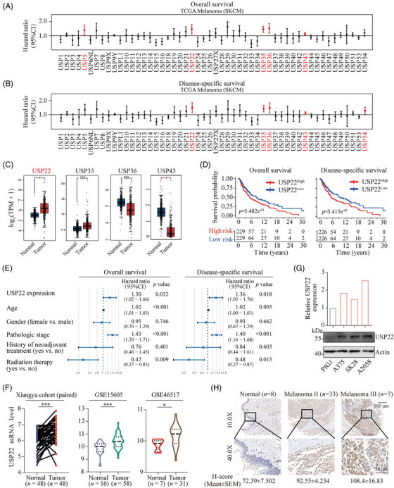
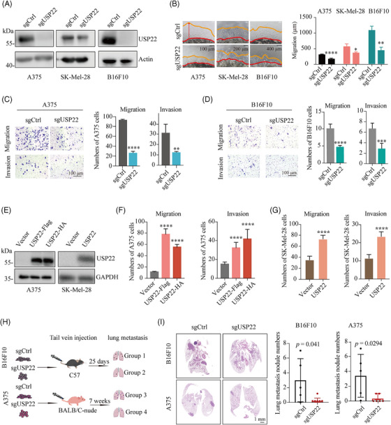
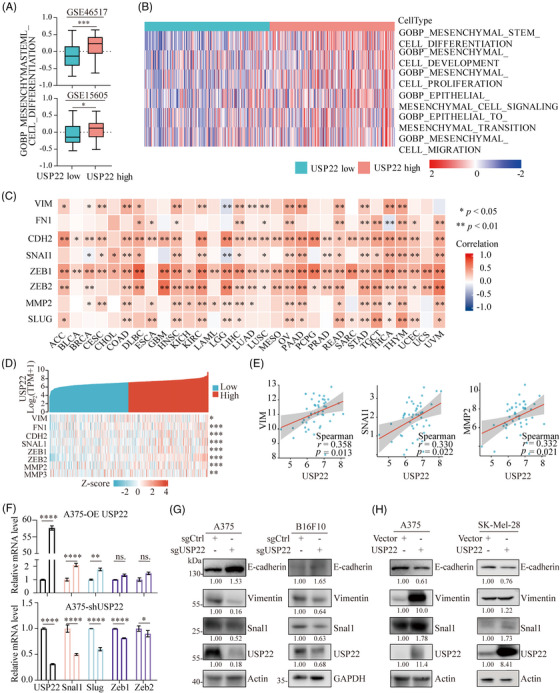
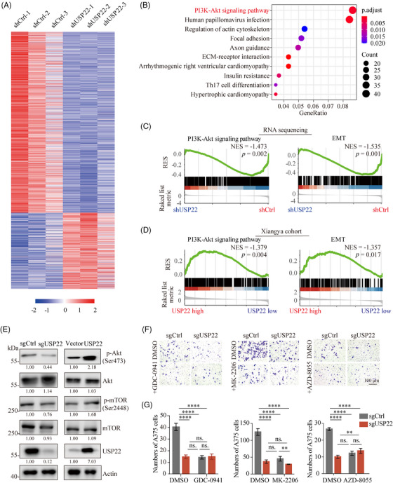
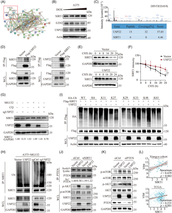
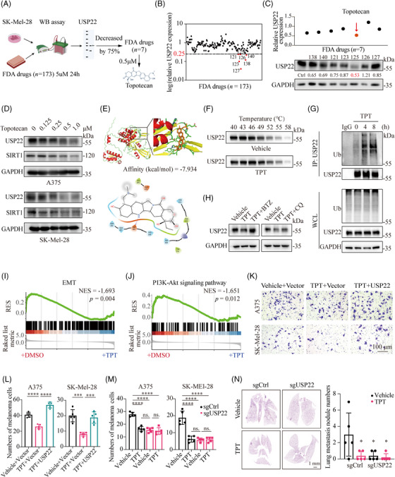
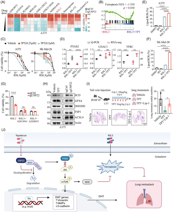
Metastasis is a major contributing factor that affects the prognosis of melanoma patients. Nevertheless, the underlying molecular mechanisms involved in melanoma metastasis are not yet entirely understood. Here, we identified ubiquitin-specific protease 22 (USP22) as a pro-oncogenic protein in melanoma through screening the survival profiles of 52 ubiquitin-specific proteases (USPs). USP22 demonstrates a strong association with poor clinical outcomes and is significantly overexpressed in melanoma. Ablation of USP22 expression remarkably attenuates melanoma migration, invasion, and epithelial-mesenchymal transition in vitro and suppresses melanoma metastasis in vivo. Mechanistically, USP22 controls melanoma metastasis through the SIRT1/PTEN/PI3K pathway. In addition, we conducted an United States Food and Drug Administration-approved drug library screening and identified topotecan as a clinically applicable USP22-targeting molecule by promoting proteasomal degradation of USP22. Finally, we found that both pharmacological and genetic silence of USP22 sensitize RSL3-induced ferroptosis through suppressing the PI3K/Akt/mTOR pathway and its downstream SCD, and ferroptosis inhibitor could partly rescued the decreased lung metastasis by topotecan in vivo. Overall, our findings reveal a prometastatic role of USP22 and identify topotecan as a potent USP22-targeting drug to limit melanoma metastasis.

Keywords: USP22; ferroptosis; melanoma; metastasis; topotecan.


Conflict of interest statement

The authors have no relevant financial or nonfinancial interests to disclose.

Figures

FIGURE 1
FIGURE 1
USP22 is overexpressed in melanoma and associated with a poor prognosis. (A and B) Univariate Cox regression analysis to reveal the association between the USP family and overall survival (A) or disease‐specific survival (B) of melanoma patients. (C) Bioinformatic analysis for the expression of USP22, USP35, USP36, USP43 expression in melanoma (n = 461) and normal skin tissues (n = 558). (D) Kaplan–Meier curves depicting overall survival or disease‐specific survival difference between USP22 high and USP22 low groups. (E) Forest plot showing the results of hazard ratio's (HR) from multivariate Cox regression analysis for the overall survival and disease‐specific survival of melanoma patients. (F) USP22 mRNA expression levels in human melanoma and normal skin data based on Xiangya cohort and two different GSE databases (GSE15605 and GSE46517). (G) USP22 protein levels in normal melanocytes (PIG1) and melanoma cell lines (A375, SK‐Mel‐28, and A2058). (H) Representative images of immunohistochemistry (IHC) staining of USP22 in tissue assay quantified with H‐score.
FIGURE 2
FIGURE 2
USP22 loss suppresses melanoma metastasis both in vitro and in vivo. (A) Western blot analysis of the indicated proteins in control (sgCtrl) and USP22‐knockout (sgUSP22) A375, SK‐Mel‐28, and B16F10 cells. (B) 3D Matrigel drop invasion assay for A375, SK‐Mel‐28, and B16F10 cells after USP22 knockout (sgUSP22). Indicated medium was exchanged every 3 days. The distances of migrated cells away from edge of the matrigel drop were measured as migration (µm) on Day 6. Experiments were triplicate repeated. Representative images and scare bars are shown. (C and D) Transwell assay quantifying the migration (without extracellular matrix) and invasive (with extracellular matrix) capacity for A375 and B16F10 cells after USP22 knockout (sgUSP22). Migrated and invaded cells were determined for 12−20 h. Five random areas were selected. (E) Western blot analysis of the indicated proteins in control (Vector) and USP22‐overexpressing A375 and SK‐Mel‐28 cells. (F and G) Transwell assay quantifying the migration and invasion for A375 (F) and SK‐Mel‐28 cells (G) after USP22 overexpression. (H and I) Schematic view and representative hematoxylin–eosin (H&E) images of lung metastasis of mice after tail vein injection of B16F10 or A375 cells with USP22‐knockout (sgUSP22) or control (sgCtrl) cells. Two‐tailed unpaired Student's t‐test was performed in (B–D, G, and I), and one‐way ANOVA was used in (F). *p < 0.05, **p < 0.01, ***p < 0.001, ****p < 0.0001.
FIGURE 3
FIGURE 3
USP22 promotes EMT activation in melanoma. (A) Correlation between USP22 expression with mesenchymal stem cell differentiational pathway based on GSE46517 and GSE15605 databases. (B) Heatmap showing USP22 expression positively associated with epithelial–mesenchymal transition (EMT)‐related pathways in TCGA‐SKCM datasets. (C and D) Heatmap showing USP22 expression positively associated with EMT‐related genes in different cancers (C) including melanoma (D) in TCGA datasets. (E) Spearman's correlation between the expression of vimentin (VIM), SNAL1, MMP2, and USP22 based on data from Xiangya cohort. (F) The mRNA expression levels of EMT‐related transcription factors in USP22‐overexpressing and USP22‐knockdown (shUSP22) A375 cells. (G and H) The protein expression levels of EMT‐related markers including E‐cadherin, vimentin and snal1 in USP22‐knockout (sgUSP22) and USP22‐overexpressing (USP22) melanoma cells. *p < 0.05, **p < 0.01, ***p < 0.001.
FIGURE 4
FIGURE 4
USP22 potentiates melanoma metastasis and EMT through activating PI3K/Akt/mTOR pathway. (A) Transcriptome sequencing was performed on USP22‐knockdown (shUSP22) and control (shCtrl) A375 cells and the results were visualized in a heatmap. (B) RNA sequencing based on KEGG pathways enrichment shows the top 10 enriched pathways in USP22‐knockdown A375 cells. (C) Gene Set Enrichment Analysis (GSEA) showing PI3K–Akt pathway and EMT signatures were enriched in control group (shCtrl) based on RNA sequencing. (D) GSEA showing PI3K–Akt pathway and EMT signatures were enriched in USP22‐high group based on Xiangya cohort. (E) The protein expression levels of Akt, p‐Akt (Ser473), mTOR, p‐mTOR (Ser2448) in USP22‐knockout (sgUSP22) and USP22‐overexpressing (USP22) A375 cells quantified by western blotting. (F and G) Transwell assay showing effect of GDC0941 (PI3Ki, 1 µM), MK2206 (Akti, 8 µM), AZD8055 (mTORi, 500 nM) on the role of USP22 silencing (sgUSP22) in A375 cell migration. Two‐way ANOVA analysis was performed in (G). Ns., not significance; ****p < 0.0001.
FIGURE 5
FIGURE 5
USP22 activates PI3K/Akt pathway via SIRT1/PTEN axis. (A) Protein–protein interactions (PPI) analysis containing USP22, USP22 binding proteins and proteins involved in modulating PI3K–Akt signal pathways. Proteins interacting with USP22 were identified from immunoprecipitation and mass spectrometry (IP‐MS) analysis. (B) Western blotting analysis showing SIRT1 and EZH2 protein expression in USP22‐inducible cells with doxycycline (0–1 µM, wedge). (C) The peak map of SIRT1 acquired from the mass spectrometry. (D) A375 cells were transfected with vectors encoding Flag‐tagged USP22 and Flag‐tagged SIRT1 respectively. Protein lysates then were harvest for immunoprecipitated with anti‐Flag beads, then immunoblotted with the SIRT1 and USP22. (E) Protein lysates of control and USP22‐overexpressing (USP22) A375 cells were treated with cycloheximide (CHX) (100 µg/mL) for the indicated time points, followed by immunoblotting with SIRT1. (F) Quantification of the data from (E) (n = 3 biologically independent samples). (G) Protein lysates of control and USP22‐knockout (sgUSP22) A375 cells were treated with DMSO, MG132 (10 µM) and CQ (20 µM) for 6 h respectively, and followed by immunoblotting with SIRT1 and USP22. (H) Protein lysates of USP22‐overexpressing (USP22) and USP22‐knockout (sgUSP22) A375 cells harvested after MG132 (10 µM) treatment (6 h) for immunoprecipitated with ubiquitin (Ub) antibody plus protein A/G beads and immunoblotted with SIRT1. (I) Immunoblot analysis of 293T cells transfected with Flag‐SIRT1, HA‐tagged ubiquitin, HA‐tagged K6‐linked ubiquitin, HA‐tagged K11‐linked ubiquitin, HA‐tagged K27‐linked ubiquitin, HA‐tagged K29‐linked ubiquitin, HA‐tagged K33‐linked ubiquitin, HA‐tagged K48‐linked ubiquitin, and HA‐tagged K63‐linked ubiquitin with or without USP22, followed by treatment with MG132 and IP with anti‐Flag beads, then detected with anti‐HA. (J) USP22‐overexpressing (USP22) and control A375 cells were transfected with SIRT1 siRNAs. Protein lysates were harvested for immunoprecipitated with PTEN antibody plus protein A/G beads and immunoblotted with acetyl‐lysine. (K) Western blot analysis of PI3K/Akt/mTOR changes in indicated control and USP22‐overexpressing (USP22) cells after transfection with PTEN siRNAs. (L) Spearman's correlation between vimentin (VIM) and SIRT1 based on data from Xiangya cohort and TCGA‐SKCM.
FIGURE 6
FIGURE 6
Identification of USP22‐targeting molecule to suppress melanoma metastasis. (A) Schematic view of western blotting‐based drug screening. (B) Screening with US FDA‐approved drug library (5 µM) for 24 h identified seven out of 173 compounds that decreased USP22 expression by 75%. (C) Screening with US FDA‐approved drug library (0.5 µM) for 24 h identified topotecan as the most efficient USP22‐targeting molecule (n = 1). (D) Western blotting showing the protein levels of USP22, SIRT1 in A375 and SK‐Mel‐28 cells treated with topotecan for 24 h. (E) Prediction of the interaction between topotecan and USP22 protein. (F) The cellular thermal shift assay (CETSA) of USP22 proteins for A375 cells treated with DMSO (Vehicle) or topotecan (TPT, 0.5 µM). (G) An IP assay was performed to verify ubiquitin (Ub) modification of USP22 in A375 cells after treatment with topotecan (TPT, 0.5 µM) for 4–8 h and MG132. (H) Western blot analysis of the USP22 expression in A375 cells following treatment with topotecan (TPT, 0.5 µM) in the absence or presence of bortezomib (BTZ; 0.2 µM) or chloroquine (CQ; 20 µM) for 24 h. (I and J) GSEA showing less enrichment of EMT (I) and PI3K–Akt pathway (J) in SK‐Mel‐28 cells treated with topotecan (TPT) for 24 h than cells treated with DMSO. (K–M) Transwell assay showing effect of topotecan (TPT, 0.5 µM) on the migration of USP22‐overexpressing (USP22) (K and L) or USP22‐knockout (sgUSP22) (M) A375 and SK‐Mel‐28 cells. (N) Representative H&E images of lung metastasis of mice in the indicated groups. Two‐way ANOVA analysis was performed in (L–N). *p < 0.05, ***p < 0.001, ****p < 0.0001.
FIGURE 7
FIGURE 7
Inhibition of USP22 sensitizes melanoma to ferroptosis. (A) Heatmap showing cell viability of USP22‐knockout (sgUSP22) and control (sgCtrl) A375 cells treated with BRAF inhibitors (LGX818, GSK2118436, PLX4032), MEK inhibitors (GDC0973, GSK1120212, MEK162), and BET inhibitor (NHWD‐870), ferroptosis inducer (RSL3), apoptosis inducer (staurosporine), necroptosis inducer (HS‐173), and cuproptosis inducer (Elesclomol‐Cu). (B) GSEA showing enrichment of ferroptosis in RSL3‐treated SK‐Mel‐28 cells after topotecan (TPT) exposure for 24 h. (C) Cell viability of A375 and SK‐Mel‐28 cells treated with RSL3 or topotecan (TPT) either alone or in combination for 24 h. (D) Real‐time PCR and RNA sequencing showing the mRNA expression of ferroptosis marker including PTGS2, CHAC1, and TFRC in SK‐Mel‐28 cells treated with RSL3 alone or in combination with topotecan (TPT). (E and F) The levels of lipid ROS detected by C11‐BODIPY 581/591 in A375 (E) and SK‐Mel‐28 cells (F) exposed to RSL3 (2.5 µM), TPT (0.5 µM) or the combination. (G) Cell viability of USP22‐knockout (sgUSP22) and control (sgCtrl) A375 cells treated with RSL3 (2.5 µM) alone or plus GDC0941 (PI3Ki, 1 µM) or AZD8055 (mTORi, 500 nM). (H) Western blotting indicated the levels of ferroptosis‐associated proteins (SCD, GPX4, DHODH, FSP1, and ACSL4) in A375 cells after treatment with topotecan (TPT, 0.5 µM) or USP22‐knockout (sgUSP22). (I) Schematic view and representative H&E images of lung metastasis of mice after tail vein injection of B16F10 cells in the indicated groups. (J) Graphical summary showing that targeting USP22 by topotecan suppresses melanoma metastasis via inhibiting EMT and sensitizes ferroptosis through SIRT1/PTEN/PI3K axis. One‐way ANOVA analysis was performed in (D–G, I). ns., no significance, *p < 0.05, **p < 0.01, ***p < 0.001, ****p < 0.0001.

References

    1. Sung H, Ferlay J, Siegel RL, et al. Global cancer statistics 2020: gLOBOCAN estimates of incidence and mortality worldwide for 36 cancers in 185 countries. CA Cancer J Clin. 2021;71(3):209‐249. - PubMed
    1. Xiong K, Qi M, Stoeger T, Zhang J, Chen S. The role of tumor‐associated macrophages and soluble mediators in pulmonary metastatic melanoma. Front Immunol. 2022;13:1000927. - PMC - PubMed
    1. Millet A, Martin AR, Ronco C, Rocchi S, Benhida R. Metastatic melanoma: insights into the evolution of the treatments and future challenges. Med Res Rev. 2017;37(1):98‐148. - PubMed
    1. Siegel RL, Miller KD, Wagle NS, Jemal A. Cancer statistics, 2023. CA Cancer J Clin. 2023;73(1):17‐48. - PubMed
    1. Villani A, Potestio L, Fabbrocini G, Troncone G, Malapelle U, Scalvenzi M. The treatment of advanced melanoma: therapeutic update. Int J Mol Sci. 2022;23(12). - PMC - PubMed