

Triangular fossa of the third cerebral ventricle - an original 3D model and morphometric study
- PMID: 39135984
- PMCID: PMC11317240
- DOI: 10.3389/fnana.2024.1398858
Triangular fossa of the third cerebral ventricle - an original 3D model and morphometric study
Abstract
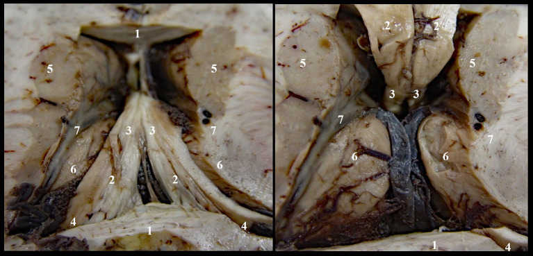
Introduction: The triangular recess (TR), also called triangular fossa or vulva cerebri, represents the anterior extension of the diencephalic ventricle, located between the anterior columns of the fornix and the anterior white commissure. Over time, this structure of the third cerebral ventricle generated many disputes. While some anatomists support its presence, others have opposite opinions, considering that it only becomes visible under certain conditions. The aim of the study is to demonstrate the tangible structure of the triangular recess. Secondly, the quantitative analysis allowed us to establish an anatomical morphometric standard, as well as the deviations from the standard.
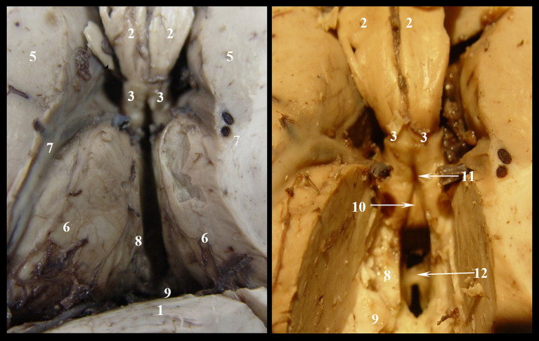
Materials and methods: Our study is both a quantitative and a qualitative evaluation of the triangular fossa. We dissected 100 non-neurological adult brains, which were fixed in 10% formaldehyde solution for 10 weeks. The samples are part of the collection of the Institute of Anatomy, "Grigore T. Popa" University of Medicine and Pharmacy, Iasi. We highlighted the triangular fossa by performing dissections in two stages at the level of the roof of the III ventricle.
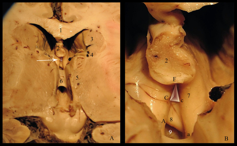
Results: The qualitative analysis is a re-evaluation of the classical data concerning the anatomy of the fossa triangularis. We proposed an original 3D model of the triangular recess in which we described a superficial part called vestibule and a deep part called pars profunda. We measured the sides of the communication between the two proposed segments, as well as the communication with the III ventricle. By applying the Heron's formula, we calculated the area of the two communications. Statistical evaluations have shown that these communications are higher than they are wide. In addition, there is a statistical difference between the surfaces of the two communications: 34.07 mm2 ± 7.01 vs. 271.43 mm2 ± 46.36 (p = 0.001).
Conclusion: The outcome of our study is both qualitative and quantitative. Firstly, we demonstrated the existence of the triangular fossa and we conceived a spatial division of this structure. Secondly, the measurements carried out establish an anatomo-morphometric norm of the triangular recess, which is useful in assessing the degree of hydrocephalus during the third endoscopic ventriculoscopy.
Keywords: Schwalbe’s fossa; anterior commissure; endoventricular navigation; third ventricle; triangular recess; ventriculoscopy; vestibulum; vulva cerebri.
Copyright © 2024 Nedelcu, Lupu, Lupu, Tepordei, Ioniuc, Stan, Vicoleanu, Haliciu, Statescu, Ursaru, Danielescu and Tarniceriu.
Conflict of interest statement
The authors declare that the research was conducted in the absence of any commercial or financial relationships that could be construed as a potential conflict of interest.
Figures


References
-
- Abdala-Vargas N. J., Cifuentes-Lobelo H. A., Ordoñez-Rubiano E., Patiño-Gomez J. G., Villalonga J. F., Lucifero A. G., et al. (2022). Anatomic variations of the floor of the third ventricle: surgical implications for endoscopic third ventriculostomy. Surg. Neurol. Int. 27:218. doi: 10.25259/SNI_404_2022 - DOI - PMC - PubMed
-
- Asghar A., Narayan R. K., Kumar P., Ravi K. S., Tubbs R. S., Patra A., et al. (2023). Absence of the interthalamic adhesion (ITA) as a neuroanatomical association or risk factor for neuropsychiatric disorders: a systemic review and meta-analysis. Indian J. Psychiatry 65, 985–994. doi: 10.4103/indianjpsychiatry.indianjpsychiatry_744_22, PMID: - DOI - PMC - PubMed
-
- Cerro Larrazabal L., Ibáñez Botella G., Ros Sanjuán Á., Ros López B., Iglesias Moroño S., Arráez Sánchez M. Á. (2023). Neuroendoscopic transventricular transchoroidal approach for access to the posterior zone of the third ventricle or pineal region. Neurosurg. Rev. 46:323. doi: 10.1007/s10143-023-02210-1 - DOI - PubMed
LinkOut - more resources
Full Text Sources
Miscellaneous