. 2024 Oct;29(10):902-920.
doi: 10.1111/gtc.13154. Epub 2024 Aug 13.

Meflin/ISLR is a marker of adipose stem and progenitor cells in mice and humans that suppresses white adipose tissue remodeling and fibrosis

Affiliations

Meflin/ISLR is a marker of adipose stem and progenitor cells in mice and humans that suppresses white adipose tissue remodeling and fibrosis

Toshikazu Ishihara et al. Genes Cells. 2024 Oct.

Abstract

Identifying specific markers of adipose stem and progenitor cells (ASPCs) in vivo is crucial for understanding the biology of white adipose tissues (WAT). PDGFRα-positive perivascular stromal cells represent the best candidates for ASPCs. This cell lineage differentiates into myofibroblasts that contribute to the impairment of WAT function. However, ASPC marker protein(s) that are functionally crucial for maintaining WAT homeostasis are unknown. We previously identified Meflin as a marker of mesenchymal stem cells (MSCs) in bone marrow and tissue-resident perivascular fibroblasts in various tissues. We also demonstrated that Meflin maintains the undifferentiated status of MSCs/fibroblasts. Here, we show that Meflin is expressed in WAT ASPCs. A lineage-tracing experiment showed that Meflin+ ASPCs proliferate in the WAT of obese mice induced by a high-fat diet (HFD), while some of them differentiate into myofibroblasts or mature adipocytes. Meflin knockout mice fed an HFD exhibited a significant fibrotic response as well as increases in adipocyte cell size and the number of crown-like structures in WAT, accompanied by impaired glucose tolerance. These data suggested that Meflin expressed by ASPCs may have a role in reducing disease progression associated with WAT dysfunction.

Keywords: Islr; Meflin; adipose stem and progenitor cells; adipose stem cells; fibrosis; glucose tolerance; immunoglobulin superfamily containing leucine‐rich repeat; mesenchymal stem cell; metabolic syndrome.


Conflict of interest statement

The authors declare no conflicts of interest.

Figures
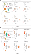
FIGURE 1
FIGURE 1
Meflin expression in ASPCs from human and mouse WAT. Single‐cell transcriptomic analyses of cells isolated from normal human (a) and mouse (b) subcutaneous and visceral WAT (GSE176067 and GSE176171). Data were examined for the expression of ISLR, which encodes Meflin, and marker genes of ASPCs (PDGFRA, CD34) and mural cells (MCAM, CSPG4). Uniform manifold approximation and projection (UMAP) plots showed specific expression of Meflin in PDGFRα+ ASPCs (arrows).
FIGURE 2
FIGURE 2
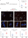
Meflin+ cells increase in the epididymal fat pads of HFD‐fed mice. (a) 10‐week‐old male C57BL/6 mice were fed either SD or HFD for 16 weeks, followed by the sacrifice of mice and analysis of tissues by qPCR and ISH. (b) Islr expression in the epididymal fat tissue of mice fed either SD or HFD was analyzed by qPCR and expressed relative to control (Actb). (c), (d) Tissues obtained from the epididymal fat pads of mice fed either SD or HFD were stained for Islr (red) and Pdgfra (green) by ISH (c), followed by quantification (d). Nuclei were visualized by DAPI staining (blue). Arrows denote Islr + cells. Boxed areas (a)–(d) were magnified in lower panels. HPF, high‐power field.
FIGURE 3
FIGURE 3
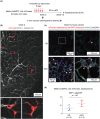
Morphology of Meflin+ cells labeled by tdTomato expression in the epididymal fat pad of Meflin‐CreERT2; LSL‐tdTomato mice. (a) Schematic diagram of the experimental protocol using Meflin‐CreERT2; LSL‐tdTomato mice. Mice were administered tamoxifen to label Meflin+ cells, followed by feeding either SD or HFD and IF for tdTomato, α‐SMA, and CD36. (b) Meflin‐CreERT2; LSL‐tdTomato mice were administered tamoxifen and were subjected to in vivo vascular staining (white) by intravenous injection of an anti‐CD31 antibody and isolectin B4 1 day after the last dose of tamoxifen. Subsequently, we isolated the epididymal fat pads. We proceeded to clearing of the tissue, and imaging of the 3D morphology of Meflin+ cells (red). Boxed areas were magnified in lower panels. (c), (d) The epididymal fat tissues of Meflin‐CreERT2; LSL‐tdTomato mice fed either SD or HFD were stained by IF for the indicated proteins (c), followed by counting the number of tdTomato+ cells (d). tdTomato fluorescence was detected by IF staining using an anti‐RFP antibody. In (c), boxed areas were magnified in adjacent panels. White and yellow arrowheads denote tdTomato+ spindle‐shaped cells and cells with a morphology of mature adipocytes, respectively. HPF, high‐power field.
FIGURE 4
FIGURE 4
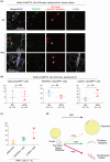
Meflin+ cells yield mature adipocytes and myofibroblasts in hypertrophic epididymal fat pads. (a)–(c) Meflin‐CreERT2; LSL‐tdTomato mice were administered tamoxifen and fed either SD or HFD for 16 weeks. The epididymal fat tissues were isolated and stained with the indicated antibodies for IF (a), followed by quantification of the number of tdTomato+ cells that exhibited an adipocyte‐like morphology and were positive for either PDGFRα or α‐SMA (b), (c). tdTomato fluorescence was detected by IF staining using an anti‐RFP antibody. Green, pink, and yellow arrowheads denote PDGFRα+tdTomato+ spindle‐shaped cells, α‐SMA+tdTomato+ spindle‐shaped cells, and cells with a morphology of mature adipocytes, respectively. (d) Schematic diagram showing the working hypothesis for the differentiation of Meflin+ ASPCs in hypertrophic WAT of mice fed HFD. Meflin+ ASPCs give rise mainly to α‐SMA+ myofibroblasts and to mature adipocytes. Some Meflin+ ASPCs proliferate, maintaining the expression of PDGFRα.
FIGURE 5
FIGURE 5
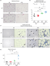
Increases in the size of adipocytes and the extent of interstitial fibrosis found in WAT of Meflin KO mice fed HFD. (a), (b) The epididymal fat tissues of wild‐type (WT) and Meflin KO mice (KO) fed either SD or HFD for 16 weeks were stained with hematoxylin and eosin (a), followed by measurement of the diameters of adipocytes and quantification (b). (c), (d) The epididymal fat tissues of wild‐type (WT) and Meflin KO mice (KO) fed either SD or HFD were subjected to Masson's Trichrome staining (c), followed by measurement of fibrotic areas (arrows) and quantification (d).
FIGURE 6
FIGURE 6
Increases in the number of CLS found in WAT of Meflin KO mice fed HFD. The epididymal fat tissues of wild‐type (WT) and Meflin KO mice (KO) fed either SD or HFD for 16 weeks were stained for the macrophage marker F4/80 by IHC (a), followed by counting the number of CLS with F4/80+ macrophages surrounding necrotic adipocytes (b). Arrows denote CLS. HPF, high‐power field.
FIGURE 7
FIGURE 7
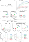
Meflin KO mice exhibited impaired glucose homeostasis compared to wild‐type mice in a condition of excessive fat accumulation in WAT. (a)–(c) Wild‐type (WT) and Meflin KO (KO) mice were fed either SD or HFD for 16 weeks (a), followed by measurement of the weights of the total body and the sums of inguinal fat pads, epidydimal fat pads, and interscapular BAT (b), (c). N.S., not significant. (d)–(g) WT and Meflin KO mice fed either SD and HFD for 16 weeks were subjected to fasting for 18 or 1 h, followed by i.p. glucose (2 g/kg) administration (IPGTT) and i.p. insulin (0.5 U/kg) administration (IPITT), respectively (d). Plasma was collected from WT and Meflin KO mice in the IPGTT test at 0 and 30 min after i.p. administration of glucose, followed by measurement of insulin and statistical quantification (e). Blood glucose levels were examined for 120 min (e), followed by the measurement of the areas under the curve (AUC) and quantification by one‐way ANOVA and the Tukey–Kramer test (f). N.S., not significant.

References

    1. Ando, R. , Shiraki, Y. , Miyai, Y. , Shimizu, H. , Furuhashi, K. , Minatoguchi, S. , Kato, K. , Kato, A. , Iida, T. , Mizutani, Y. , Ito, K. , Asai, N. , Mii, S. , Esaki, N. , Takahashi, M. , & Enomoto, A. (2024). Meflin is a marker of pancreatic stellate cells involved in fibrosis and epithelial regeneration in the pancreas. The Journal of Pathology, 262(1), 61–75. 10.1002/path.6211 - DOI - PubMed
    1. Barrett, R. L. , & Puré, E. (2020). Cancer‐associated fibroblasts and their influence on tumor immunity and immunotherapy. eLife, 9, e57243. 10.7554/eLife.57243 - DOI - PMC - PubMed
    1. Berry, R. , & Rodeheffer, M. S. (2013). Characterization of the adipocyte cellular lineage in vivo. Nature Cell Biology, 15(3), 302–308. 10.1038/ncb2696 - DOI - PMC - PubMed
    1. Buechler, M. B. , Pradhan, R. N. , Krishnamurty, A. T. , Cox, C. , Calviello, A. K. , Wang, A. W. , Yang, Y. A. , Tam, L. , Caothien, R. , Roose‐Girma, M. , Modrusan, Z. , Arron, J. R. , Bourgon, R. , Müller, S. , & Turley, S. J. (2021). Cross‐tissue organization of the fibroblast lineage. Nature, 593(7860), 575–579. 10.1038/s41586-021-03549-5 - DOI - PubMed
    1. Cattaneo, P. , Mukherjee, D. , Spinozzi, S. , Zhang, L. , Larcher, V. , Stallcup, W. B. , Kataoka, H. , Chen, J. , Dimmeler, S. , Evans, S. M. , & Guimarães‐Camboa, N. (2020). Parallel lineage‐tracing studies establish fibroblasts as the prevailing in vivo adipocyte progenitor. Cell Reports, 30(2), 571–582.e2. 10.1016/j.celrep.2019.12.046 - DOI - PubMed

MeSH terms