Randomized Controlled Trial
. 2024 Aug 1;65(10):21.
doi: 10.1167/iovs.65.10.21.

Choroidal Changes During and After Discontinuing Long-Term 0.01% Atropine Treatment for Myopia Control

Affiliations
Randomized Controlled Trial

Choroidal Changes During and After Discontinuing Long-Term 0.01% Atropine Treatment for Myopia Control

Samantha Sze-Yee Lee et al. Invest Ophthalmol Vis Sci. .

Abstract

Purpose: Few studies have explored choroidal changes after cessation of myopia control. This study evaluated the choroidal thickness (ChT) and choroidal vascularity index (CVI) during and after discontinuing long-term low-concentration atropine eye drops use for myopia control.

Methods: Children with progressive myopia (6-16 years; n = 153) were randomized to receive 0.01% atropine eye drops or a placebo (2:1 ratio) instilled daily over 2 years, followed by a 1-year washout (no eye drop use). Optical coherence tomography imaging of the choroid was conducted at the baseline, 2-year (end of treatment phase), and 3-year (end of washout phase) visits. The main outcome measure was the subfoveal ChT. Secondary measures include the CVI.

Results: During the treatment phase, the subfoveal choroids in both treatment and control groups thickened by 12-14 µm (group difference P = 0.56). During the washout phase, the subfoveal choroids in the placebo group continued to thicken by 6.6 µm (95% confidence interval [CI] = 1.7 to 11.6), but those in the atropine group did not change (estimate = -0.04 µm; 95% CI = -3.2 to 3.1). Participants with good axial eye growth control had greater choroidal thickening than the fast-progressors during the treatment phase regardless of the treatment group (P < 0.001), but choroidal thickening in the atropine group's fast-progressors was not sustained after stopping eye drops. CVI decreased in both groups during the treatment phase, but increased in the placebo group after treatment cessation.

Conclusions: On average, compared to placebo, 0.01% atropine eye drop treatment did not cause a differential rate of change in ChT during treatment, but abrupt cessation of long-term 0.01% atropine eye drops may disrupt normal choroidal thickening in children.


Conflict of interest statement

Disclosure: S.S.-Y. Lee, None; G. Lingham, Ocumetra (E); A. Clark, None; S.A. Read, None; D. Alonso-Caneiro, None D.A. Mackey, Novartis (C)

Figures

Figure 1.
Figure 1.
Choroidal thickness imaging and measurement. (A) Horizontal and vertical 30 degrees B-scans centered on the fovea; (B) thickness were estimated at each subfield of the early treatment of diabetic retinopathy grid which comprises a central 0.5mm radius circle centered on the fovea (C0), and the superior (S), inferior (I), temporal (T), and nasal (N) segments of the inner (between 0.5 and 1.5 mm radii circles) and outer (between 1.5 and 3.0 mm radii circles) rings; (C) machine-learning program segmentation of the retinal pigmented epithelium (green line) and chorioscleral interface (blue line).
Figure 2.
Figure 2.
Binarization of cross-sectional choroidal scans. (A) The original grey-scale scan was manipulated such that (B) the naturally curved choroid is flattened to (C) allow binarization, whereas the non-choroidal areas were cropped out. The white and black areas of the binarized images corresponds to the stromal and luminal areas, respectively. The choroidal vascularity index is the ratio of the luminal area to the total choroidal area within the cross-sectional scan.
Figure 3.
Figure 3.
Sample size and retention. aReasons for withdrawal: Three did not like diagnostic or study drops; four had difficulty adhering to the treatment regimen; three had difficulty attending appointments + concern of receiving placebo; two were uncontactable; one relocated; three wanted to seek myopia treatment (atropine or orthokeratology) privately; two were uncontactable; and four cited personal reasons/did not provide reason. bReasons for withdrawal: One sought atropine and orthokeratology eye drops privately, and one was uncontactable.
Figure 4.
Figure 4.
Figure numbers indicate the estimated marginal mean change in subfoveal choroidal measures from baseline. Error bars represent standard errors.
Figure 5.
Figure 5.
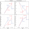
Estimated marginal mean change in choroidal thickness (µm) and 95% confidence interval during the 2-year treatment phase (top panel) and the 1-year washout phase (bottom panel). The placebo and atropine groups are shown in the left and middle columns, respectively. Blue numbers indicate significant thickening of the choroid during either phase at P < 0.05. Group difference in the amount of change is shown in the right column. The yellow grids indicate significant difference between groups at P < 0.05. Figures corrected for age, baseline value, and time of imaging.
Figure 6.
Figure 6.
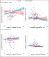
Myopia progression against change in subfoveal choroidal thickness (ChT) during treatment phase (top panel) and washout phase (bottom panel).
Figure 7.
Figure 7.
Figure numbers indicate the estimated marginal mean change (±1 standard error) change in subfoveal choroidal measures from baseline according to treatment group and axial elongation.

References

    1. Lee SS, Alonso-Caneiro D, Lingham G, et al. .. Choroidal thickening during young adulthood and baseline choroidal thickness predicts refractive error change. Invest Ophthalmol Vis Sci . 2022; 63(5): 34. - PMC - PubMed
    1. Read SA, Alonso-Caneiro D, Vincent SJ, Collins MJ.. Longitudinal changes in choroidal thickness and eye growth in childhood. Invest Ophthalmol Vis Sci . 2015; 56(5): 3103–3112. - PubMed
    1. Nickla DL, Wallman J.. The multifunctional choroid. Prog Retin Eye Res . 2010; 29(2): 144–168. - PMC - PubMed
    1. Prousali E, Dastiridou A, Ziakas N, Androudi S, Mataftsi A.. Choroidal thickness and ocular growth in childhood. Surv Ophthalmol . 2021; 66(2): 261–275. - PubMed
    1. Wang D, Chun RK, Liu M, et al. .. Optical defocus rapidly changes choroidal thickness in schoolchildren. PLoS One . 2016; 11(8): e0161535. - PMC - PubMed

Publication types

MeSH terms

LinkOut - more resources