. 2024 Sep 4;481(17):1125-1142.
doi: 10.1042/BCJ20240255.

Inhibitors identify an auxiliary role for mTOR signalling in necroptosis execution downstream of MLKL activation

Affiliations

Inhibitors identify an auxiliary role for mTOR signalling in necroptosis execution downstream of MLKL activation

Sarah E Garnish et al. Biochem J. .

Abstract

Necroptosis is a lytic and pro-inflammatory form of programmed cell death executed by the terminal effector, the MLKL (mixed lineage kinase domain-like) pseudokinase. Downstream of death and Toll-like receptor stimulation, MLKL is trafficked to the plasma membrane via the Golgi-, actin- and microtubule-machinery, where activated MLKL accumulates until a critical lytic threshold is exceeded and cell death ensues. Mechanistically, MLKL's lytic function relies on disengagement of the N-terminal membrane-permeabilising four-helix bundle domain from the central autoinhibitory brace helix: a process that can be experimentally mimicked by introducing the R30E MLKL mutation to induce stimulus-independent cell death. Here, we screened a library of 429 kinase inhibitors for their capacity to block R30E MLKL-mediated cell death, to identify co-effectors in the terminal steps of necroptotic signalling. We identified 13 compounds - ABT-578, AR-A014418, AZD1480, AZD5363, Idelalisib, Ipatasertib, LJI308, PHA-793887, Rapamycin, Ridaforolimus, SMI-4a, Temsirolimus and Tideglusib - each of which inhibits mammalian target of rapamycin (mTOR) signalling or regulators thereof, and blocked constitutive cell death executed by R30E MLKL. Our study implicates mTOR signalling as an auxiliary factor in promoting the transport of activated MLKL oligomers to the plasma membrane, where they accumulate into hotspots that permeabilise the lipid bilayer to cause cell death.

Keywords: mechanistic target of rapamycin; necroptosis; protein kinase; pseudokinase; signalling.


Conflict of interest statement

All authors contribute, or have contributed, to a project developing necroptosis pathway inhibitors in collaboration with Anaxis Pharma Pty Ltd. J.M.M. has received research funding from Anaxis Pharma Pty Ltd.

Figures

Figure 1.
Figure 1.. RIPK3 deletion does not inhibit R30E MLKL-mediated cell death.
(A,B) Evaluation of necroptotic signalling by wild-type and R30E full-length human MLKL in MLKL−/− and MLKL−/− RIPK3−/− HT29 cells. Human MLKL expression was induced with doxycycline (Dox; 100 ng/ml) and cell death was measured in the presence or absence of a necroptotic stimulus (TNF, Smac mimetic, IDN-6556; TSI). Cell death was quantified as a percentage of SYTOX Green-positive cells using IncuCyte SX5 live cell imaging. MLKL−/− RIPK3−/− HT29 independent lines were assayed in n = 2 for each of two independent cell lines for a total of n = 4 independent experiments. One MLKL−/− HT29 cell line was assayed in n = 2 and the other in n = 3, for a total of n = 5 independent experiments. Data are plotted as mean ± SEM. (C) MLKL−/− or MLKL−/− RIPK3−/− HT29 cells were stimulated with doxycycline (Dox; 100 ng/ml overnight) to induce the expression of wild-type or R30E human MLKL. Protein levels of MLKL and RIPK3, both unphosphorylated and phosphorylated forms, were detected by immunoblotting in the presence or absence of necroptotic stimulation (TSI; 4 h). Data are representative of duplicate independent experiments. (D,E) MLKL−/− and MLKL−/− RIPK3−/− HT29 cells stably transduced with MLKLR30E were treated with doxycycline (Dox) in the presence of RIPK3 kinase inhibitor, GSK′872, at increasing concentrations. IncuCyte SX5 imaging was used to quantify the percentage of cell death (SYTOX Green-positive cells) every hour for 48 h. Data are plotted as mean ± SEM of n = 3. One independent cell line transduced with MLKLR30E was assayed in n = 3 independent experiments.
Figure 2.
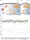
Figure 2.. Screen identifies 13 kinase inhibitors that block R30E MLKL-mediated cell death.
(A) Schematic of the necroptosis pathway. TNF (T) stimulates the TNFR1; cIAP1/2 activity is blocked with Smac mimetic (S); and the pan-caspase inhibitor, IDN-6556 (I), blocks caspase-8 activity. This leads to the formation of the necrosome, subsequent phosphorylation and activation of MLKL. Activated MLKL oligomerizes and traffics to biological membranes where its accumulation facilitates membrane permeabilization. (B) The activated R30E MLKL mutant (highlighted by red star) oligomerizes and initiates cell death in the absence of TSI stimulation or RIPK3 activation enabling a screen for inhibitors of necroptosis downstream of MLKL activation. The skull and crossbones image (Mycomorphbox_Deadly.png; by Sven Manguard) is used via a Creative Commons Attribution-Share Alike 4.0 license. (C) Phenotypic screen of 429 kinase inhibitors to identify rescue of R30E MLKL constitutive cell death. MLKL−/− HT29 cells were stimulated with 100 ng/ml doxycycline to induce expression of R30E MLKL. Following 16 h of induction, cells were treated with 1 µM compound. IncuCyte SX5 imaging was used to quantify the percentage of cell death by determining the number SYTOX Green-positive cells (dead cells) relative to the number of DRAQ5-positive cells (total cell number) at 24 h post-stimulation. Mean of technical duplicates for each compound is plotted as fold decrease in percentage cell death. GSK′872 depicted in pink, with mean±SD of n = 11 independent biological replicates. Positive hits that displayed a fold decrease in R30E MLKL-mediated cell death ≥0.2027 (dotted line; GSK′872 — 1 SD) are depicted in yellow. (D) Fifty positive hit compounds were re-screened for their capacity to inhibit R30E MLKL cell death. MLKL−/− HT29 cells were stimulated with 100 ng/ml doxycycline to induce expression of R30E MLKL and stimulated with 1 µM of compound simultaneously. IncuCyte SX5 imaging was used to quantify the percentage of cell death (SYTOX Green-positive cells) at 24 h post-stimulation. Data plotted as fold decrease between R30E MLKL-mediated cell death in the absence and presence of the compound. Plotted data represent one independent replicate. Dotted line (0.6948) represents the population mean (0.388) plus 1 standard deviation (0.307). Thirteen identified compounds that displayed a fold decrease in R30E MLKL-mediated cell death ≥0.6948 (dotted line) are coloured.
Figure 3.
Figure 3.. PHA-793887, Ridaforolimus, Temsirolimus, Rapamycin and ABT-578 exhibit concentration-independent inhibition of R30E MLKL death.
(AM) Evaluation of R30E MLKL constitutive death inhibition in MLKL−/−HT29 cells, following simultaneous stimulation with varying compound concentrations (0.25–4 µM) and doxycycline (100 ng/ml) to induce MLKL expression. (N) Evaluation of R30E MLKL constitutive death inhibition in MLKL−/− RIPK3−/− HT29 cells, following simultaneous stimulation with compound (1 µM) and doxycycline (100 ng/ml) to induce MLKL expression. Cell death was quantified over 44 h as percentage by determining the number SYTOX Green-positive cells (dead cells) relative to the number of DRAQ5-positive cells (total cell number) using IncuCyte SX5 live cell imaging. Cells were assayed in n = 3 (G, N), n = 4 (H, I, M), n = 5 (A, B, C, D, F, K, L) or n = 6 (E, J) independent experiments with data plotted as mean ± SEM.
Figure 4.
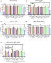
Figure 4.. PHA-793887, Rapamycin and Ridaforolimus inhibit constitutive cell death induced by MLKL-USP21.
(AC) Evaluation of compound inhibition of necroptotic signalling in wild-type HT29 cells stimulated with necroptotic stimulus. Wild-type HT29 cells were treated with 1 µM of compound and (A) TSI (TNF, Smac mimetic, IDN-6556), (B) TSQ (TNF, Smac mimetic, QVD-OPh), or (C) LSI (LPS, Smac mimetic, IDN-6556). Cell death was quantified at 24 h as a percentage by determining the number SYTOX Green-positive cells (dead cells) relative to the number of DRAQ5-positive cells (total cell number) using IncuCyte SX5 live cell imaging. Cells were assayed in n = 4 independent experiments with data plotted as mean ± SEM. (D) MLKLWT expression was induced with doxycycline (100 ng/ml) in MLKL−/− HT29 cells treated with TSI (TNF, Smac mimetic, IDN-6556) and 1 µM of compound. (E) Evaluation of compound inhibition on MLKL-USP21 constitutive cell death. MLKL−/− HT29 cells were treated with doxycycline (100 ng/ml) to induce the expression of MLKL-USP21 and stimulated with 1 µM of the respective compound. Cell death was quantified at 48 h as a percentage by determining the number SYTOX Green-positive cells (dead cells) relative to the number of DRAQ5-positive cells (total cell number) using IncuCyte SX5 live cell imaging. Cells were assayed in n = 4 (E) or n = 5 (D) independent experiments with data plotted as mean ± SEM. P-value calculated using an unpaired, two-tailed t-test. *P < 0.05, **P < 0.01, ***P < 0.001, ****P < 0.0001.
Figure 5.
Figure 5.. Compounds inhibit the association of R30E MLKL oligomers with the membrane.
MLKL−/− HT29 cells stably transduced with MLKLWTor MLKLR30E were treated with doxycycline (Dox; 100 ng/ml) only or doxycycline plus necroptotic stimulus (Dox, TSI) in the presence or absence of kinase inhibitors (1 µM). Membrane fractions were resolved by BN-PAGE and fractionation was verified by probing for VDAC via SDS–PAGE. Images are representative of n = 2 independent experiments.
Figure 6.
Figure 6.. Inhibiting the mTOR pathway blocks R30E MLKL membrane translocation and cell death.
Our data argue for a role for mTOR signalling in promoting R30 MLKL-mediated cell death. Inhibition of mTOR kinase activity or upstream activators of mTOR block R30E MLKL membrane translocation and killing. The skull and crossbones image (Mycomorphbox_Deadly.png; by Sven Manguard) is used via a Creative Commons Attribution-Share Alike 4.0 license.

Similar articles

References

    1. Nakano, H., Murai, S. and Moriwaki, K. (2022) Regulation of the release of damage-associated molecular patterns from necroptotic cells. Biochem. J. 479, 677–685 10.1042/BCJ20210604 - DOI - PubMed
    1. Fletcher-Etherington, A., Nobre, L., Nightingale, K., Antrobus, R., Nichols, J., Davison, A.J.et al. (2020) Human cytomegalovirus protein pUL36: a dual cell death pathway inhibitor. Proc. Natl Acad. Sci. U.S.A. 117, 18771–18779 10.1073/pnas.2001887117 - DOI - PMC - PubMed
    1. Kitur, K., Wachtel, S., Brown, A., Wickersham, M., Paulino, F., Penaloza, H.F.et al. (2016) Necroptosis promotes Staphylococcus aureus clearance by inhibiting excessive inflammatory signaling. Cell Rep. 16, 2219–2230 10.1016/j.celrep.2016.07.039 - DOI - PMC - PubMed
    1. Liu, Z., Nailwal, H., Rector, J., Rahman, M.M., Sam, R., McFadden, G.et al. (2021) A class of viral inducer of degradation of the necroptosis adaptor RIPK3 regulates virus-induced inflammation. Immunity 54, 247–258.e7. 10.1016/j.immuni.2020.11.020 - DOI - PMC - PubMed
    1. Pearson, J.S., Giogha, C., Muhlen, S., Nachbur, U., Pham, C.L., Zhang, Y.et al. (2017) Espl is a bacterial cysteine protease effector that cleaves RHIM proteins to block necroptosis and inflammation. Nat. Microbiol. 2, 16258 10.1038/nmicrobiol.2016.258 - DOI - PMC - PubMed

Publication types

MeSH terms