. 2024 Aug 13;81(1):347.
doi: 10.1007/s00018-024-05389-8.

Novel CDKL5 targets identified in human iPSC-derived neurons

Affiliations

Novel CDKL5 targets identified in human iPSC-derived neurons

Sean Massey et al. Cell Mol Life Sci. .

Erratum in

Abstract

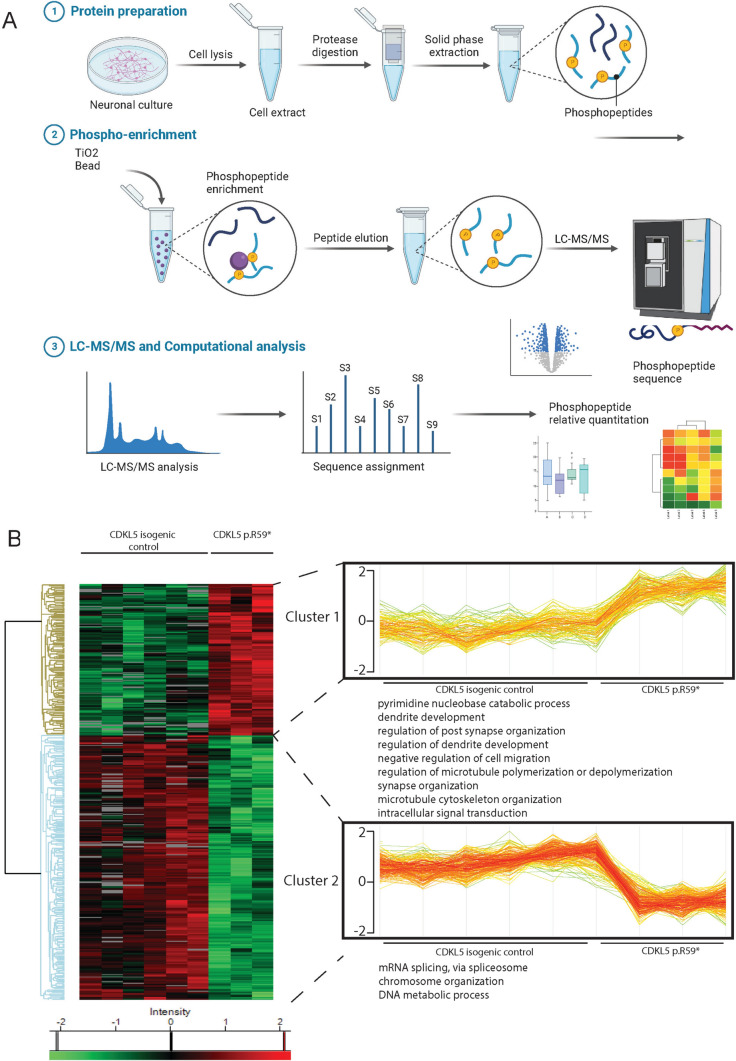
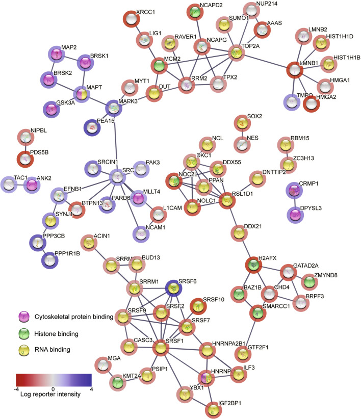
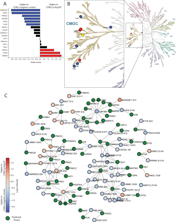
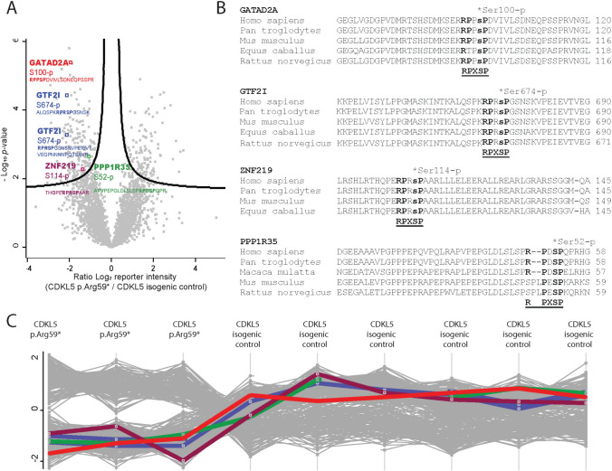
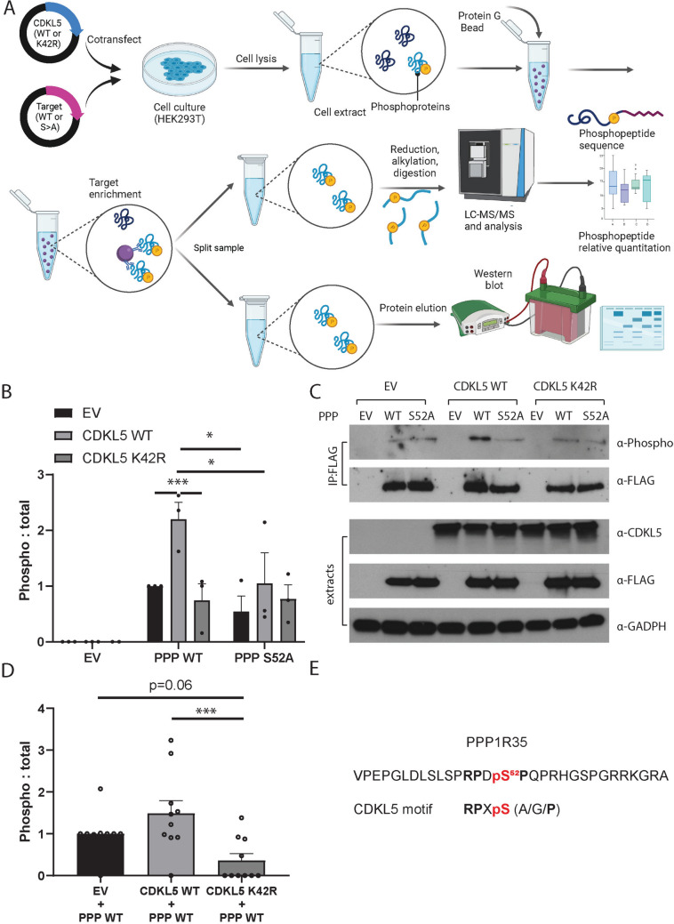
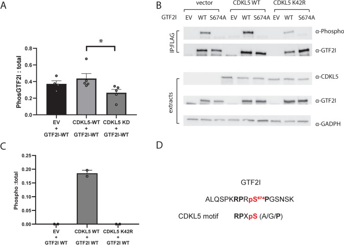
CDKL5 Deficiency Disorder (CDD) is a debilitating epileptic encephalopathy disorder affecting young children with no effective treatments. CDD is caused by pathogenic variants in Cyclin-Dependent Kinase-Like 5 (CDKL5), a protein kinase that regulates key phosphorylation events in neurons. For therapeutic intervention, it is essential to understand molecular pathways and phosphorylation targets of CDKL5. Using an unbiased phosphoproteomic approach we identified novel targets of CDKL5, including GTF2I, PPP1R35, GATAD2A and ZNF219 in human iPSC-derived neuronal cells. The phosphoserine residue in the target proteins lies in the CDKL5 consensus motif. We validated direct phosphorylation of GTF2I and PPP1R35 by CDKL5 using complementary approaches. GTF2I controls axon guidance, cell cycle and neurodevelopment by regulating expression of neuronal genes. PPP1R35 is critical for centriole elongation and cilia morphology, processes that are impaired in CDD. PPP1R35 interacts with CEP131, a known CDKL5 phospho-target. GATAD2A and ZNF219 belong to the Nucleosome Remodelling Deacetylase (NuRD) complex, which regulates neuronal activity-dependent genes and synaptic connectivity. In-depth knowledge of molecular pathways regulated by CDKL5 will allow a better understanding of druggable disease pathways to fast-track therapeutic development.

Keywords: CDKL5 deficiency disorder; GTF2I; Kinase; Neurodevelopmental disorder; PPP1R35; Phosphoproteomics; Phosphorylation.


Conflict of interest statement

The authors have no relevant financial or non-financial interests to disclose.

Figures

Fig. 1
Fig. 1
Phosphoproteomic workflow and analysis in CDKL5 human neurons. A Workflow for phosphoproteomic analysis in neurons. Neurons from CDKL5 p.(Arg59*) and CDKL5 isogenic controls were cultured in parallel for phosphoproteomic analysis. Neurons were lysed and protein extracts were subject to trypsin digest, peptides were extracted and phosphopeptides were enriched from each sample using TiO2 chromatography. Peptides were separated on a nano-HPLC and analysed by quantitative MS on an Orbitrap Mass Spectrometer. Data was analysed with MaxQuant software and visualised in Perseus software. Source data for the Fig. is available in Supplemental Materials and online. B Hierarchical heatmap clustering (z-score normalized) of significantly changed proteins from two-sided students T-test (BH FDR < 0.05, S0 = 0.1; n = 6 CDKL5 isogenic control neurons, n = 3 CDKL5 p.(Arg59*) neurons) between the CDKL5 p.(Arg59*) and CDKL5 isogenic control showing phosphosites identified by phosphoproteomic analysis. Horizontal tree indicates 9 independent samples. Vertical tree indicates the 454 significant phosphosites identified. Phosphorylation proteins were separated into two clusters, and each cluster was interrogated by Gene Ontology (GO) analysis. Two of the proteins clusters showing shared GO clusters were bracketed and individual samples were plotted as profile plots of log2-transformed normalized intensities for phosphoproteins in CDKL5 p.(Arg59*) and CDKL5 isogenic controls
Fig. 2
Fig. 2
Global phosphoproteomic STRING network analysis in CDKL5 human neurons. STRING analysis (http://www.string-db.org) derived protein–protein interaction networks for phosphoproteins that are differentially regulated in CDKL5 p.(Arg59*) compared to CDKL5 isogenic control neurons. The network nodes represent proteins. Log reporter intensity is from CDKL5 p.(Arg59*)/CDKL5 isogenic control data presented in volcano plot in Fig. 4A, so blue represents higher in CDKL5 p.(Arg59*) and red represents higher in CDKL5 isogenic control. Reporter intensity is indicated by a blue or red circle around the protein name. Interaction score in STRING is set at the highest confidence (0.900), and major gene ontology groups highlighted in the centre of the circle, with cytoskeletal proteins (pink centre), histone-binding proteins (green centre) and RNA binding proteins (yellow centre) indicated
Fig. 3
Fig. 3
Kinome profiling and kinase-substrate interaction in CDKL5 neurons. Phosphoproteomic data was analysed in Phosphomatic.com to determine disrupted kinase activity in CDKL5 neurons. A KSEA analysis provides information on signalling cascades that are altered in CDD subsequent to pathogenic variants in CDKL5. KSEA analysis identifies kinases that are associated with phosphorylation sites in a data set that substantially differ in abundance between two treatment groups. The KSEA Z-score assesses the statistical significance of inferred activities, by normalizing the total log-fold change of substrates with the standard deviation of the log-fold changes of all sites in the dataset. Blue bars represent enrichment of the phospho-sites in CDKL5 isogenic control neurons (Z-scores less than 0) and red bars represent enrichment of the phospho-sites in CDKL5 p.(Arg59*) neurons (Z-score greater than 0). B Kinases from KSEA were mapped onto the human kinase map and circles represent enrichment of kinases that phosphorylate phospho-sites from KSEA analysis. Blue circles are higher in CDKL5 isogenic control neurons and red circles are higher in CDKL5 p.(Arg59*) neurons. CDKL5 is indicated in yellow. C The phosphoproteomic network displays specific relationships between the substrates in CDKL5 isogenic controls compared to CDKL5 p.(Arg59*) neurons and known upstream kinases based on data from PhosphoSitePlus and Signor. Network diagrams represent substrates of the active data group as coloured circles (blue is higher in CDKL5 isogenic control and red is higher in CDKL5 p.(Arg59*) neurons) and known upstream kinases as green circles. Colour-coding of substrates represents mean fold-change between the two groups
Fig. 4
Fig. 4
Identification of potential specific CDKL5 phosphotargets in CDKL5 human neurons. A Label free quantitative phospho-MS (LFQPMS) of phosphorylated CDKL5 neuronal proteins (left = higher in wild-type), highlighting sites containing the CDKL5 consensus motif. Volcano plot shows significantly changed proteins from Perseus analysis (BH FDR < 0.05, S0 = 0.1; n = 6 CDKL5 isogenic control neurons, n = 3 CDKL5 p.(Arg59*) neurons). B Conservation of phosphorylated sites in proteins containing the CDKL5 motif. C Phosphorylation level of targets in each sample were consistent between replicate samples. Each coloured line represents phosphorylation levels of the four targets identified in volcano plot in Fig. 4A (PPP1R35 in green, GTF2I in blue, GATAD2A in red and ZNF219 in purple)
Fig. 5
Fig. 5
Orthogonal validation approach to demonstrate that CDKL5 phosphorylates PPP1R35 at Ser52 in human cells. A HEK293T cells were co-transfected with CDKL5 WT or CDKL5 K42R and PPP1R35 (PPP) WT or PPP1R35 S52A, where serine is converted to an alanine. Cells were then lysed and the target was immunoprecipitated with an antibody against the epitope FLAG tag. Immunoprecipitated proteins were split between quantitative phospho-MS and phospho-western blotting. B Anti‐FLAG immunoprecipitates were probed with Anti-MAPK/CDK -Phospho (top panel), and anti-FLAG (bottom panel). Empty vector (EV) lysates were used as a negative control. Quantification of three independent experiments showed a significant increase in the phosphorylation levels of PPP1R35 WT when co-expressed with CDKL5 WT compared to either empty vector or CDKL5 kinase dead (K42R) mutant. Site-directed mutagenesis (PPP1R35 S52A) did reduce overall phosphorylation levels when comparing PPP1R35 WT to PPP1R35 S52A. Data is representative of 3 independent samples from 3 independent experiments. Data is mean ± SEM. *P < 0.05, **P < 0.01. One-way ANOVA with Dunnett’s multiple comparisons test. C Lysate extract sets from co-transfection experiments were probed with anti-CDKL5 (top panel), anti-FLAG (middle panel) and anti-GAPDH (bottom panel). The expression patterns indicate that the co-transfection experiments were successful. Three independent experiments were completed, and one representative experiment is shown. D Phosphorylation of PPP1R35 WT was higher in HEK293T cells co-transfected with CDKL5 WT compared to co-transfected with CDKL5 K42R or empty vector. Phospho-peptides were quantified using LC–MS/MS intensity of phosphopeptides and was normalised to the label-free quantification of PPP1R35. Data is representative of 10 independent samples from six independent experiments. Data is mean ± SEM. ***P < 0.001. One-way ANOVA with Sidak’s multiple comparisons test. E Phosphopeptide detected from PPP1R35 that is phosphorylated by CDKL5
Fig. 6
Fig. 6
Orthogonal validation approach to demonstrate that CDKL5 phosphorylates GTF2I at Ser674 in human cells. A Quantification of three independent experiments showed a significant difference in the phosphorylation levels in GTF2I when co-expressed with CDKL5 WT compared to either empty vector (EV) or CDKL5 kinase dead (K42R) mutant. Site-directed mutagenesis of GTF2I at Ser674 to Ala (SA) completely abolished phosphorylation levels and was not quantifiable. Data is representative of 5 independent samples from 5 independent experiments. Data is mean ± SEM. *P < 0.05. B Lysate extract sets from co-transfection experiments were probed with anti-CDKL5 (top panel), anti-GTF2I (middle panel) and anti-GAPDH (bottom panel). Five independent experiments were completed and the expression patterns indicate that the co-transfection experiments were successful. Data is mean ± SEM. *P < 0.05. One-way ANOVA with Dunnett’s multiple comparisons test. C Phosphorylation of GTF2I WT was only detected in HEK293T cells co-transfected with CDKL5 WT compared to co-transfected with CDKL5 K42R or empty vector. Phospho-peptides were quantified using LC–MS/MS intensity of phosphopeptides was normalised to the label-free quantification of GTF2I. The phosphorylation event was detected in two of six independent experiments. D Phosphopeptide detected from GTF2I that is phosphorylated by CDKL5

References

    1. Kawahata I, Fukunaga K (2023) Protein kinases and neurodegenerative diseases. Int J Mol Sci. 10.3390/ijms24065574 10.3390/ijms24065574 - DOI - PMC - PubMed
    1. Olson HE, Demarest ST, Pestana-Knight EM, Swanson LC, Iqbal S, Lal D et al (2019) Cyclin-dependent kinase-like 5 deficiency disorder: clinical review. Pediatr Neurol 97:18–25. 10.1016/j.pediatrneurol.2019.02.015 10.1016/j.pediatrneurol.2019.02.015 - DOI - PMC - PubMed
    1. Weaving LS, Christodoulou J, Williamson SL, Friend KL, McKenzie OL, Archer H et al (2004) Mutations of CDKL5 cause a severe neurodevelopmental disorder with infantile spasms and mental retardation. Am J Hum Genet 75(6):1079–1093. 10.1086/426462 10.1086/426462 - DOI - PMC - PubMed
    1. Tao J, Van Esch H, Hagedorn-Greiwe M, Hoffmann K, Moser B, Raynaud M et al (2004) Mutations in the X-linked cyclin-dependent kinase-like 5 (CDKL5/STK9) gene are associated with severe neurodevelopmental retardation. Am J Hum Genet 75(6):1149–1154. 10.1086/426460 10.1086/426460 - DOI - PMC - PubMed
    1. Arora S, Goodall S, Viney R, Einfeld S (2020) Societal cost of childhood intellectual disability in Australia. J Intellect Disabil Res 64(7):524–537. 10.1111/jir.12732 10.1111/jir.12732 - DOI - PubMed

MeSH terms

Substances

Supplementary concepts

LinkOut - more resources