Review
. 2024 Nov;26(11):1153-1162.
doi: 10.1007/s11886-024-02114-6. Epub 2024 Aug 13.

Recent Advances in Positron Emission Tomography Radiotracers to Image Cardiac Amyloidosis

Affiliations
Review

Recent Advances in Positron Emission Tomography Radiotracers to Image Cardiac Amyloidosis

Ardel J Romero Pabón et al. Curr Cardiol Rep. 2024 Nov.

Abstract

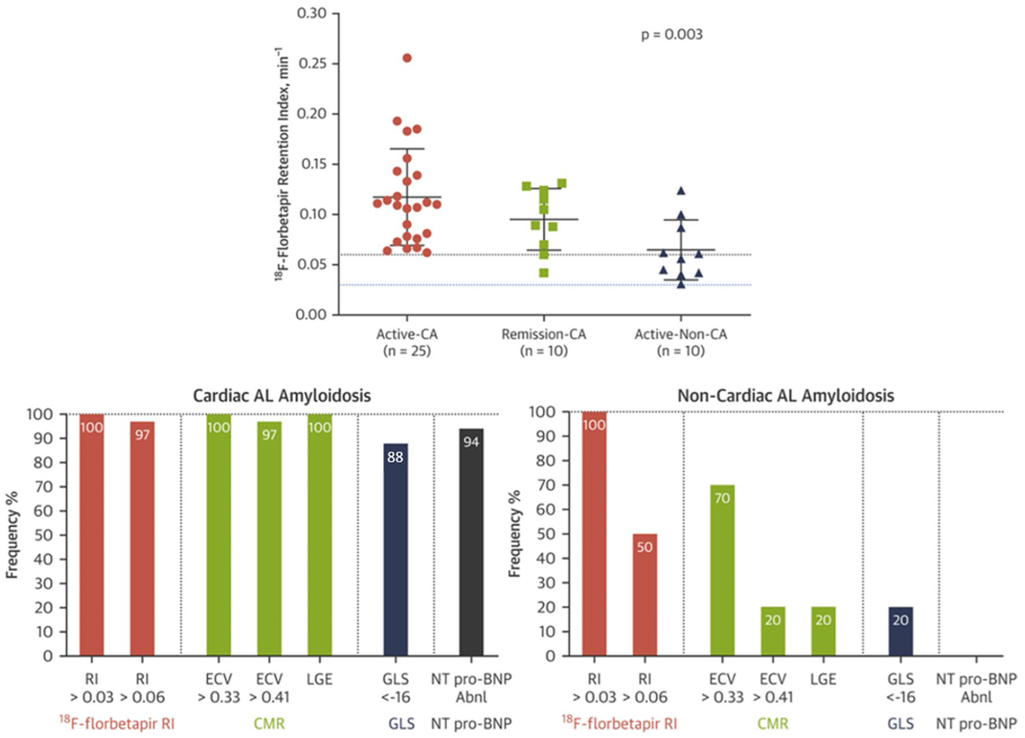
Cardiac amyloidosis includes a group of protein-misfolding diseases characterized by fibril accumulation within the extracellular space of the myocardium and cardiac dysfunction. Cardiac amyloidosis has high mortality. Emerging radionuclide techniques have helped us to better understand disease pathogenesis, prognostication, and treatment response in cardiac amyloidosis. PURPOSE OF REVIEW: To review recent advances in molecular imaging of cardiac amyloidosis using amyloid PET radiotracers. RECENT FINDINGS: Multiple single center studies have shown that amyloid PET radiotracers allow definitive diagnosis and quantification of cardiac amyloid burden. These amyloid targeting tracers may provide means to improve early disease detection, risk stratification and treatment monitoring. Amyloid PET imaging may inform definitive imaging-based diagnosis for therapeutic decisions, risk stratification, and treatment monitoring. More research in unselected cohorts of patients with suspected cardiac amyloidosis is needed to optimize the clinical implementation of amyloid PET imaging.

Keywords: Cardiac Amyloidosis; Positron Emission Tomography; Radiotracers.


Conflict of interest statement

Conflict of Interests The authors declare no competing interests.

Figures

Fig. 1
Fig. 1
A Molecular structures of various amyloid radiotracers. B and C the synthetic polypeptide 124I-evuzamitide binds to the negatively charged glycosaminoglycans present in amyloid fibrils. Figure A was adapted from Uzuegbunam et al. [59] [Creative Commons CC BY 4.0 license]. Figures B and C were provided courtesy of Dr. Jonathan Wall, University of Tennessee)
Fig. 2
Fig. 2
18F-florbetapir pulmonary tracer uptake. 18F-florbetapir PET/CT images of three patients with a no significant tracer uptake, b diffuse mild uptake, and c diffuse intense uptake, respectively. Of note, CT average lung densities did not differ among patients with different degrees of tracer uptake, and the control group (bottom row, d). Additionally, the rate of tracer washout was slowest in subjects with intense uptake and fastest among controls (bottom row, e). (Reprinted with permission from Khor et al. [46]; permission conveyed through Copyright Clearance Center, Inc.)
Fig. 3
Fig. 3
Imaging findings in different groups of patients with AL amyloidosis. The top row shows the distribution of cardiac MRI ECV values in patients systemic AL amyloidosis with cardiac involvement (active-CA), without cardiac involvement by conventional criteria (active-non-CA), and patients with cardiac involvement in remission for at least 1 year (remission-CA). The dotted blue line represents the cutoff value to diagnose the presence or absence of CA while the dotted black line represents the upper threshold of normal observed in healthy controls. The p-value listed in the figure is the cross-group comparison. The bottom row shows the prevalence of abnormal indexes of cardiac amyloid deposition in these groups. Cardiac AL Amyloidosis = active-CA and remission-CA. Non-Cardiac AL Amyloidosis = active-non-CA. (Reprinted with permission from Cuddy et al. [48], with permission from Elsevier.)
Fig. 4
Fig. 4
Myocardial (top 2 rows) and systemic organ (bottom row) uptake of 18F-florbetapir and 124I-evuzamitide in patients with various forms of amyloidosis. Liver uptake is physiologic with 18F-florbetapir imaging. Amyloid PET tracers bind to various types of amyloid fibrils and can image amyloid deposition in the cardiac and extracardiac tissues. Apo-A-IV = Apolipoprotein AIV. (Reprinted with permission from Dorbala et al. [11].)
Fig. 5
Fig. 5
Kaplan-Meier curves for MACE and all-cause death based on left ventricular amyloid burden. Left ventricular amyloid burden was assessed by tertiles of 18F-florbetapir uptake quantified as percentage of the injected dose. The p-values in each graph correspond to the log-rank tests performed. LV = Left ventricular. %ID = percentage of the injected dose. MACE = Major Adverse Cardiovascular Events (all-cause mortality, heart failure hospitalization or cardiac transplantation). (Reprinted with permission from Clerc et al. [51], with permission from Elsevier.)

References

    1. Buxbaum JN, Dispenzieri A, Eisenberg DS, et al. Amyloid nomenclature 2022: Update, novel proteins, and recommendations by the International Society of Amyloidosis (ISA) Nomenclature Committee. Amyloid. 2022;29:213–9. - PubMed
    1. Falk RH, Alexander KM, Liao R, Dorbala S. AL (light-chain) cardiac amyloidosis: A review of diagnosis and therapy. J Am Coll Cardiol. 2016;68:1323–41. - PubMed
    1. Ruberg FL, Grogan M, Hanna M, Kelly JW, Maurer MS. Transthyretin Amyloid Cardiomyopathy: JACC State-of-the-Art Review. J Am Coll Cardiol. 2019;73:2872–91. - PMC - PubMed
    1. Merlini G, Bellotti V. Molecular mechanisms of amyloidosis. N Engl J Med. 2003;349:583–96. - PubMed
    1. Dorbala S, Cuddy S, Falk RH. How to image cardiac amyloidosis: A practical approach. JACC Cardiovasc Imaging. 2020;13:1368–83. - PMC - PubMed

MeSH terms