. 2024 Aug 20;121(34):e2406519121.
doi: 10.1073/pnas.2406519121. Epub 2024 Aug 13.

Phase separation of PML/RARα and BRD4 coassembled microspeckles governs transcriptional dysregulation in acute promyelocytic leukemia

Affiliations

Phase separation of PML/RARα and BRD4 coassembled microspeckles governs transcriptional dysregulation in acute promyelocytic leukemia

Yi Zhang et al. Proc Natl Acad Sci U S A. .

Abstract

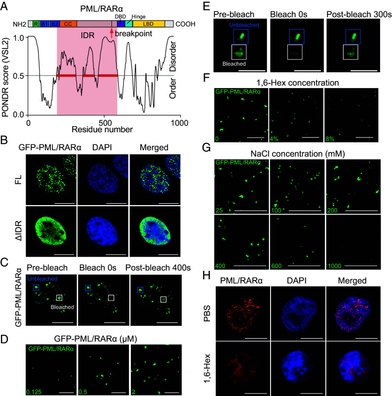
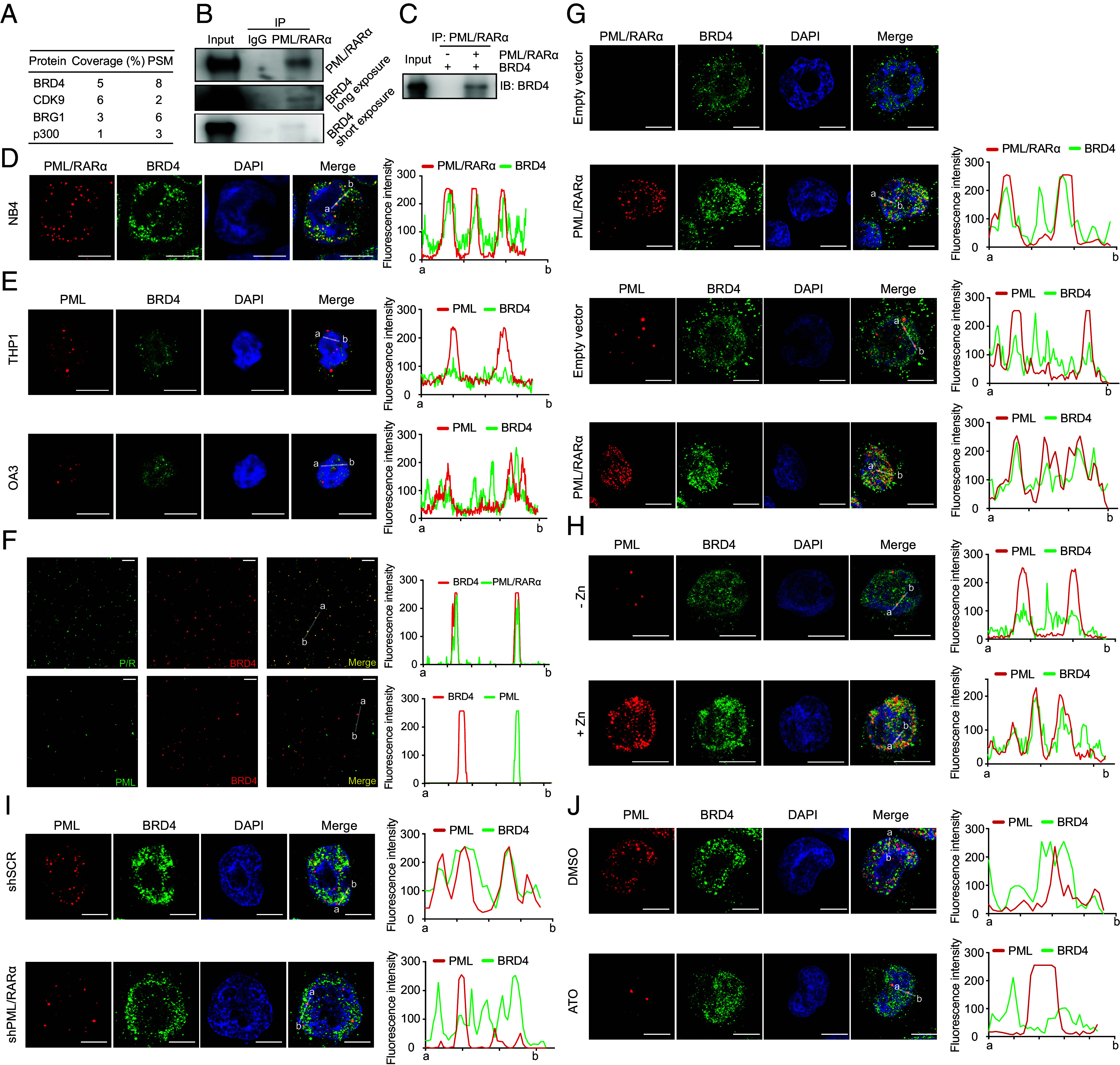
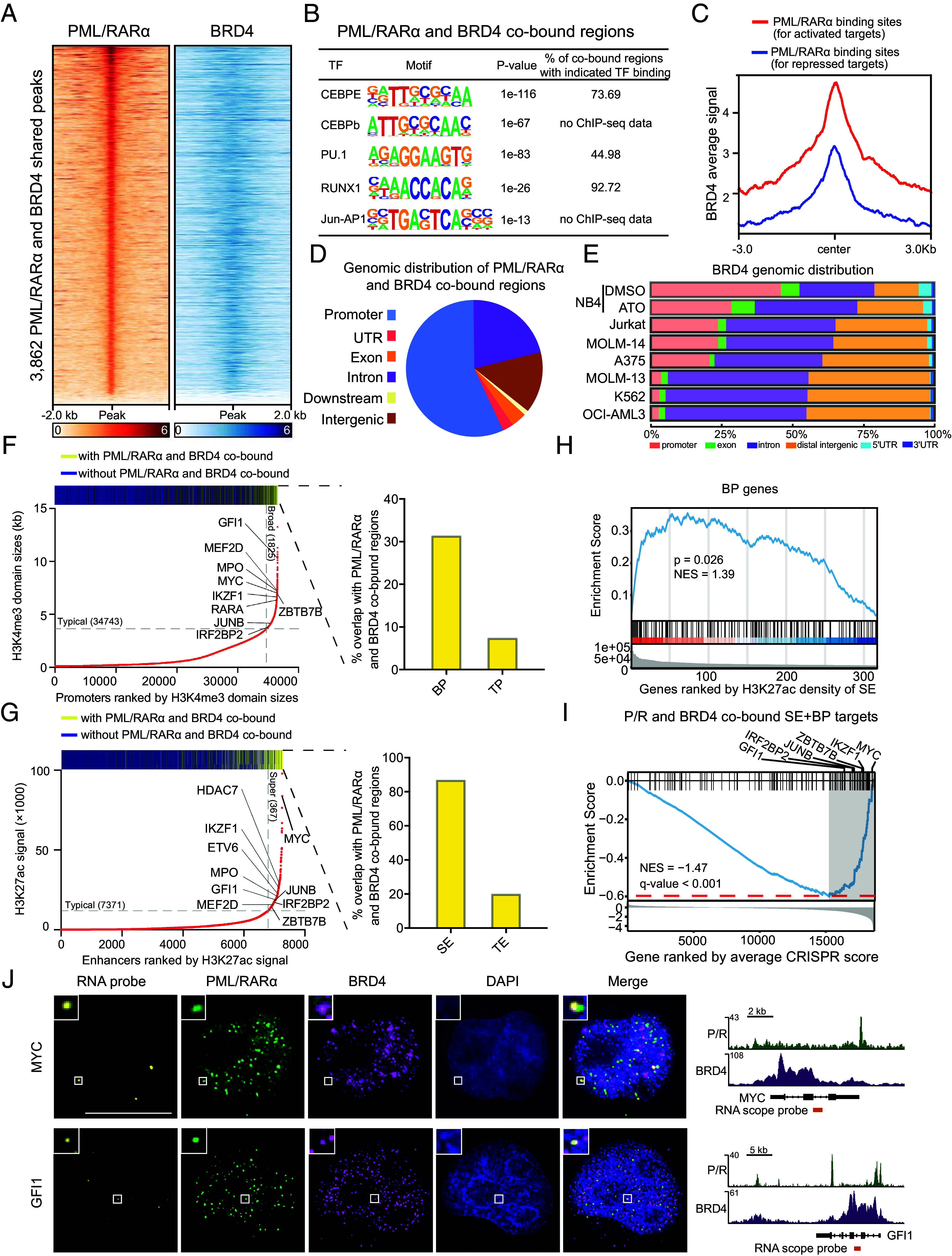
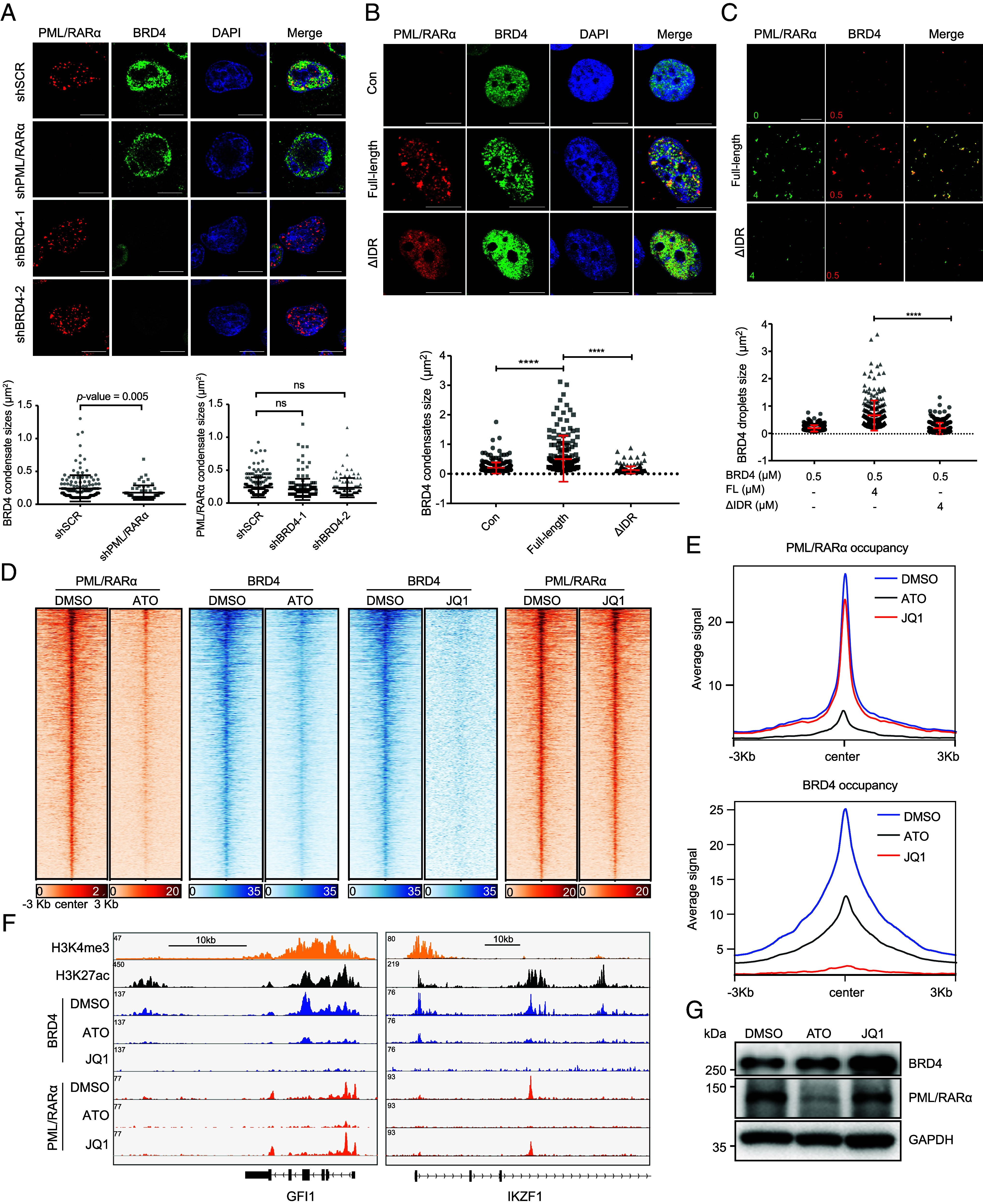
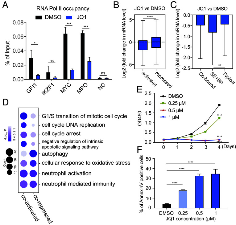
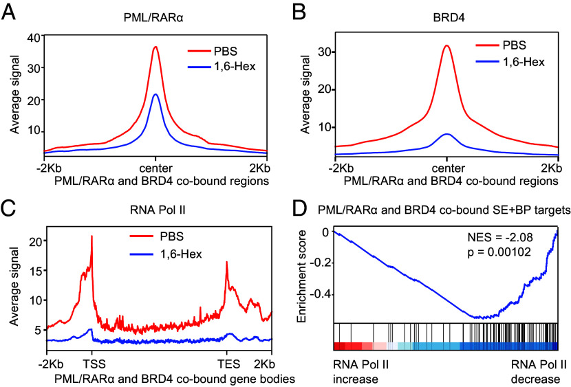
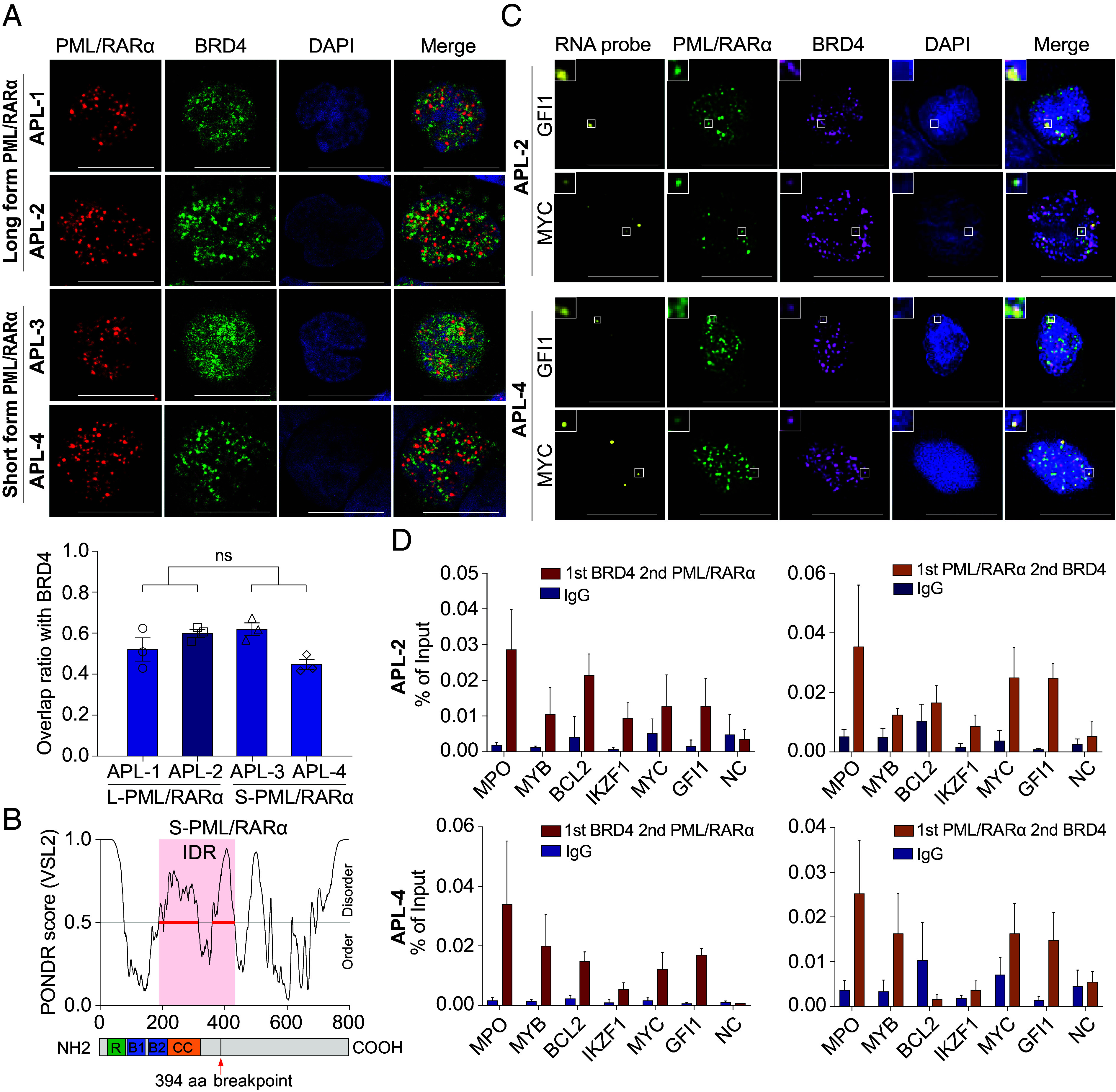
In acute promyelocytic leukemia (APL), the promyelocytic leukemia-retinoic acid receptor alpha (PML/RARα) fusion protein destroys PML nuclear bodies (NBs), leading to the formation of microspeckles. However, our understanding, largely learned from morphological observations, lacks insight into the mechanisms behind PML/RARα-mediated microspeckle formation and its role in APL leukemogenesis. This study presents evidence uncovering liquid-liquid phase separation (LLPS) as a key mechanism in the formation of PML/RARα-mediated microspeckles. This process is facilitated by the intrinsically disordered region containing a large portion of PML and a smaller segment of RARα. We demonstrate the coassembly of bromodomain-containing protein 4 (BRD4) within PML/RARα-mediated condensates, differing from wild-type PML-formed NBs. In the absence of PML/RARα, PML NBs and BRD4 puncta exist as two independent phases, but the presence of PML/RARα disrupts PML NBs and redistributes PML and BRD4 into a distinct phase, forming PML/RARα-assembled microspeckles. Genome-wide profiling reveals a PML/RARα-induced BRD4 redistribution across the genome, with preferential binding to super-enhancers and broad-promoters (SEBPs). Mechanistically, BRD4 is recruited by PML/RARα into nuclear condensates, facilitating BRD4 chromatin binding to exert transcriptional activation essential for APL survival. Perturbing LLPS through chemical inhibition (1, 6-hexanediol) significantly reduces chromatin co-occupancy of PML/RARα and BRD4, attenuating their target gene activation. Finally, a series of experimental validations in primary APL patient samples confirm that PML/RARα forms microspeckles through condensates, recruits BRD4 to coassemble condensates, and co-occupies SEBP regions. Our findings elucidate the biophysical, pathological, and transcriptional dynamics of PML/RARα-assembled microspeckles, underscoring the importance of BRD4 in mediating transcriptional activation that enables PML/RARα to initiate APL.

Keywords: BRD4; PML/RARα; acute promyelocytic leukemia; liquid–liquid phase separation; transcriptional dysregulation.


Conflict of interest statement

Competing interests statement:The authors declare no competing interest.

Figures

Fig. 1.
Fig. 1.
PML/RARα undergoes LLPS. (A) Schematic diagram illustrating the IDR domain of the long-form PML/RARα protein, derived from PML/RARα bcr1 fusion. The exact coordinates of RBCC domains, IDR domain, DBD domain, Hinge domain, and LBD domain were indicated: Ring domain (49 to 104 aa), B1 box (124 to 166 aa), B2 box (183 to 229 aa), coiled-coil domain (230 to 368 aa), IDR domain (205 to 589 aa), DBD domain (590 to 640 aa), Hinge domain (646 to 691 aa), and LBD domain (692 to 911 aa). The breakpoint of PML/RARα was also indicated with a red arrow (552 aa). (B) Representative confocal images illustrating the presence of microspeckles for the full-length (FL) PML/RARα and diffuse distribution for the IDR domain deletion mutant in transfected U2OS cells. (Scale bar, 10 μm.) (C) Representative images of FRAP assays in living cells. (Scale bar, 10 μm.) (D) Representative images of droplet formation at different protein concentrations. (E) Representative images of FRAP assay in cellular extracts. (Scale bar, 3 μm.) (F) Representative images of droplet formation assays at different 1,6-Hex concentrations. (G) Representative images of droplet formation assays at different salt concentrations. (H) Immunofluorescence images demonstrating the sensitivity of PML/RARα microspeckles to 1,6-Hex in the nucleus. PML/RARα was immunostained using a customized antibody in NB4 cells treated with 8% 1,6-Hex (Lower) or PBS (Upper) for 2 min.
Fig. 2.
Fig. 2.
Colocalization with BRD4 is a unique characteristic of PML/RARα-assembled microspeckles rather than PML NBs. (A) Top PML/RARα-interacting partners enriched over IgG and associated with phase-separated transcriptional hubs. PSM for the peptide spectrum match and Coverage% for the percentage of the protein sequence covered by the identified peptide in MS. (B) The interaction between endogenous PML/RARα and BRD4 in NB4 cells. (C) The interaction between ectopically expressed PML/RARα and BRD4 in HEK-293T cells. (D) Colocalization of PML/RARα microspeckles with BRD4 in NB4 cells. (Scale bar, 10 μm.) (Right) fluorescence intensity of PML/RARα and BRD4 from a to b. (E) Absence of colocalization between PML NBs and BRD4 in non-APL cell lines THP1 and OCI-AML3. (Scale bar, 10 μm.) (F) Overlap of PML/RARα droplets with BRD4 droplets in cellular extracts. In contrast, absence of colocalization between PML droplets and BRD4 droplets. (Scale bar, 10 μm.) (G) Ectopic expression of PML/RARα leading to the disruption of PML NBs and redistribution of BRD4 into PML/RARα-assembled condensates in U937 cells. (H) De novo expression of PML/RARα induced by ZnSO4 leading to the disruption of PML NBs and redistribution of BRD4 into microspeckles in PR9 cells. (I and J) Depletion of PML/RARα resulting in the restoration of PML NBs and BRD4 puncta in NB4 cells. PML/RARα downregulation was achieved using shRNA (I) or ATO (J) degradation.
Fig. 3.
Fig. 3.
PML/RARα and BRD4 coassembled condensates tend to occupy on SEs and BPs, targeting genes crucial for APL leukemogenesis. (A) Heatmap showing PML/RARα and BRD4 cobinding in 3,862 genomic regions. (B) Enrichment of hematopoietic master TFs motif in PML/RARα and BRD4 cobound regions. (C) Higher binding signals of BRD4 at PML/RARα directly activated genes than PML/RARα directly repressed genes (30). (D) Distribution of PML/RARα and BRD4 cobinding sites in NB4 cells. (E) Reduced binding of BRD4 to promoter regions in the absence of PML/RARα. NB4 was PML/RARα positive cell line and K562, OCI-AML3, MOLM-13, MOLM-14, A375, and Jurkat were PML/RARα negative cell lines. (F) Preferential cobinding of PML/RARα and BRD4 to BPs compared to typical promoters (TPs) in NB4 cells, with a fourfold higher likelihood. The bar above depicts the distribution of PML/RARα and BRD4 cobinding among promoters, in which yellow represents the peaks with PML/RARα and BRD4 cobinding and blue for the peaks without PML/RARα and BRD4 cobinding. The bar graph shows the percentages of PML/RARα and BRD4 cobinding within BP regions and TP regions. (G) Preferential cobinding of PML/RARα and BRD4 to SEs over fourfold more likely than typical enhancers (TEs) in NB4 cells. (H) GSEA showing the enrichment of BP genes among genes most likely targeted by SEs with PML/RARα and BRD4 cobinding. (I) GSEA showing the tendency of PML/RARα and BRD4 cobound SEBP genes to be essential for the survival of APL cells. Representative genes at the leading edge are labeled. (J) PML/RARα and BRD4 IF with concurrent RNA fluorescence in situ hybridization (RNA-FISH) demonstrating coassembly of PML/RARα and BRD4 condensates at MYC and GFI1gene loci. (Right) depiction of MYC and GFI1 gene loci, PML/RARα and BRD4 ChIP-seq, and location of RNA-FISH probes.
Fig. 4.
Fig. 4.
Recruitment of BRD4 within PML/RARα-assembled condensates facilitates the chromatin targeting of BRD4. (A) Immunofluorescence of PML/RARα and BRD4 upon PML/RARα or BRD4 knockdown in NB4 cells. (Scale bar, 10 μm.) Low, analysis were performed on 6 to 10 cells, with ns denoting no significance. (B) Immunofluorescence of FL PML/RARα or ΔIDR-PML/RARα and BRD4 in transiently transfected U2OS cells. (Scale bar, 10 μm.) Low, analysis performed on 6 to 10 cells. ****P < 0.0001 (two-tailed Student’s t test). (C) Representative images of droplet formation assays of red fluorescent protein tagged BRD4 (RFP-BRD4) at 0.5 μM mixed with FL GFP-PML/RARα or ΔIDR mutant GFP-PML/RARα at 4 μM each. Low, quantification of the area of BRD4 droplets. ****P < 0.0001 (two-tailed Student’s t test). (D) ChIP-seq signal of PML/RARα and BRD4 on their cobound regions in NB4 cells treated with DMSO, ATO, or JQ1. (E) Metaplots showing the average BRD4 (Top) or PML/RARα (Bottom) ChIP-seq signals at PML/RARα and BRD4 cobound regions in NB4 cells treated with DMSO, ATO, or JQ1. (F) IGV tracks displaying the indicated ChIP-seq signals at GFI1 and IKZF1 in NB4 cells. (G) PML/RARα degradation had no influence on the protein levels of BRD4 in NB4 cells.
Fig. 5.
Fig. 5.
Blockage of BRD4 activity hinders PML/RARα-mediated transcriptional activation and impairs APL leukemogenesis. (A) Decreased RNA pol II binding signals on PML/RARα and BRD4 cobound SE+BP gene bodies upon BRD4 inhibition. NC, negative control. *P < 0.05; ***P < 0.001; ns no significance. (B) Significant downregulation of PML/RARα directly activated targets (n = 424) upon JQ1 treatment and a lesser impact on PML/RARα directly repressed genes (n = 363). ****P < 0.0001 (two-tailed Student’s t test). (C) Much more reduced mRNA levels of SE+BP genes upon BRD4 suppression compared to TP genes. Typical genes are defined as those nearest to TEs and promoters, and SE+BP genes for those nearest to SEBPs. (D) Functional enrichment analysis revealing significant GO terms among PML/RARα and BRD4 coregulated genes. (E and F) BRD4 inhibition decreased proliferation and induced apoptosis in APL cells. Cell proliferation ability was measured by the CCK8 assay (E). Apoptosis (F) was analyzed on day 2 after DMSO or JQ1 treatment. ****P < 0.0001; ns for no significance.
Fig. 6.
Fig. 6.
Disrupting LLPS abolishes PML/RARα and BRD4 chromatin coverage and suppresses transcription of their target genes. (A and B) Metaplots illustrating the reduction of average PML/RARα (A) or BRD4 (B) ChIP-seq signals at PML/RARα and BRD4 cobound regions in NB4 cells following treatment with 1,6-Hex. (C) Metaplots demonstrating the attenuation of average RNA Pol II ChIP-seq signals at PML/RARα and BRD4 cobound gene bodies in NB4 cells following 1,6-Hex treatment. TSS, transcription start site. TES, transcription end site. (D) GSEA indicates a high enrichment for PML/RARα and BRD4 cobound SEBP targets among genes exhibiting the most significant decrease in RNA Pol II occupancy after 1,6-Hex treatment. Enrichment score profile is shown, together with the position of SEBP target genes. Genes are ranked by their log2 fold change in RNA Pol II ChIP-seq density within the gene body.
Fig. 7.
Fig. 7.
Coassembly of both long- and short-type PML/RARα with BRD4 to form condensates at SEBP regions on APL blast cells. (A) Colocalization of both types of PML/RARα and BRD4 in primary APL blasts assessed by coimmunostaining. (Scale bar, 10 μm.) Low, the overlap ratio of BRD4 with PML/RARα condensates in APL blast cells expressing long-form or short form of PML/RARα, respectively. Analysis was conducted on 10 cells. (B) Schematic diagram illustrating the IDR domain of the short-type PML/RARα. The exact coordinate of the IDR domain was indicated: 205 to 431 aa. The breakpoint of PML/RARα was also indicated with a red arrow (394 aa). (C) IF with concurrent RNA-FISH demonstrating coassembly of both types of PML/RARα with BRD4 forming condensates at MYC and GFI1 gene loci in primary APL blasts. (D) Validation of the co-occupancy of PML/RARα and BRD4 on the same regulatory genomic regions in APL blasts through re-ChIP assays. NC, negative control.

Similar articles

Cited by

References

    1. Mitrea D. M., Mittasch M., Gomes B. F., Klein I. A., Murcko M. A., Modulating biomolecular condensates: A novel approach to drug discovery. Nat. Rev. Drug. Discov. 21, 841–862 (2022). - PMC - PubMed
    1. Xu C., Kim A., Corbin J. M., Wang G. G., Onco-condensates: Formation, multi-component organization, and biological functions. Trends Cancer 9, 738–751 (2023), 10.1016/j.trecan.2023.05.006. - DOI - PMC - PubMed
    1. Wei M., Huang X., Liao L., Tian Y., Zheng X., SENP1 decreases RNF168 phase separation to promote DNA damage repair and drug resistance in colon cancer. Cancer Res. 83, 2908–2923 (2023), 10.1158/0008-5472.CAN-22-4017. - DOI - PubMed
    1. Dyck J. A., et al. , A novel macromolecular structure is a target of the promyelocyte-retinoic acid receptor oncoprotein. Cell 76, 333–343 (1994). - PubMed
    1. Weis K., et al. , Retinoic acid regulates aberrant nuclear localization of PML-RAR alpha in acute promyelocytic leukemia cells. Cell 76, 345–356 (1994). - PubMed

MeSH terms

Substances

LinkOut - more resources