. 2024 Sep 20;19(9):1888-1895.
doi: 10.1021/acschembio.4c00237. Epub 2024 Aug 13.

Targeted Degradation of Protein Kinase A via a Stapled Peptide PROTAC

Affiliations

Targeted Degradation of Protein Kinase A via a Stapled Peptide PROTAC

Matthew K Whittaker et al. ACS Chem Biol. .

Abstract

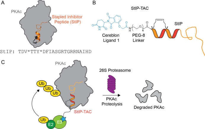
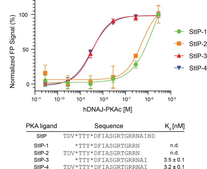
Proteolysis-targeting chimeras (PROTACs) are bifunctional molecules that bind and recruit an E3 ubiquitin ligase to a targeted protein of interest, often through the utilization of a small molecule inhibitor. To expand the possible range of kinase targets that can be degraded by PROTACs, we sought to develop a PROTAC utilizing a hydrocarbon-stapled peptide as the targeting agent to bind the surface of a target protein of interest. In this study, we describe the development of a proteolysis-targeting chimera, dubbed Stapled Inhibitor Peptide - PROTAC or StIP-TAC, linking a hydrocarbon-stapled peptide with an E3 ligase ligand for targeted degradation of Protein Kinase A (PKA). This StIP-TAC molecule stimulated E3-mediated protein degradation of PKA, and this effect could be reversed by the addition of the proteasomal inhibitor MG-132. Further, StIP-TAC treatment led to a significant reduction in PKA substrate phosphorylation. Since many protein targets of interest lack structural features that make them amenable to small molecule targeting, development of StIP-TACs may broaden the potential range of protein targets using a PROTAC-mediated proteasomal degradation approach.


Conflict of interest statement

The authors declare no competing financial interest.

Figures

Figure 1
Figure 1
Schematic of PROTAC design and activity. (A) Schematic of StIP binding to PKA along with parent sequence. * = (S)-2-(4-pentenyl)alanine. (B) Schematic of PROTAC structure. StIP-derived peptide is conjugated to thalidomide-derived Cereblon Ligand 1 via a PEG-8 linker. (C) Proposed mechanism of action of PKI-derived PROTAC molecule. StIP-derived POI ligand binds PKA and recruits E3 ligase complex via Cereblon Ligand 1, resulting in ubiquitination (Ub) of PKA and subsequent targeted degradation by the proteasome. E2 = E2 ligase, E3 = E3 ligase.
Figure 2
Figure 2
Direct binding measurements of fluorescein-labeled truncated peptides to PKA catalytic subunit determined by fluorescence polarization. StIP-3 and StIP-4 demonstrate low nanomolar binding to PKA with truncated sequences compared to the parent peptide sequence, StIP. * = location where (S)-2-(4-pentenyl)alanine is incorporated into the peptide sequence to form the hydrocarbon staple. Mean values and standard deviation of three independent measurements are given. n.d. = not determined. Both StIP-T3 and StIP-T4 were found to have low-nanomolar affinities.
Figure 3
Figure 3
StIP-TAC treatment induces PKA degradation. (A) Representative Western blot of cells treated with vehicle control, StIP-TAC, and StIP-TAC + SAINT Protein reagent (Lipid) for 5 h (n = 3) with or without a 30 min forskolin (FSK) stimulation. Cells treated with StIP-TAC in conjunction with the SAINT Protein lipid delivery formulation demonstrate reduced PKA protein levels. (B) Densitometric quantification of three independent Western blots (n = 3) demonstrates a statistically significant reduction in PKA protein levels for cells treated with StIP-TAC and SAINT Protein reagent. No significant difference in PKA levels was detected between forskolin-stimulated and unstimulated treatments. Quantification was performed via Li-COR Image Studio. PKA bands for each treatment were normalized to the GAPDH loading control and compared to the DMSO control treatment. ***p < 0.001; ns, not significant as assessed by one-way ANOVA and Bonferroni’s multiple comparisons test. Error bars represent standard deviation. (C) Representative Western blot of cells treated with 10 μM proteasomal inhibitor MG-132 with vehicle control or StIP-TAC with SAINT protein reagent for 5 h (n = 3). Cells treated with StIP-TAC and SAINT protein demonstrate a reduction in PKA protein levels that are rescued with the addition of proteasomal inhibitor. (D) Densitometric quantification of three independent Western blots (n = 3) demonstrating a statistically significant reduction in PKA protein levels in cells treated with StIP-TAC and SAINT Protein reagent that is rescued to near-baseline levels upon proteasomal inhibition. Quantification was performed in Li-COR Image Studio. PKA bands for each treatment were normalized to the GAPDH loading control and compared to the DMSO control treatment. **p = 0.003; ns, not significant as assessed by one-way ANOVA and Bonferroni’s multiple comparisons test.
Figure 4
Figure 4
StIP-TAC treatment inhibits phosphorylation of PKA substrates. (A) Representative Western blot of cells treated with vehicle control, H89, StIP-TAC, or StIP-TAC + SAINT Protein reagent for 5 h with or without 30 min stimulation with 50 μM forskolin (n = 3). Cells treated with H89 and StIP-TAC alongside SAINT protein display a reduction in fold change of total phosphorylated PKA substrate levels after forskolin stimulation. (B) Densitometric quantification of Western blots for three independent experiments (n = 3) demonstrating a statistically significant reduction in total PKA substrate phosphorylation in cells treated with H89 and StIP-TAC with SAINT Protein reagent. Quantification was performed via Li-COR Image Studio. *** p < 0.001; ns, not significant as assessed by one-way ANOVA and Bonferroni’s multiple comparisons test. Error bars represent standard deviation. (C) Representative Western blot image of select PKA phosphorylated substrates at approximately 55, 60, 80, and 100 kDa bands, demonstrating a prominent reduction in phosphorylation after forskolin stimulation upon treatment with StIP-TAC and SAINT Protein reagent (n = 3). (D) Densitometric quantification of Western blots from three separate experiments (n = 3), demonstrating a statistically significant reduction in phosphorylation of the approximately 55, 60, 80, and 100 kDa substrate bands for cells treated with StIP-TAC and SAINT Protein reagent. Quantification was performed via Li-COR Image Studio. ***p < 0.001; ns, not significant as assessed by one-way ANOVA and Bonferroni’s multiple comparisons test. Error bars represent standard deviation.

References

    1. Sakamoto K. M.; Kim K. B.; Kumagai A.; Mercurio F.; Crews C. M.; Deshaies R. J. Protacs: chimeric molecules that target proteins to the Skp1-Cullin-F box complex for ubiquitination and degradation. Proc. Natl. Acad. Sci. U. S. A. 2001, 98 (15), 8554–8559. 10.1073/pnas.141230798. - DOI - PMC - PubMed
    1. Gao H.; Sun X.; Rao Y. PROTAC Technology: Opportunities and Challenges. ACS Med. Chem. Lett. 2020, 11 (3), 237–240. 10.1021/acsmedchemlett.9b00597. - DOI - PMC - PubMed
    1. Jin J.; Wu Y.; Chen J.; Shen Y.; Zhang L.; Zhang H.; Chen L.; Yuan H.; Chen H.; Zhang W.; Luan X. The peptide PROTAC modality: a novel strategy for targeted protein ubiquitination. Theranostics 2020, 10 (22), 10141–10153. 10.7150/thno.46985. - DOI - PMC - PubMed
    1. Hines J.; Gough J. D.; Corson T. W.; Crews C. M. Posttranslational protein knockdown coupled to receptor tyrosine kinase activation with phosphoPROTACs. Proc. Natl. Acad. Sci. U. S. A. 2013, 110 (22), 8942–7. 10.1073/pnas.1217206110. - DOI - PMC - PubMed
    1. Ma D.; Zou Y.; Chu Y.; Liu Z.; Liu G.; Chu J.; Li M.; Wang J.; Sun S. Y.; Chang Z. A cell-permeable peptide-based PROTAC against the oncoprotein CREPT proficiently inhibits pancreatic cancer. Theranostics 2020, 10 (8), 3708–3721. 10.7150/thno.41677. - DOI - PMC - PubMed

Publication types

MeSH terms

Substances

LinkOut - more resources