. 2024 Nov 4;84(21):3629-3639.
doi: 10.1158/0008-5472.CAN-23-3574.

Targeting BCL2 with Venetoclax Enhances the Efficacy of the KRASG12D Inhibitor MRTX1133 in Pancreatic Cancer

Affiliations

Targeting BCL2 with Venetoclax Enhances the Efficacy of the KRASG12D Inhibitor MRTX1133 in Pancreatic Cancer

Jeffrey H Becker et al. Cancer Res. .

Abstract

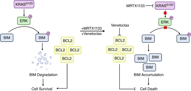
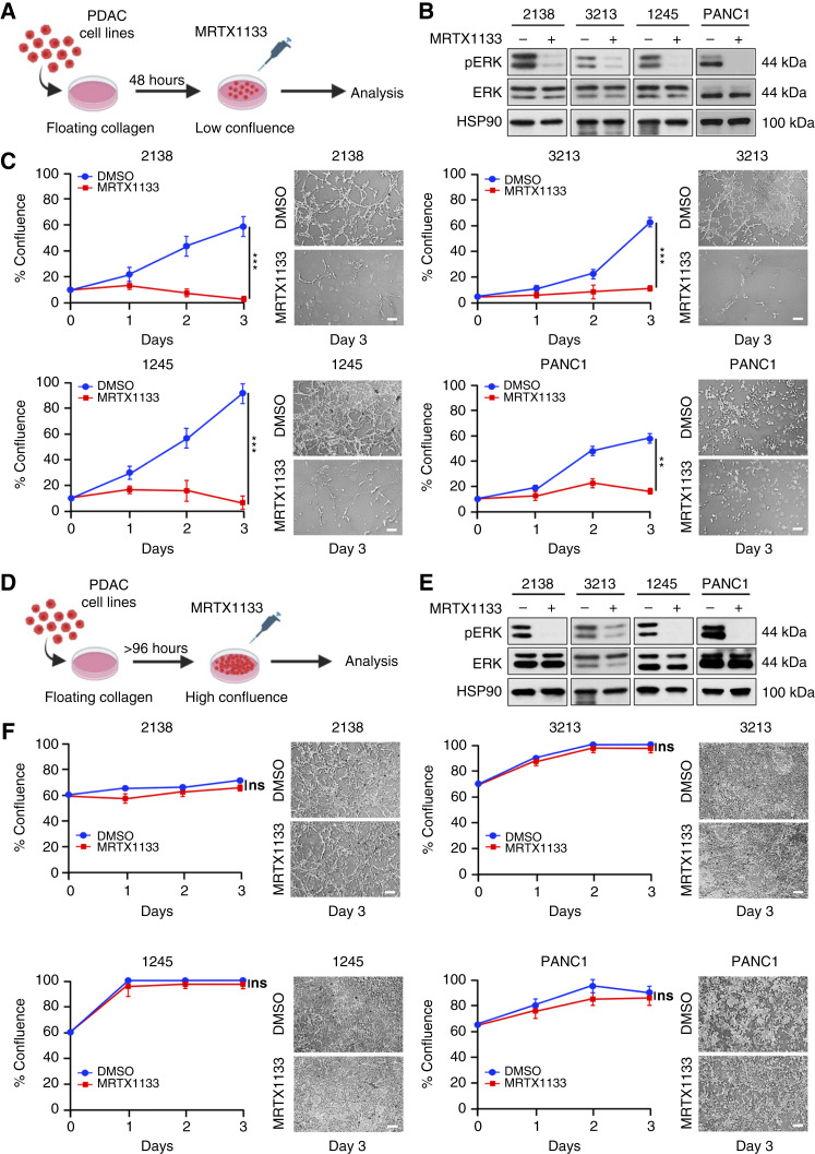
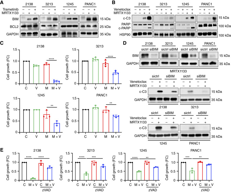
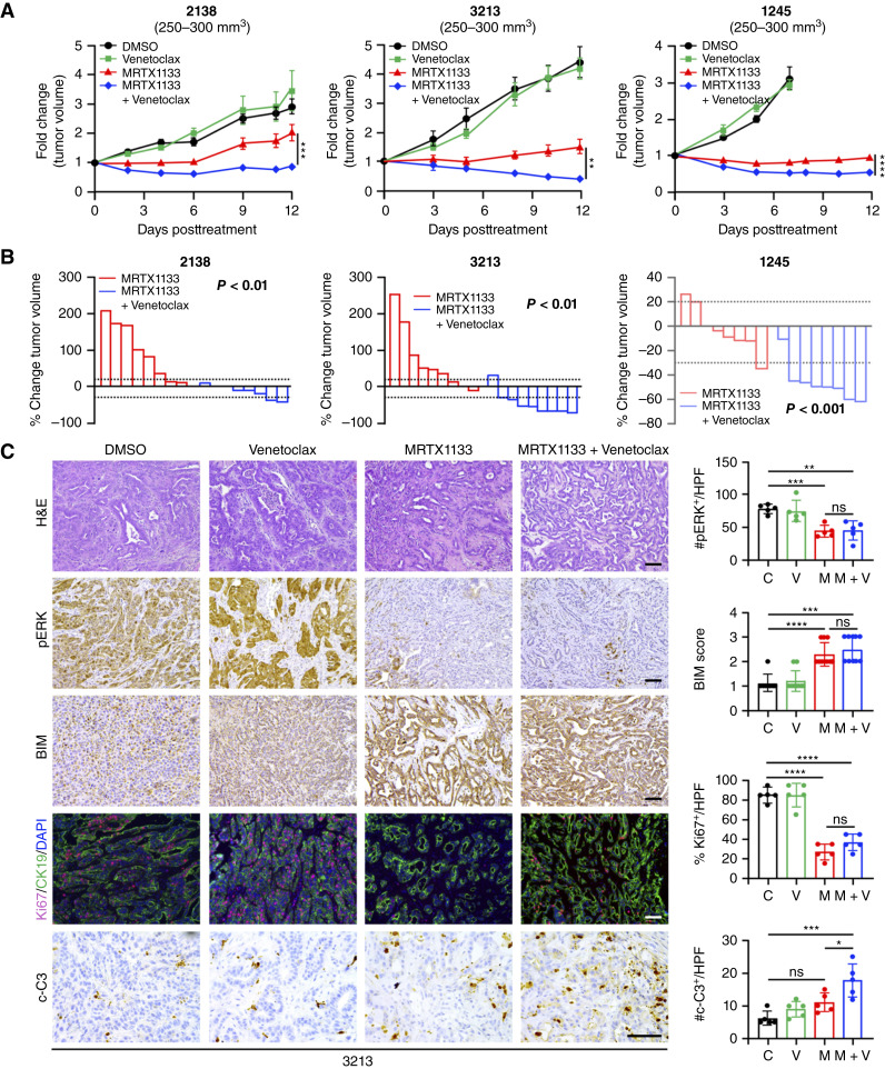
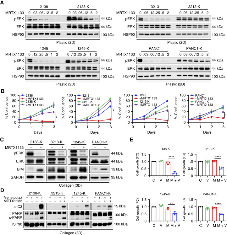
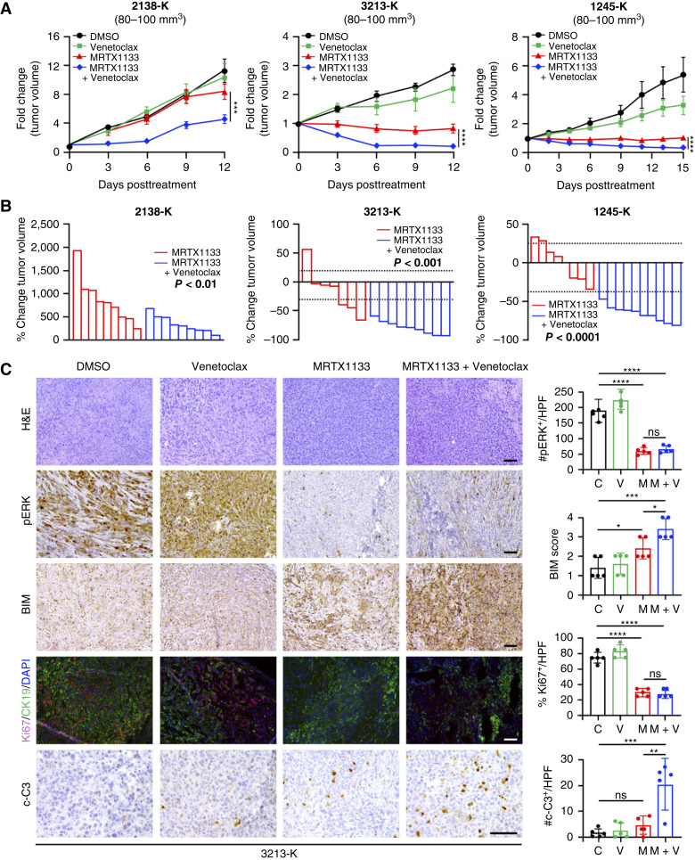
MRTX1133 is currently being evaluated in patients with pancreatic ductal adenocarcinoma (PDAC) tumors harboring a KRASG12D mutation. Combination strategies have the potential to enhance the efficacy of MRTX1133 to further promote cell death and tumor regression. In this study, we demonstrated that MRTX1133 increased the levels of the proapoptotic protein BIM in PDAC cells and conferred sensitivity to the FDA-approved BCL2 inhibitor venetoclax. Combined treatment with MRTX1133 and venetoclax resulted in cell death and growth suppression in 3D cultures. BIM was required for apoptosis induced by the combination treatment. Consistently, BIM was induced in tumors treated with MRTX1133, and venetoclax enhanced the efficacy of MRTX1133 in vivo. Venetoclax could also resensitize MRTX1133-resistant PDAC cells to MRTX1133 in 3D cultures, and tumors established from resistant cells responded to the combination of MRTX1133 and venetoclax. These results provide a rationale for the clinical testing of MRTX1133 and venetoclax in patients with PDAC. Significance: The combination of MRTX1133 and the FDA-approved drug venetoclax promotes cancer cell death and tumor regression in pancreatic ductal adenocarcinoma, providing rationale for testing venetoclax with KRASG12D inhibitors in patients with pancreatic cancer.


Conflict of interest statement

No disclosures were reported.

Figures

None
Graphical abstract
Figure 1.
Figure 1.
MRTX1133 inhibits the growth of PDAC cells in “low”- but not “high”-confluent collagen cultures. A, PDAC cells (1 × 10⁵) were grown in floating collagen gels (1.2 mg/mL) for 48 hours to generate “low”-confluent cultures. Depiction of “low”-confluent floating collagen cultures. B, PDAC cell lines (2138, 3213, 1245, PANC1) grown in “low”-confluent collagen cultures were treated with MRTX1133 (0.5 µmol/L) for 8 hours, and the effect on ERK1/2 phosphorylation was analyzed by Western blotting. Blots are representative of at least three biological replicates. C, PDAC cell lines grown in “low”-confluent collagen cultures were treated with DMSO or 0.5 µmol/L MRTX1133 for 72 hours. The cells were imaged at baseline and after 1, 2, and 3 days of treatment, and the relative growth was quantified. Error bars, ± SD; n = 3, unpaired t test. **, P < 0.01; ***, P < 0.001. Scale bar, 100 μm. D, PDAC cells (1 × 10⁵) were grown in floating collagen gels (1.2 mg/mL) for 96 to 120 hours to generate “high-confluent” cultures. Depiction of “high-confluent” floating collagen cultures. E, PDAC cell lines grown in “high-confluent” collagen cultures were treated with MRTX1133 (0.5 µmol/L) for 8 hours, and the effect on ERK1/2 phosphorylation was analyzed by Western blotting. Blots are representative of at least three biological replicates. F, PDAC cell lines grown in “high-confluent” collagen cultures were treated with DMSO or 0.5 µmol/L MRTX1133 for 72 hours. The cells were imaged at baseline and after 1, 2, and 3 days of treatment, and the relative growth was quantified. Error bars, ± SD; n = 3, unpaired t test. ns, not significant. Scale bar, 100 μm.
Figure 2.
Figure 2.
BCL2 inhibitor venetoclax enhances the efficacy of MRTX1133 in “high”-confluent collagen cultures. A, PDAC cells (2138, 3213, 1245, and PANC1) in “high”-confluent collagen cultures were treated with MRTX1133 (0.5 µmol/L) or the MEK1/2 inhibitor trametinib (0.1 µmol/L) for 8 hours. The effect on BIM and BCL2 was analyzed by Western blotting. Blots are representative of at least three biological replicates. B, PDAC cells in “high”-confluent collagen cultures were cotreated with venetoclax (2.5 µmol/L) and MRTX1133 (0.5 µmol/L) for 8 hours, and the effect on cell death (c-C3, cleaved-PARP) was analyzed. Blots are representative of at least three biological replicates. C, “High”-confluent collagen cultures of PDAC cells were cotreated with venetoclax (V; 2.5 µmol/L) and MRTX1133 (M; 0.5 µmol/L) for 48 hours, and the effect on cell growth was analyzed. Error bars, ± SD; n = 3 to 4. One-way ANOVA, followed by the Tukey multiple comparison test. D, PDAC cells were transfected with control siRNA or siRNA against BIM for 72 hours, plated in collagen, and treated with MRTX1133 (0.5 µmol/L) for 8 hours. The effect on BIM was analyzed using Western blotting. The transfected cells were also cotreated with venetoclax (2.5 µmol/L) and MRTX1133 (0.5 µmol/L) for 8 hours, and the effect on cell death (c-C3) was analyzed. Blots are representative of three biological replicates. E, “High”-confluent collagen cultures of PDAC cells were cotreated with venetoclax (2.5 µmol/L) and MRTX1133 (0.5 µmol/L) for 48 hours with or without the pan-caspase inhibitor zVAD-FMK (zVAD; 10 µmol/L), and the effect on cell growth was analyzed. Error bars, ± SD, n = 3. One-way ANOVA, followed by the Tukey multiple comparison test. **, P < 0.01; ***, P < 0.001; ****, P < 0.0001.
Figure 3.
Figure 3.
Venetoclax enhances the efficacy of MRTX1133 in vivo. The 2138, 3213, and 1245 KPC cells (2.5 × 10⁴) were implanted via subcutaneous injection into the flanks of C57BL/6 mice. Once animals developed 250 to 300 mm3 tumors, they were treated with vehicle control (DMSO), venetoclax (15 mg/kg daily), MRTX1133 (30 mg/kg 2×/day), or the combination of venetoclax and MRTX1133. A, Tumor size was measured 2 to 3 times/week by caliper, and tumor volume was calculated using the formula V = (W2 × L)/2 and normalized to tumor volume at the start of treatment. Error bars, ± SEM, n = 8 to 9 per treatment group. Significance between the MRTX1133-treated group and the combination treatment group was determined by two-way ANOVA with the Sidak multiple comparisons test. Changes in tumor volume between the dotted lines (−30%, +20%) were considered as stable disease. B, Relative tumor volumes at study endpoints were compared with tumors at the start of treatment, and change in tumor volume was calculated. Unpaired t test. C, 3213 tumors at the study endpoints were hematoxylin and eosin (H&E)–stained and stained for pERK, BIM, the proliferation marker Ki67, the epithelial marker CK19, and the apoptosis marker c-C3. Error bars, ± SD. One-way ANOVA, followed by the Tukey multiple comparison test. *, P < 0.05; **, P < 0.01; ***, P < 0.001; ****, P < 0.0001; ns, not significant. Scale bars, 100 μm. C, DMSO; V, venetoclax; M, MRTX1133.
Figure 4.
Figure 4.
MRTX1133-resistant cells respond to the combination of MRTX1133 and venetoclax in collagen cultures. PDAC (2138, 3213, 1245, and PANC1) cells were treated over 6 to 8 weeks with increasing concentrations of MRTX1133 to 2 µmol/L or greater to generate PDAC-K cells. A, PDAC and PDAC-K cells growing on tissue culture plastic were treated with increasing concentrations of MRTX1133 for 8 hours, and ERK activation was evaluated by Western blotting. Blots are representative of three biological replicates. B, PDAC and PDAC-K cells grown in “low”-confluent collagen cultures were treated with DMSO or 2 µmol/L MRTX1133 for 72 hours. The cells were imaged at baseline and after 1, 2, and 3 days of treatment, the relative growth was quantified. Error bars, ± SD, n = 3. Unpaired t test. C, PDAC-K cells growing in “high”-confluent collagen cultures were treated with MRTX1133 (2 µmol/L), and the effect on pERK and BIM protein levels was analyzed by Western blotting. Blots are representative of three biological replicates. D, PDAC-K cells in “high”-confluent collagen cultures were treated with venetoclax (2.5 µmol/L) and MRTX1133 (2.0 µmol/L) for 8 hours, and the effect on cell death (c-C3 and cleaved-PARP) was analyzed. Blots are representative of at least three biological replicates. E, PDAC-K cells growing in “high”-confluent collagen cultures were cotreated with venetoclax (V; 2.5 µmol/L) and MRTX1133 (M; 2.0 µmol/L) for 48 hours, and the effect on cell growth was analyzed. Error bars, ± SD, n = 3. One-way ANOVA, followed by the Tukey multiple comparison test. *, P < 0.05; **, P < 0.01; ****, P < 0.0001.
Figure 5.
Figure 5.
Tumors established from KPC-K cells respond to the combination of MRTX1133 and venetoclax. The 2138-K, 3213-K, and 1245-K KPC cells (2.5 × 10⁴) were implanted via subcutaneous injection into the flanks of C57BL/6 mice. Once animals developed ∼80 to 100 mm3 tumors, they were treated with vehicle control (DMSO), venetoclax (15 mg/kg daily), MRTX1133 (30 mg/kg 2×/day), or the combination of venetoclax and MRTX1133. A, Tumor size was measured 2 to 3 times/week by caliper, and tumor volume was calculated using the formula V = (W2 × L)/2 and normalized to tumor volume at the start of treatment. Error bars, ± SEM, n = 7–9 per treatment group. Significance between the MRTX1133-treated group and the combination treatment group was determined by two-way ANOVA with the Sidak multiple comparisons test. Changes in tumor volume between the dotted lines (−30%, +20%) were considered as stable disease. B, Relative tumor volumes at study endpoints were compared with tumors at the start of treatment, and change in tumor volume was calculated. Unpaired t test. C, 3213-K tumors at the study endpoints were hematoxylin and eosin (H&E)–stained and stained for pERK, BIM, the proliferation marker Ki67, the epithelial marker CK19, and the apoptosis marker c-C3. Error bars, ± SD. One-way ANOVA, followed by the Tukey multiple comparison test. *, P < 0.05; **, P < 0.01; ***, P < 0.001; ****, P < 0.0001; ns, not significant. Scale bars, 100 μm. C, DMSO; V, venetoclax; M, MRTX1133.

Similar articles

Cited by

References

    1. Nevala-Plagemann C, Hidalgo M, Garrido-Laguna I. From state-of-the-art treatments to novel therapies for advanced-stage pancreatic cancer. Nat Rev Clin Oncol 2020;17:108–23. - PubMed
    1. Siegel RL, Giaquinto AN, Jemal A. Cancer statistics, 2024. CA Cancer J Clin 2024;74:12–49. - PubMed
    1. Principe DR, Kamath SD, Korc M, Munshi HG. The immune modifying effects of chemotherapy and advances in chemo-immunotherapy. Pharmacol Ther 2022;236:108111. - PMC - PubMed
    1. Collins MA, Bednar F, Zhang Y, Brisset J-C, Galbán S, Galbán CJ, et al. . Oncogenic Kras is required for both the initiation and maintenance of pancreatic cancer in mice. J Clin Invest 2012;122:639–53. - PMC - PubMed
    1. Simanshu DK, Nissley DV, McCormick F. RAS proteins and their regulators in human disease. Cell 2017;170:17–33. - PMC - PubMed

MeSH terms