. 2024 Aug 13;7(1):179.
doi: 10.1038/s42004-024-01262-8.

Activity of botulinum neurotoxin X and its structure when shielded by a non-toxic non-hemagglutinin protein

Affiliations

Activity of botulinum neurotoxin X and its structure when shielded by a non-toxic non-hemagglutinin protein

Markel Martínez-Carranza et al. Commun Chem. .

Abstract

Botulinum neurotoxins (BoNTs) are the most potent toxins known and are used to treat an increasing number of medical disorders. All BoNTs are naturally co-expressed with a protective partner protein (NTNH) with which they form a 300 kDa complex, to resist acidic and proteolytic attack from the digestive tract. We have previously identified a new botulinum neurotoxin serotype, BoNT/X, that has unique and therapeutically attractive properties. We present the cryo-EM structure of the BoNT/X-NTNH/X complex and the crystal structure of the isolated NTNH protein. Unexpectedly, the BoNT/X complex is stable and protease-resistant at both neutral and acidic pH and disassembles only in alkaline conditions. Using the stabilizing effect of NTNH, we isolated BoNT/X and showed that it has very low potency both in vitro and in vivo. Given the high catalytic activity and translocation efficacy of BoNT/X, low activity of the full toxin is likely due to the receptor-binding domain, which presents very weak ganglioside binding and exposed hydrophobic surfaces.


Conflict of interest statement

P.S. and M.D. are inventors on patents regarding BoNT/X. M.B., D.B., M.E., J.P., S.D., and F.H. are employees of Ipsen. The authors declare no additional conflict of interest.

Figures

Fig. 1
Fig. 1. Activity of BoNT/X.
Rat cortical neurons were exposed to varying concentrations of recombinant full-length BoNT/X (filled circles) or LHNX (open circles). After 24 h of incubation cells were lysed, and lysates analyzed for VAMP2 (a) and VAMP4 (b) by Western blots (error bars represent standard deviation from n = 3 replicates). c Mouse phrenic nerve hemidiaphragm (mPNHD) preparations were exposed to 10 pM BoNT/X, BoNT/A, and BoNT/B, 100 pM LHN/X, and muscle contraction responses to indirect stimulation recorded until contraction was no longer detectable or to 180 min (n = 3). d BoNT/A (6 pg) induced flaccid paralysis when injected into the right gastrocnemius muscle of mice in DAS assays, whereas injection of BoNT/X (1 µg) did not induce muscle paralysis. e LHN/X (40 µg) was injected to the right gastrocnemius muscle of mice (n = 3). The limb developed partial paralysis after 24 h (DAS Score = 2 out of 4) and the toes showed a reduced ability to spread following a startle stimulus. LHN/X that had not been activated with trypsin served as a control. A representative image of paralysis obtained from injection of 40 µg is shown and experiment data are shown in Supplementary Table 4.
Fig. 2
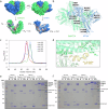
Fig. 2. Cryo-EM structure of BoNT/X in complex with NTNH/X.
a Surface view of the BoNT/X (green)—NTNH (blue) complex. b Cartoon view of the different domains in each protein. BoNT/X domains are colored in green shades, NTNH/X domains are colored in blue shades. c Local resolution estimates (Å) for the cryo-EM map of the M-PTC/X. d cryo-EM map around the LC active site. e cryo-EM map around the SxWY ganglioside-binding motif. f cryo-EM map around the HC patch rich in aromatic residues.
Fig. 3
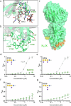
Fig. 3. Structure of the BoNT/X light chain.
a HN and LC domains from the BoNT/X-NTNH/X cryo-EM structure (green). The belt region of HN surrounding the LC is highlighted in orange. b crystal structure of LC/F (PDB ID 3FIE, colored in red) bound to a VAMP substrate-like inhibitor (purple) superimposed to HN-LC/X (green). c, d crystal structure of LC/X (PDB ID 6F47, colored in magenta) superimposed to the cryo-EM structure of HN-LC/X (green). Noteworthy differences in secondary structure are highlighted in (d).
Fig. 4
Fig. 4. BoNT/X-NTNH/X complex stability.
a BoNT/X-NTNH/X interface; BoNT/X is shown in green and NTNH/X in blue. Top: interfacing residues are highlighted in different shades of green and blue. Bottom: residues at the interface of the complex are colored according to electrostatic potential (negative electrostatic potential is colored in shades of red, and positive in shades of blue). b Electrostatic interactions across the BoNT/X-NTNH/X interface. c Size-exclusion chromatograms from the M-PTC/X pH stability experiments. The A280nm response for the chromatograms at pH 5.5 and 6.5 was divided by the factor of two to normalize it to the other chromatograms, where the signal was lower. d Flexible linker between HN and HC followed by a small helical region leading to the HC (residues 876–895). e, f Limited proteolysis assay for examining the stability of the BoNT/X-NTNH/X complex at different pH conditions. BoNT/X (marked as X), NTNH/X (marked as N), and BoNT/X-NTNH/X complex (marked as XN) were incubated with trypsin at the indicated pH conditions and then analyzed by SDS-PAGE and visualized by Coomassie blue staining. BoNT/X alone and NTNH/X alone were degraded at all pH conditions. BoNT/X-NTNH/X is largely resistant to trypsin at pH 5.0, 6.0, and 7.5, but not at pH 8.0. Trypsin treatment is able to cleave the linker between LC and HC of BoNT/X within the BoNT/X-NTNH/X complex, thus BoNT/X showed as two separate bands (LC/X and HC/X) after the disulfide bond connecting the LC and HC is reduced. One contaminant is present in purified BoNT/X and another is present in purified BoNT/X-NTNH/X complex.
Fig. 5
Fig. 5. Investigation of potential ganglioside binding by BoNT/X.
a Alignment of HC/X (green) and GD1a-bound HC/A (in grey, PDB ID 5TPC, GD1a shown in grey sticks), showing the conserved ganglioside-binding SxWY motif residues and (b) the same alignment showing the exposed region rich in aromatic residues. Location of the ganglioside-binding motif (yellow) and the hydrophobic patch (orange) is shown in the context of Hc/X in (c). Binding of BoNT/X (green), and HC/A (grey) to GT1b (d), GD1a (e), GM1 (f), and GD1b (g), error bars represent standard deviation from n = 3 replicates. Glycobloc schematic representation is shown for each ganglioside.

References

    1. Dong, M., Masuyer, G. & Stenmark, P. Botulinum and tetanus neurotoxins. Annu. Rev. Biochem.88, 811–837 (2019). 10.1146/annurev-biochem-013118-111654 - DOI - PMC - PubMed
    1. Rossetto, O., Pirazzini, M. & Montecucco, C. Botulinum neurotoxins: genetic, structural and mechanistic insights. Nat. Rev. Microbiol.12, 535–549 (2014). 10.1038/nrmicro3295 - DOI - PubMed
    1. Zhang, S. et al. Identification and characterization of a novel botulinum neurotoxin. Nat. Commun.8, 14130 (2017). 10.1038/ncomms14130 - DOI - PMC - PubMed
    1. Kakinuma, H., Maruyama, H., Takahashi, H., Yamakawa, K. & Nakamura, S. The first case of type B infant botulism in Japan. Pediatr. Int.38, 541–543 (1996).10.1111/j.1442-200X.1996.tb03542.x - DOI - PubMed
    1. Kozaki, S. et al. Characterization of Clostridium botulinum type B neurotoxin associated with infant botulism in Japan. Infect. Immun.66, 4811–4816 (1998). 10.1128/IAI.66.10.4811-4816.1998 - DOI - PMC - PubMed

LinkOut - more resources