Meta-Analysis
. 2024 Aug 13;14(1):18791.
doi: 10.1038/s41598-024-69612-z.

Eyelid dermatitis in patch-tested adult patients: a systematic review with a meta-analysis

Affiliations
Meta-Analysis

Eyelid dermatitis in patch-tested adult patients: a systematic review with a meta-analysis

Elena Borzova et al. Sci Rep. .

Abstract

Eyelid dermatitis (ED) affects a cosmetically significant area and leads to patients' distress. Despite ongoing and recent research efforts, ED remains a multidisciplinary problem that needs further characterization. We aimed to evaluate the atopic eyelid dermatitis (AED) frequency in ED patients and to perform their clinical profiling. PubMed databases were searched from 01.01.1980 till 01.02.2024 to PRISMA guidelines using a search strategy: (eyelid OR periorbital OR periocular) AND (dermatitis or eczema). Studies with patch-tested ED patients were included. Proportional meta-analysis was performed using JBI SUMARI software. We included 65 studies across Europe, North America, Asia and Australia, with a total of 21,793 patch-tested ED patients. AED was reported in 27.5% (95% CI 0.177, 0.384) of patch-tested ED patients. Isolated ED was noted in 51.6% (95% CI 0.408, 0.623) of 8453 ED patients with reported lesion distribution, including 430 patients with isolated AED. Our meta-analysis demonstrated that the AED frequency in patch-tested ED patients exceeded the previous estimate of 10%. Isolated AED was noted in adult patients, attending contact allergy clinics. Future studies are needed to elucidate the global prevalence and natural history of isolated AED in adults.


Conflict of interest statement

EB serves on the WAO Committee on Atopic Dermatitis, received honoraria for educational lectures from Novartis and Sanofi and research funding from GSK, outside the submitted work. EB provided pro bono consulting to Kimialys (France) outside the submitted work. EB received research funding from the British Skin Foundation (2009) for her research, outside the submitted work. ES received honoraria for lectures from Dr. Reddy’s Laboratories Ltd., as well as support for attending congresses from Galderma and Drs. Reddy’s Laboratories Ltd. as a lecturer. AB is working on her PhD research project on atopic eyelid dermatitis supervised by ES and EB.

Figures

Figure 1
Figure 1
Clinical presentations in patients with atopic eyelid dermatitis. Legend: The representative images of three patients with adult-onset atopic eyelid dermatitis (AED) (Patients 1–3), who are under care at the Department of Dermatology and Venereology (Head—Prof. Olga Olisova), I.M. Sechenov First Moscow State Medical University: patient 1 (A,D,H), aged 32 (an AED onset at 30 years), patient 2 (B,E,I), aged 37 (an AED onset at 37 years), patient 3, (C,F,J) aged 31 (an AED onset at 21 years). Facial skin imaging was carried out using the AI face recognition technology, 8-spectrum imaging technology and deep quantitative analysis by Capricorn Intelligent Imager ZMLH01 (Beijing Sincoheren S&T Development, Beijing, China). The high-definition images were acquired in a negatively polarized UV light (365 nm, A-F), white light (RGB, range 420–620 nm, H-J). AED patients presented with symmetrical, scaly erythematous periorbital areas with a hyperpigmentation and a lichenification, particularly pronounced at the medial aspects of the upper and lower eyelids. Informed consents from the AED patients for a publication of these images were obtained.
Figure 2
Figure 2
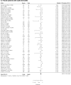
(A-Q) Forest plots for demographic and clinical characteristics of patients with eyelid dermatitis. Legend: The proportional meta-analyses [random-effects] were carried out for female patients (A) and patients aged 40 years or older (B) in the studies including the patients with eyelid dermatitis. The proportion of atopic eyelid dermatitis in the studies with patients with eyelid dermatitis (C) was estimated at 27.5% (95% CI 0.177, 0.384). The differential diagnosis of eyelid dermatitis includes eyelid allergic contact dermatitis (D), seborrheic dermatitis (E), psoriasis (F), and irritant contact dermatitis (G). HN represent proportional meta-analyses for contact sensitizations to nickel (positive patch tests and clinically relevant reactions), gold, thimerosal, neomycin, and corticosteroids (budesonide and toxicortol), respectively. Clinically, the proportion of isolated eyelid dermatitis (O) of 8453 ED patients was 51.6% [95% CI 0.408, 0.623] whereas the proportion of isolated atopic eyelid dermatitis (P) of the patients with atopic dermatitis with an eyelid involvement was 26.9% [95% CI 0.117, 0.449].
Figure 2
Figure 2
(A-Q) Forest plots for demographic and clinical characteristics of patients with eyelid dermatitis. Legend: The proportional meta-analyses [random-effects] were carried out for female patients (A) and patients aged 40 years or older (B) in the studies including the patients with eyelid dermatitis. The proportion of atopic eyelid dermatitis in the studies with patients with eyelid dermatitis (C) was estimated at 27.5% (95% CI 0.177, 0.384). The differential diagnosis of eyelid dermatitis includes eyelid allergic contact dermatitis (D), seborrheic dermatitis (E), psoriasis (F), and irritant contact dermatitis (G). HN represent proportional meta-analyses for contact sensitizations to nickel (positive patch tests and clinically relevant reactions), gold, thimerosal, neomycin, and corticosteroids (budesonide and toxicortol), respectively. Clinically, the proportion of isolated eyelid dermatitis (O) of 8453 ED patients was 51.6% [95% CI 0.408, 0.623] whereas the proportion of isolated atopic eyelid dermatitis (P) of the patients with atopic dermatitis with an eyelid involvement was 26.9% [95% CI 0.117, 0.449].

Similar articles

References

    1. Beltrani, V. S. Eyelid dermatitis. Curr. Allergy Asthma Rep.1, 380–388 (2001). 10.1007/s11882-001-0052-0 - DOI - PubMed
    1. Feser, A., Plaza, T., Vogelgsang, L. & Mahler, V. Periorbital dermatitis—a recalcitrant disease: Causes and differential diagnoses. Br. J. Dermatol.159, 858–863 (2008). 10.1111/j.1365-2133.2008.08790.x - DOI - PubMed
    1. Ayala, F. et al. Eyelid dermatitis: An evaluation of 447 patients. Am. J. Contact Dermat.14, 69–74 (2003). 10.1097/01634989-200306000-00004 - DOI - PubMed
    1. Warshaw, E. M. et al. Eyelid dermatitis in patients referred for patch testing: Retrospective analysis of North American Contact Dermatitis Group data, 1994–2016. J Am Acad Dermatol.84, 953–964 (2021). 10.1016/j.jaad.2020.07.020 - DOI - PubMed
    1. Herbst, R. A., Uter, W., Pirker, C., Geier, J. & Frosch, P. J. Allergic and non-allergic periorbital dermatitis: Patch test results of the Information Network of the Departments of Dermatology during a 5-year period. Contact Dermat.51, 13–19 (2004). 10.1111/j.0105-1873.2004.00334.x - DOI - PubMed

LinkOut - more resources