

CT-based surrogate parameters for MRI-based disc height and endplate degeneration in the lumbar spine
- PMID: 39138416
- PMCID: PMC11323600
- DOI: 10.1186/s12880-024-01395-1
CT-based surrogate parameters for MRI-based disc height and endplate degeneration in the lumbar spine
Abstract
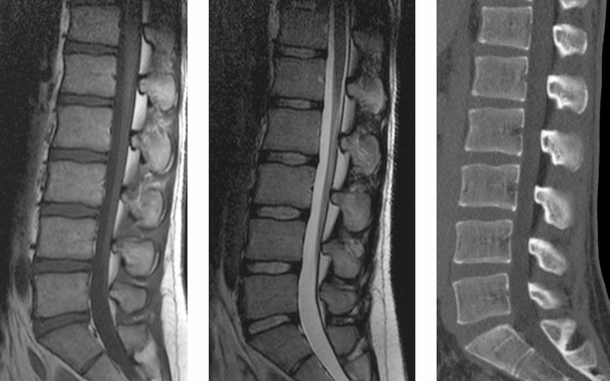
Purpose: This study investigated potential use of computed tomography (CT)-based parameters in the lumbar spine as a surrogate for magnetic resonance imaging (MRI)-based findings.
Methods: In this retrospective study, all individuals, who had a lumbar spine CT scan and MRI between 2006 and 2012 were reviewed (n = 198). Disc height (DH) and endplate degeneration (ED) were evaluated between Th12/L1-L5/S1. Statistics consisted of Spearman correlation and univariate/multivariable regression (adjusting for age and gender).
Results: The mean CT-DH increased kranio-caudally (8.04 millimeters (mm) at T12/L1, 9.17 mm at L1/2, 10.59 mm at L2/3, 11.34 mm at L3/4, 11.42 mm at L4/5 and 10.47 mm at L5/S1). MRI-ED was observed in 58 (29%) individuals. CT-DH and MRI-DH had strong to very strong correlations (rho 0.781-0.904, p < .001). MRI-DH showed higher absolute values than CT-DH (mean of 1.76 mm). There was a significant association between CT-DH and MRI-ED at L2/3 (p = .006), L3/4 (p = .002), L4/5 (p < .001) and L5/S1 (p < .001). A calculated cut-off point was set at 11 mm.
Conclusions: In the lumbar spine, there is a correlation between disc height on CT and MRI. This can be useful in trauma and emergency cases, where CT is readily available in the lack of an MRI. In addition, in the middle and lower part of the lumbar spine, loss of disc height on CT scans is associated with more pronounced endplate degeneration on MRIs. If the disc height on CT scans is lower than 11 mm, endplate degeneration on MRIs is likely more pronounced.
Level and design: Level III, a retrospective study.
Keywords: CT; Disc degeneration; Disc height; Endplate degeneration; MRI.
© 2024. The Author(s).
Conflict of interest statement
The authors declare no competing interests.
Figures


Similar articles
-
[Correlation of lumbar disc degeneration and spinal-pelvic sagittal balance].Zhonghua Yi Xue Za Zhi. 2013 Apr 16;93(15):1123-8. Zhonghua Yi Xue Za Zhi. 2013. PMID: 23902878 Chinese.
-
The effects of age, sex, ethnicity, and spinal level on the rate of intervertebral disc degeneration: a review of 1712 intervertebral discs.Spine (Phila Pa 1976). 2011 Aug 1;36(17):1333-9. doi: 10.1097/BRS.0b013e3181f2a177. Spine (Phila Pa 1976). 2011. PMID: 21217432 Free PMC article.
-
Non-invasive quantification of age-related changes in the vertebral endplate in rats using in vivo DCE-MRI.J Orthop Surg Res. 2017 Nov 9;12(1):169. doi: 10.1186/s13018-017-0669-x. J Orthop Surg Res. 2017. PMID: 29121960 Free PMC article.
-
Hybrid Bone Single Photon Emission Computed Tomography Imaging in Evaluation of Chronic Low Back Pain: Correlation with Modic Changes and Degenerative Disc Disease.World Neurosurg. 2017 Aug;104:816-823. doi: 10.1016/j.wneu.2017.03.107. Epub 2017 Apr 2. World Neurosurg. 2017. PMID: 28377243
-
Endplate degeneration observed on magnetic resonance imaging of the lumbar spine: correlation with pain provocation and disc changes observed on computed tomography diskography.Spine (Phila Pa 1976). 2002 Oct 15;27(20):2274-8. doi: 10.1097/00007632-200210150-00017. Spine (Phila Pa 1976). 2002. PMID: 12394906 Clinical Trial.
Cited by
-
[Biomechanical study of lumbar vertebra during gait cycle in adolescent idiopathic scoliosis].Sheng Wu Yi Xue Gong Cheng Xue Za Zhi. 2025 Jun 25;42(3):601-609. doi: 10.7507/1001-5515.202410010. Sheng Wu Yi Xue Gong Cheng Xue Za Zhi. 2025. PMID: 40566784 Free PMC article. Chinese.
References
MeSH terms
LinkOut - more resources
Full Text Sources
Medical
