. 2024 Aug 13;24(1):392.
doi: 10.1186/s12890-024-03207-2.

Immunologic features of nontuberculous mycobacterial pulmonary disease based on spatially resolved whole transcriptomics

Affiliations

Immunologic features of nontuberculous mycobacterial pulmonary disease based on spatially resolved whole transcriptomics

Jaemoon Koh et al. BMC Pulm Med. .

Abstract

Background: The immunologic features of nontuberculous mycobacterial pulmonary disease (NTM-PD) are largely unclear. This study investigated the immunologic features of NTM-PD using digital spatial profiling techniques.

Methods: Lung tissues obtained from six patients with NTM-PD between January 1, 2006, and December 31, 2020, at Seoul National University Hospital were subjected to RNA sequencing. Cores from the peribronchial areas were stained with CD3, CD68, and DNASyto13, and gene expression at the whole-transcriptome level was quantified using PCR amplification and Illumina sequencing. Lung tissues from six patients with bronchiectasis collected during the same period were used as controls. The RNA sequencing results were validated using immunohistochemistry (IHC) in another cohort (30 patients with NTM-PD and 15 patients with bronchiectasis).

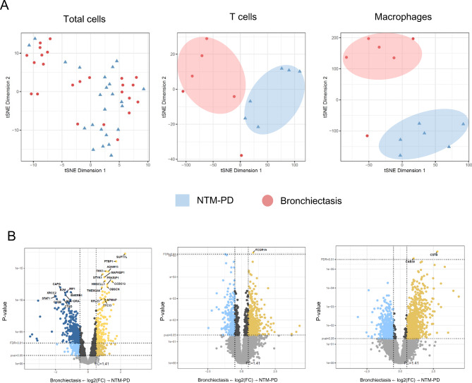
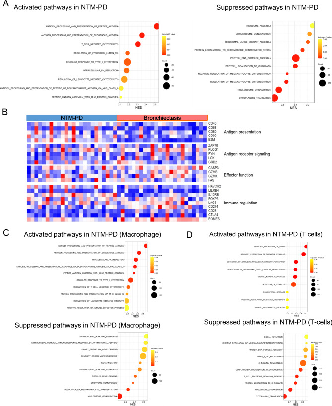
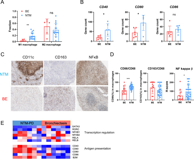
Results: NTM-PD exhibited distinct gene expression patterns in T cells and macrophages. Gene set enrichment analysis revealed that pathways related to antigen presentation and processing were upregulated in NTM-PD, particularly in macrophages. Macrophages were more prevalent and the expression of genes associated with the M1 phenotype (CD40 and CD80) was significantly elevated. Although macrophages were activated in the NTM-PD group T cell activity was unaltered. Notably, expression of the costimulatory molecule CD28 was decreased in NTM-PD. IHC analysis showed that T cells expressing Foxp3 or TIM-3, which facilitate the regulatory functions of T cells, were increased.

Conclusions: NTM-PD exhibits distinct immunologic signatures characterized by the activation of macrophages without T cell activation.

Keywords: Lung disease; M1 phenotype; Macrophage activation; Nontuberculous mycobacteria.


Conflict of interest statement

Dr. JJ Yim has served as the overall or institutional principal investigator for clinical trials related to non-tuberculous mycobacterial pulmonary disease sponsored by LigaChem Biosciences, Insmed and AN2 Therapeutics. Additionally, he has received several drugs free of charge as a principal investigator for previous trials related to tuberculosis from Pfizer, Otsuka, and Yuhan.

Figures

Fig. 1
Fig. 1
Overview of digital spatial profiling (DSP) workflow. (a) Schematic of study participants. Lung specimens were obtained from six patients undergoing surgical resection for nontuberculous mycobacterial pulmonary disease (NTM-PD) and six patients with bronchiectasis. Lesion samples from the peribronchial areas were collected and used to create tissue microarray (TMA) slides for DSP analysis. (b) Representative images of a multi-label immunofluorescence scan of a TMA sample and representative immunofluorescence staining features of each marker. CD3 (red), CD68 (yellow), DNA (DAPI), and a merged staining image are shown. (c) Representative images of peribronchial areas in patients with NTM-PD and bronchiectasis. RNA sequencing analyses were performed using CD3- and CD68-positive cells from each region-of-interest
Fig. 2
Fig. 2
Identification of differentially expressed genes. (a) Principal component analysis plots of RNA sequencing data illustrating RNA expression patterns of total cells, T cells, and macrophages in lung tissues from patients with NTM-PD or bronchiectasis. (b) Volcano plots of gene expression changes in total cells, T cells, and macrophages between NTM-PD and bronchiectasis. Yellow dots indicate genes upregulated in NTM-PD, while dark blue dots indicate genes upregulated in bronchiectasis samples (p < 0.05)
Fig. 3
Fig. 3
Pathways enriched in NTM-PD and bronchiectasis. (a–d) Data from total immune cells between NTM-PD and bronchiectasis. (a) Dot plot of gene set enrichment analysis (GSEA) displaying activation and inactivation of significantly enriched signaling pathways in total cells from NTM-PD compared with bronchiectasis. (b) Expression pattern of genes involving antigen presentation, antigen receptor signaling, effector function, and immune regulation in total cells. (c–d) Dot plots of GSEA displaying activation and inactivation of significantly enriched signaling pathways in macrophages (c) and T cells (d) from NTM-PD compared with bronchiectasis
Fig. 4
Fig. 4
Distribution of immune cells according to disease and validation. (a) Stacked bar charts comparing proportions of immune cell subsets in peribronchial area between NTM-PD and bronchiectasis. (b) Comparison of the relative abundance of lymphoid and myeloid cells between groups. (c) Schematic overview of TMA construction for validation cohort. TMAs were constructed using samples from the peribronchial areas of patients with NTM-PD and bronchiectasis, respectively, and immunohistochemical staining for immune cell markers was performed. (d) Representative images of immunohistochemical staining for CD3 (T-lymphocyte marker) and CD68 (macrophage marker) in the two groups. (e) Bar graphs showing mean number of CD3+ lymphocytes and CD68+ macrophages between the groups
Fig. 5
Fig. 5
Macrophages exhibit dominant M1 phenotype in NTM-PD. (a) Bar graph showing proportions of M1 and M2 macrophages between NTM-PD and bronchiectasis. (b) Boxplots comparing levels of CD80, CD40, and CD86 between the groups. (c) Representative images of immunohistochemical staining for CD68, CD11c, CD163, and NF-κB in the two groups. (d) Plots showing proportions of M1 macrophage (CD11c+ per CD68+ cells), M2 macrophage (CD163+ per CD68+ cells), and H-score of NF-κB between the groups. (e) Heatmap of genes involving transcription regulation and antigen presentation in macrophages from the two groups
Fig. 6
Fig. 6
Absence of T cell activation following macrophage activation in NTM-PD. (a) Comparison of CD8, CD4, and regulatory T cell proportions in RNA sequencing data between NTM-PD and bronchiectasis. (b) Heatmap of genes involving transcription regulation, antigen presentation, effector function, and immune regulation in T cells from the two groups. (c) Expression of CD28, EOMES, GZMB, FOXP3, LAG3, and ITGAE determined using RNA sequencing in the two groups. (d) Representative images of immunohistochemical staining for CD8, CD103, Foxp3, PD-1, TIM-3, and granzyme β in the two groups. (e) Plots showing the ratio of CD103+ to CD8+ T lymphocytes, number of Foxp3+ or PD-1+ T lymphocytes, ratio of TIM-3+ to CD8 T+ lymphocytes, and H-score for granzyme β between the groups

Similar articles

References

    1. Griffith DE, Aksamit T, Brown-Elliott BA, Catanzaro A, Daley C, Gordin F, et al. An official ATS/IDSA statement: diagnosis, treatment, and prevention of nontuberculous mycobacterial diseases. Am J Respir Crit Care Med. 2007;175(4):367–416. 10.1164/rccm.200604-571ST - DOI - PubMed
    1. Lee H, Myung W, Lee E-M, Kim H, Jhun BW. Mortality and prognostic factors of nontuberculous mycobacterial infection in Korea: a population-based comparative study. Clin Infect Dis. 2021;72(10):e610–9. 10.1093/cid/ciaa1381 - DOI - PubMed
    1. Kim J-Y, Kwak N, Yim J-J, editors. The rise in prevalence and related costs of nontuberculous mycobacterial diseases in South Korea, 2010–2021. Open Forum Infect Dis. 2022; 9(12):ofac649. - PMC - PubMed
    1. Orme IM, Ordway DJ. Host response to nontuberculous mycobacterial infections of current clinical importance. Infect Immun. 2014;82(9):3516–22. 10.1128/IAI.01606-13 - DOI - PMC - PubMed
    1. Shu C-C, Wu M-F, Pan S-W, Wu T-S, Lai H-C, Lin M-C. Host immune response against environmental nontuberculous mycobacteria and the risk populations of nontuberculous mycobacterial lung disease. J Formos Med Assoc. 2020;119:S13–22. 10.1016/j.jfma.2020.05.001 - DOI - PubMed

MeSH terms