. 2024 Aug 13;30(1):123.
doi: 10.1186/s10020-024-00883-y.

Progesterone (P4) ameliorates cigarette smoke-induced chronic obstructive pulmonary disease (COPD)

Affiliations

Progesterone (P4) ameliorates cigarette smoke-induced chronic obstructive pulmonary disease (COPD)

Bin Xie et al. Mol Med. .

Abstract

Background: Chronic obstructive pulmonary disease (COPD) is a chronic inflammatory lung disease associated with high morbidity and mortality worldwide. Oxidative injury and mitochondrial dysfunction in the airway epithelium are major events in COPD progression.

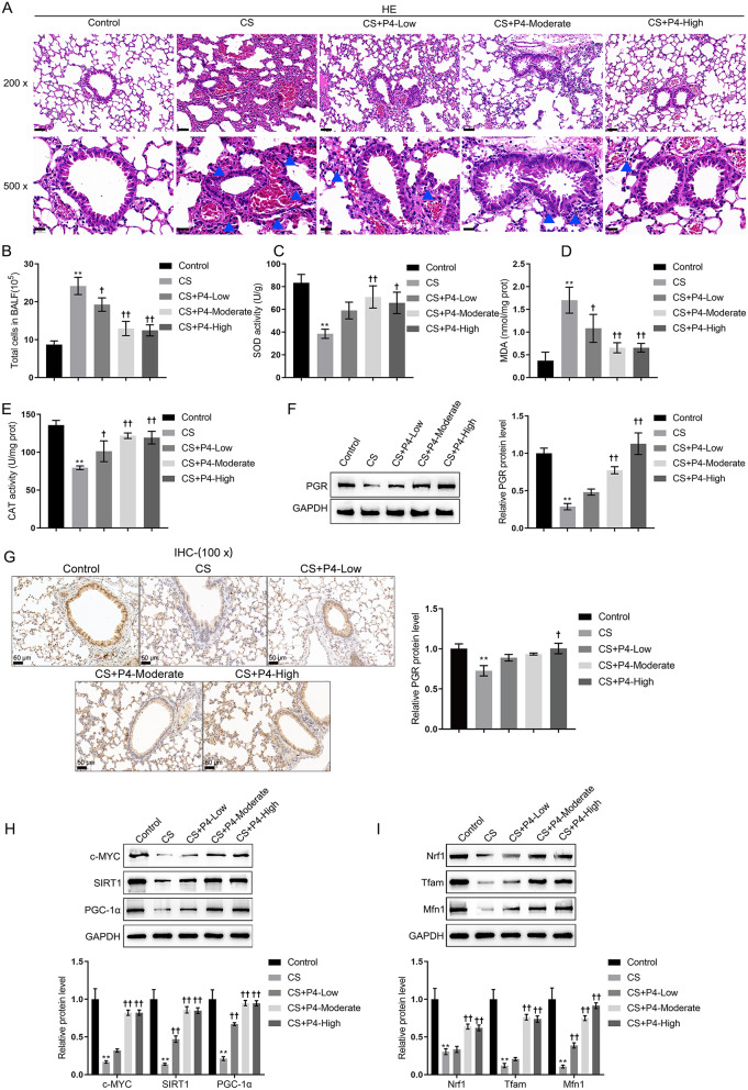
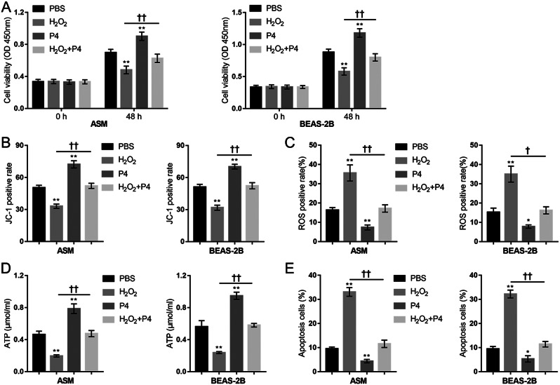
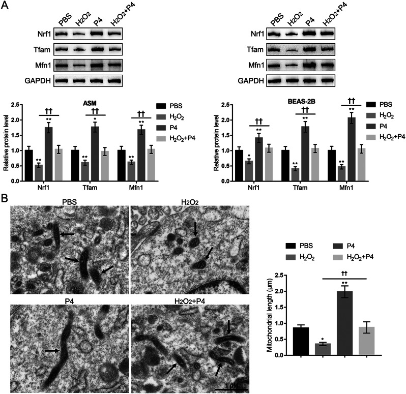
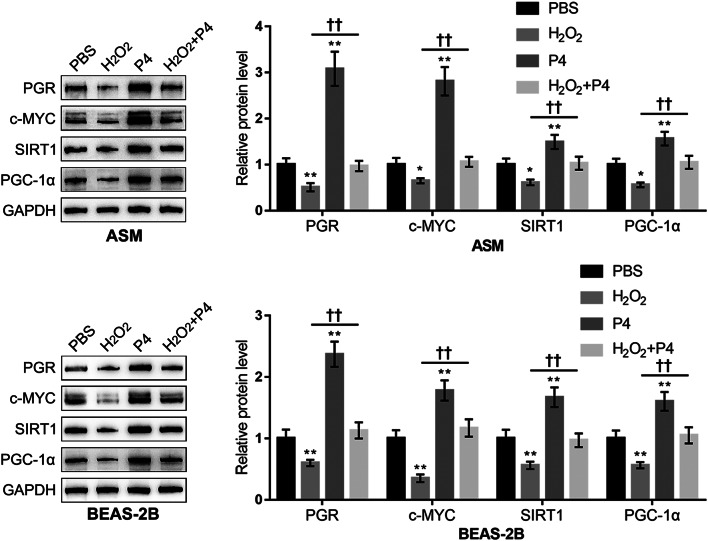
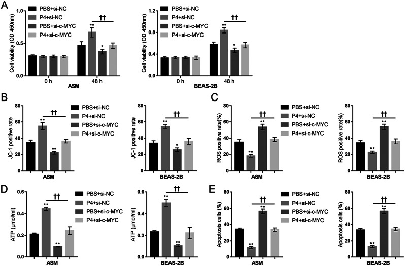
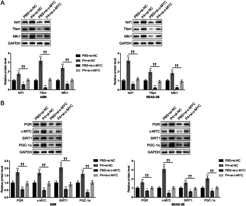
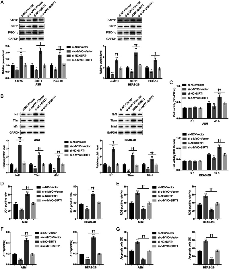
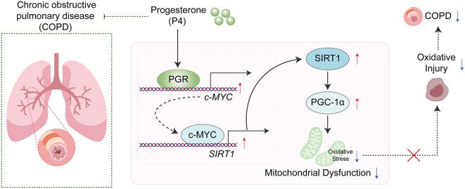
Methods and results: The therapeutic effects of Progesterone (P4) were investigated in vivo and in vitro in this study. In vivo, in a cigarette smoke (CS) exposure-induced COPD mouse model, P4 treatment significantly ameliorated CS exposure-induced physiological and pathological characteristics, including inflammatory cell infiltration and oxidative injury, in a dose-dependent manner. The c-MYC/SIRT1/PGC-1α pathway is involved in the protective function of P4 against CS-induced COPD. In vitro, P4 co-treatment significantly ameliorated H2O2-induced oxidative injury and mitochondrial dysfunctions by promoting cell proliferation, increasing mitochondrial membrane potential, decreasing ROS levels and apoptosis, and increasing ATP content. Moreover, P4 co-treatment partially attenuated H2O2-caused inhibition in Nrf1, Tfam, Mfn1, PGR-B, c-MYC, SIRT1, and PGC-1α levels. In BEAS-2B and ASM cells, the c-MYC/SIRT1 axis regulated P4's protective effects against H2O2-induced oxidative injury and mitochondrial dysfunctions.

Conclusion: P4 activates the c-MYC/SIRT1 axis, ameliorating CS-induced COPD and protecting both airway epithelial cells and smooth muscle cells against H2O2-induced oxidative damage. PGC-1α and downstream mitochondrial signaling pathways might be involved.

Keywords: Airway epithelium; Chronic obstructive pulmonary disease (COPD); Mitochondrial dysfunction; Oxidative injury; Progesterone (P4).


Conflict of interest statement

Not applicable.

Figures

Fig. 1
Fig. 1
Progesterone (P4) protected CS-induced COPD mouse model. (A) The histological changes of lung tissues from control, CS, CS + P4-low, -moderate, and -high (inhalation of (0.03, 0.1, and 0.3 mg/L P4) group mice were observed by HE staining; the blue arrows indicated the infiltration of inflammatory cells. The scale bar is 50 μm for 200x and 20 μm for 500x. (B) The total cell number in BALF was counted. n = 6. (C-E) The level of MDA and the activity of SOD and CAT in mouse lung tissues homogenate were determined. (F-G) The protein levels of PGR-B in mouse lung tissues were determined using Immunoblotting and IHC staining. The scale bar is 50 μm. The relative expression of PGR-B was shown in the right panel of G. (H) and (I) The protein levels of c-MYC, SIRT1, PGC-1α, Nrf1, Tfam, and Mfn1 using Immunoblotting. n = 3. ** p < 0.01 compared to control group, † p < 0.05, †† p < 0.01 compared to CS group
Fig. 2
Fig. 2
P4 protects lung epithelial cells and smooth muscle cells from H2O2 -induced oxidative injury. BEAS-2B and ASM cells were divided into four groups: PBS (control), H2O2, P4, and H2O2 + P4; cells were treated or co-treated as described and examined for cell viability using MTT assay (A); mitochondrial membrane potential by JC-1 staining (B); mitochondrial ROS by MitoSOX Green followed by Flow cytometry (C); ATP content using ATP Detection Kit (D); the apoptosis rate using Annexin-V/PI staining (E). N = 3, * p < 0.05, ** p < 0.01 compared to PBS group, † p < 0.05, †† p < 0.01 compared to H2O2 group
Fig. 3
Fig. 3
P4 modulates mitochondrial function-related proteins expression and mitochondrial length. (A) the protein levels of Nrf1, Tfam, and Mfn1 in ASM and BEAS-2B cells were determined using Immunoblotting. (B) the mitochondrial morphology changes in ASM cells were observed by TEM. N = 3, * p < 0.05, ** p < 0.01 compared to PBS group, †† p < 0.01 compared to H2O2 group
Fig. 4
Fig. 4
Changes in the c-MYC/SIRT1/PGC-1α cascades in P4 protection against oxidative injury. BEAS-2B and ASM cells were divided into the aforementioned four groups and the protein levels of PGR-B, c-MYC, SIRT1, and PGC-1α were determined using Immunoblotting. N = 3, * p < 0.05, ** p < 0.01 compared to PBS group, †† p < 0.01 compared to H2O2 group
Fig. 5
Fig. 5
c-MYC knockdown attenuated P4 protection against oxidative injury. BEAS-2B and ASM cells were transfected with si-c-MYC, co-treated with P4, exposed to H2O2, and examined for cell viability using MTT assay (A); mitochondrial membrane potential by JC-1 staining (B); mitochondrial ROS by MitoSOX Green followed by Flow cytometry (C); ATP content using ATP Detection Kit (D); the apoptosis rate using Annexin-V/PI staining (E). N = 3, * p < 0.05, ** p < 0.01 compared to PBS + si-NC group, †† p < 0.01 compared to PBS + si-c-MYC group
Fig. 6
Fig. 6
c-MYC knockdown attenuated P4 induced activation of c-MYC/SIRT1/PGC-1α pathway under oxidative injury. BEAS-2B and ASM cells were transfected with si-c-MYC, co-treated with P4, and exposed to H2O2. (A) the protein levels of Nrf1, Tfam, and Mfn1 using Immunoblotting; (B) the protein levels of PGR-B, c-MYC, SIRT1, and PGC-1α were determined using Immunoblotting. N = 3, * p < 0.05, ** p < 0.01 compared to PBS + si-NC group, †† p < 0.01 compared to PBS + si-c-MYC group
Fig. 7
Fig. 7
Dynamic effects of the c-MYC knockdown/SIRT1 overexpression axis on PGC-1α and its downstream proteins expression under H2O2 induced oxidative injury. BEAS-2B and ASM cells were co-transfected with si-c-MYC and SIRT1-overexpressing vector (SIRT1), exposed to H2O2, and examined for the protein levels of c-MYC, SIRT1, and PGC-1α were determined using Immunoblotting (A); the protein levels of Nrf1, Tfam, and Mfn1 using Immunoblotting (B); the cell proliferation using MTT assay (C); mitochondrial membrane potential by JC-1 staining (D); mitochondrial ROS by MitoSOX Green followed by Flow cytometry (E); ATP content using ATP Detection Kit (F); the apoptosis rate using Annexin-V/PI staining (G). N = 3, * p < 0.05, ** p < 0.01 compared to si-NC + Vector group, † p < 0.05, †† p < 0.01 compared to PBS + si-c-MYC + Vector group
Fig. 8
Fig. 8
The schematic diagram of the protective role of progesterone in COPD

Similar articles

Cited by

References

    1. Abohalaka R. Bronchial epithelial and airway smooth muscle cell interactions in health and disease. Heliyon. 2023;9(9):e19976. 10.1016/j.heliyon.2023.e19976 - DOI - PMC - PubMed
    1. Afonso V, Champy R, Mitrovic D, Collin P, Lomri A. Reactive oxygen species and superoxide dismutases: role in joint diseases. Joint Bone Spine. 2007;74(4):324–9. 10.1016/j.jbspin.2007.02.002 - DOI - PubMed
    1. Ahuja P, Zhao P, Angelis E, Ruan H, Korge P, Olson A, et al. Myc controls transcriptional regulation of cardiac metabolism and mitochondrial biogenesis in response to pathological stress in mice. J Clin Invest. 2010;120(5):1494–505. 10.1172/JCI38331 - DOI - PMC - PubMed
    1. Ambhore NS, Kalidhindi RSR, Sathish VJLIH, Disease VI. Sex-steroid signaling in lung diseases and inflammation. 2021:243 – 73. - PMC - PubMed
    1. Baillargeon J, Urban RJ, Zhang W, Zaiden MF, Javed Z, Sheffield-Moore M, et al. Testosterone replacement therapy and hospitalization rates in men with COPD. Chronic Resp Dis. 2019;16:1479972318793004. - PMC - PubMed

MeSH terms