. 2024 Aug 13;23(1):165.
doi: 10.1186/s12943-024-02075-y.

Novel humanized monoclonal antibodies against ROR1 for cancer therapy

Affiliations

Novel humanized monoclonal antibodies against ROR1 for cancer therapy

Rong Wei et al. Mol Cancer. .

Abstract

Background: Overexpression of receptor tyrosine kinase-like orphan receptor 1 (ROR1) contributes to cancer cell proliferation, survival and migration, playing crucial roles in tumor development. ROR1 has been proposed as a potential therapeutic target for cancer treatment. This study aimed to develop novel humanized ROR1 monoclonal antibodies and investigate their anti-tumor effects.

Methods: ROR1 expression in tumor tissues and cell lines was analyzed by immunohistochemistry and flow cytometry. Antibodies from mouse hybridomas were humanized by the complementarity-determining region (CDR) grafting technique. Surface plasmon resonance spectroscopy, ELISA assay and flow cytometry were employed to characterize humanized antibodies. In vitro cellular assay and in vivo mouse experiment were conducted to comprehensively evaluate anti-tumor activity of these antibodies.

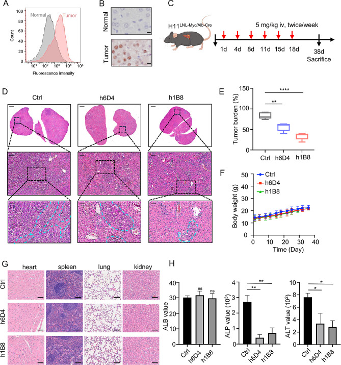
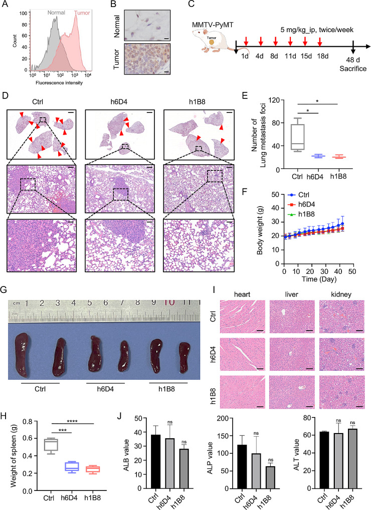
Results: ROR1 exhibited dramatically higher expression in lung adenocarcinoma, liver cancer and breast cancer, and targeting ROR1 by short-hairpin RNAs significantly inhibited proliferation and migration of cancer cells. Two humanized ROR1 monoclonal antibodies were successfully developed, named h1B8 and h6D4, with high specificity and affinity to ROR1 protein. Moreover, these two antibodies effectively suppressed tumor growth in the lung cancer xenograft mouse model, c-Myc/Alb-cre liver cancer transgenic mouse model and MMTV-PyMT breast cancer mouse model.

Conclusions: Two humanized monoclonal antibodies targeting ROR1, h1B8 and h6D4, were successfully developed and exhibited remarkable anti-tumor activity in vivo.

Keywords: Affinity; Humanized antibody; ROR1; Specificity.


Conflict of interest statement

Y.P., R.W., X.L., and J.L. have filed patents related to this study. The other authors declare no competing interests.

Figures

Fig. 1
Fig. 1
Targeting ROR1 effectively inhibits the proliferation and migration of cancer cells. (A) Representative immunohistochemistry images and H-score of ROR1 expression from tissue microarrays of lung, liver, and breast cancer samples. N0: no regional lymph nodes metastasis; N1: regional lymph nodes metastasis. H-score was quantified by inForm software. Scale bar, 50 μm. (B-C) Flow cytometric analysis of ROR1 expression in lung, liver, and breast cancer cells (B), as well as in ROR1-knockdown tumor cells (C). (D-E) Effects of ROR1 knockdown on colony formation (D) and migration (E) of LM3, A549 and MDA-MB-231 cells. Scale bar, 250 μm
Fig. 2
Fig. 2
Development and characterization of humanized anti-ROR1 antibody. (A) Flow cytometry was used to detect the binding ability of murine anti-ROR1 mAbs (purified from hybridoma supernatants) to ROR1 protein on the surface of MDA-MB-231 cells. (B) Coomassie brilliant blue staining (left) and immunoblotting (right) of purified recombinant extracellular domain of ROR1 protein. (C) ELISA assays show the binding of murine anti-ROR1 antibodies to recombinant extracellular domain of ROR1 protein. (D) Tumor growth curves of H1299-ROR1 xenograft in mice treated with control, 1B8, and 6D4 antibodies. (E) Agarose gel analysis of 5’RACE products from 1B8 and 6D4 hybridoma. H, heavy chain; L, light chain. (F) Schematic representation of humanized anti-ROR1 antibodies. CDR sequences of 1B8 and 6D4 were defined and inserted into the FR sequence of selected human IgG1. The light chain of 6D4 contains a revertant mutation (marked by red color). (G) Flow cytometric analyses show the binding of 24 humanized antibody variants to ROR1 proteins on cell surface. (H) SPR analyses of h1B8 and h6D4 antibodies. (I) The purity of h1B8 and h6D4 antibodies were examined by SDS-PAGE under non-reduced (left) and reduced (right) conditions followed by Coomassie brilliant blue staining. Label 1 represents h1B8; label 2 represents h6D4. (J) Size exclusion chromatograms of purified h1B8 and h6D4 antibodies
Fig. 3
Fig. 3
In vitro characterization of humanized h1B8 and h6D4 antibodies. (A) Flow cytometric analyses show the binding ability of h1B8 and h6D4 antibodies to ROR1+ or ROR1 cells. (B) Internalization analyses of h1B8 and h6D4 antibodies into cancer cells. Cells were incubated with antibodies at 4 °C to inhibit internalization or at 37 °C to induce internalization before flow cytometric analysis. (C) Effects of h1B8 and h6D4 antibodies on cell viability. Cells were cultured with different concentrations of anti-ROR1 antibodies for 72 h, and then subjected to MTT assays. (D) Effects of h1B8 and h6D4 antibodies on cell growth. Cancer cells was treated with 100 µg/mL of antibodies for certain times and then subjected to colony formation assays. (E) Representative images of Transwell migration assays in cells with or without incubation of h1B8 and h6D4 antibodies (100 µg/mL). (F) Immunoblotting analyses show the effects of h1B8 and h6D4 antibodies on AKT signaling in different cancer cells upon anti-ROR1 antibody treatment (100 µg/mL)
Fig. 4
Fig. 4
Humanized h1B8 and h6D4 antibodies suppress tumor growth in lung cancer xenograft mice. (A) A549 cells were subcutaneously injected into nude mice to establish lung cancer xenograft model. 14 days later, mice were administrated by intraperitoneal injection of anti-ROR1 antibodies or vehicle control (5 mg/kg, twice a week, totally six times). (B-E) Tumor growth curves (B), tumor pictures (C), tumor weights (D), and body weights (E) from A549 xenograft mice treated with or without anti-ROR1 antibodies. (F) The ratios of tumor weights to body weights of each group. (G) Representative H&E staining images of major mouse organs. Scale bar, 50 μm. (H) Biochemical analyses of alanine transaminase (ALT), albumin (ALB), and alkaline phosphatase (ALP) in the blood from different mice
Fig. 5
Fig. 5
Humanized h1B8 and h6D4 antibodies alleviate HCC progression in transgenic mice. (A-B) Flow cytometric analysis (A) and immunohistochemistry assays (B) were conducted to examine ROR1 protein expression in HCC tumor tissues. Scale bar, 50 μm. (C) Schematic representation of animal experiments. Mice were administrated by anti-ROR1 antibodies (5 mg/kg) or the vehicle control via tail-vein injection (twice a week, totally six times). (D) Representative H&E staining images of livers from each group (tumors are indicated by the blue dashed line). Scale bar: Upper, 1 mm; Middle, 100 μm; Lower, 50 μm. (E-F) Tumor burden (E) and body weights (F) of mice treated with or without anti-ROR1 antibodies. (G) H&E staining images of major mouse organs. Scale bar, 50 μm. (H) Biochemical analyses of ALT, ALB, and ALP in the blood from different mice
Fig. 6
Fig. 6
Humanized h1B8 and h6D4 antibodies inhibit lung metastasis in the MMTV-PyMT breast cancer model. (A-B) ROR1 protein expression in breast tumors of mice was determined by flow cytometry (A) and immunohistochemistry assays (B). Scale bar, 50 μm. (C) Schematic illustration of animal study. MMTV-PyMT mice were treated with anti-ROR1 antibodies (5 mg/kg) or the vehicle control, with injections given (twice a week, totally six times). (D) Representative H&E staining images of lung metastatic nodules from different groups of mice. Scale bar: Upper, 1 mm; Middle, 100 μm; Lower, 50 μm. (E-F) Lung metastatic foci (E) and body weights (F) of mice treated with or without anti-ROR1 antibodies. (G-H) Representative pictures (G) and weights (H) of spleens of mice treated with or without anti-ROR1 antibodies. (I) H&E staining images of major mouse organs. Scale bar, 50 μm. (J) Biochemical analyses of ALT, ALB, and ALP in the blood from different mice

References

    1. Green JL, Kuntz SG, Sternberg PW. Ror receptor tyrosine kinases: orphans no more. Trends Cell Biol. 2008;18:536–44. 10.1016/j.tcb.2008.08.006 - DOI - PMC - PubMed
    1. Matsuda T, Nomi M, Ikeya M, Kani S, Oishi I, Terashima T, et al. Expression of the receptor tyrosine kinase genes, Ror1 and Ror2, during mouse development. Mech Dev. 2001;105:153–6. 10.1016/S0925-4773(01)00383-5 - DOI - PubMed
    1. Balakrishnan A, Goodpaster T, Randolph-Habecker J, Hoffstrom BG, Jalikis FG, Koch LK, et al. Analysis of ROR1 protein expression in human cancer and normal tissues. Clin Cancer Res. 2017;23:3061–71. 10.1158/1078-0432.CCR-16-2083 - DOI - PMC - PubMed
    1. Cui B, Ghia EM, Chen L, Rassenti LZ, DeBoever C, Widhopf GF 2, et al. High-level ROR1 associates with accelerated disease progression in chronic lymphocytic leukemia. Blood. 2016;128:2931–40. 10.1182/blood-2016-04-712562 - DOI - PMC - PubMed
    1. Zheng YZ, Ma R, Zhou JK, Guo CL, Wang YS, Li ZG, et al. ROR1 is a novel prognostic biomarker in patients with lung adenocarcinoma. Sci Rep. 2016;6:36447. 10.1038/srep36447 - DOI - PMC - PubMed

Substances

LinkOut - more resources