. 2024 Aug 13;22(1):758.
doi: 10.1186/s12967-024-05527-7.

miR-876-3p is a tumor suppressor on 9p21 that is inactivated in melanoma and targets ERK

Affiliations

miR-876-3p is a tumor suppressor on 9p21 that is inactivated in melanoma and targets ERK

Vladimir Bezrookove et al. J Transl Med. .

Abstract

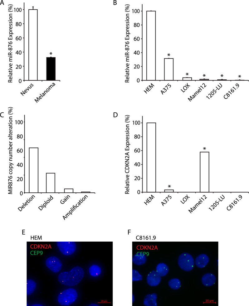
Background: While melanomas commonly harbor losses of 9p21, on which CDKN2A resides, the presence of additional tumor suppressor elements at this locus is incompletely characterized. Here we assess the expression levels and functional role of microRNA-876-3p (miR-876), whose gene also maps to 9p21.

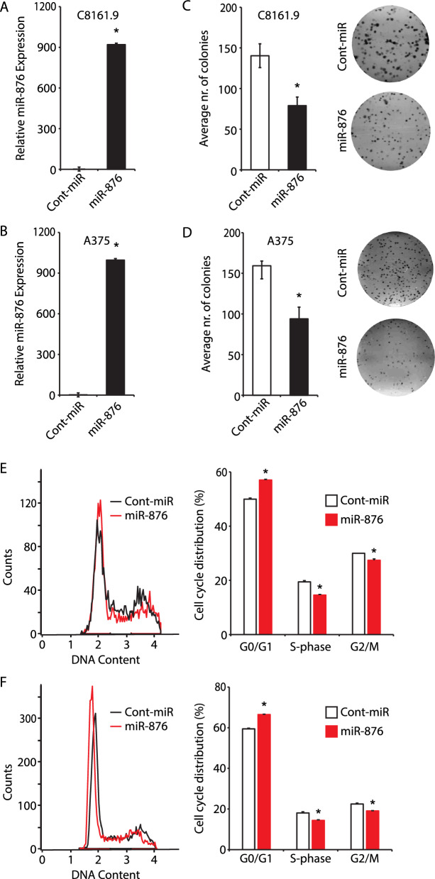
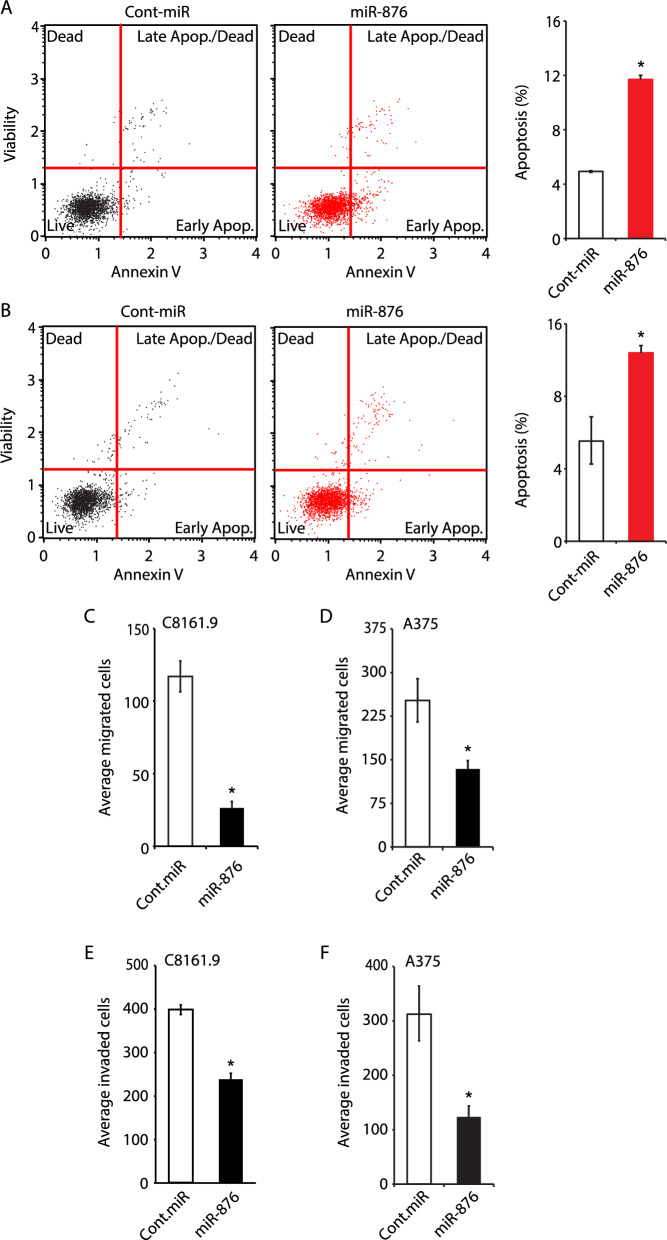
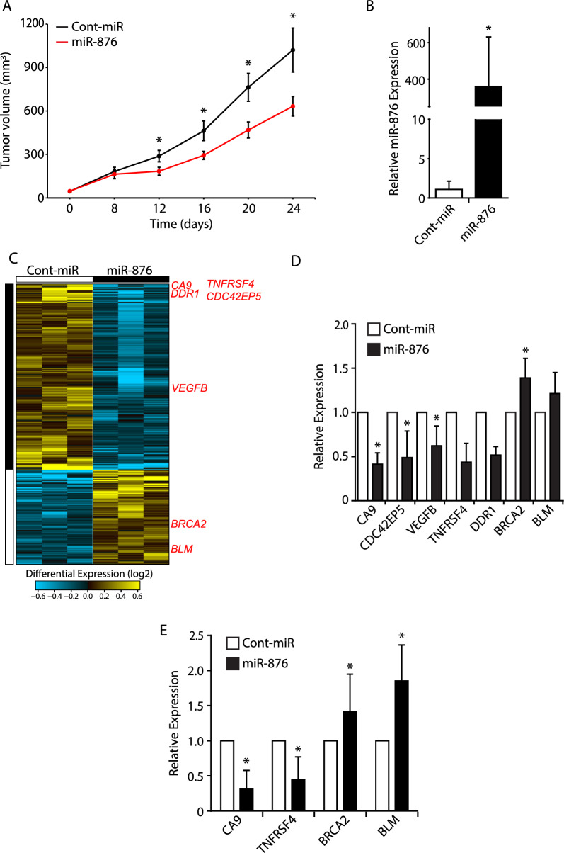
Methods: Expression of miR-876 was assessed in human tissues and cell lines using quantitative miRNA reverse transcriptase polymerase chain reaction (qRT-PCR). MIR876 copy number was determined in The Cancer Genome Atlas (TCGA) melanoma cohort. The consequences of regulation of miR-876 expression were assessed on melanoma cell colony formation, migration, invasion, apoptosis, cell cycle progression, and drug sensitivity in culture, and on in vivo tumor growth in a xenograft model. Genome-wide transcriptomic changes induced by miR-876 overexpression were determined using RNA sequencing (RNA-Seq).

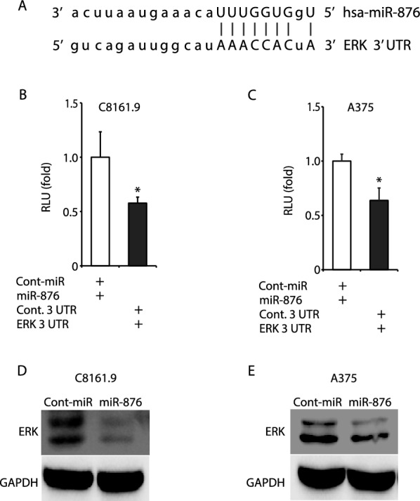
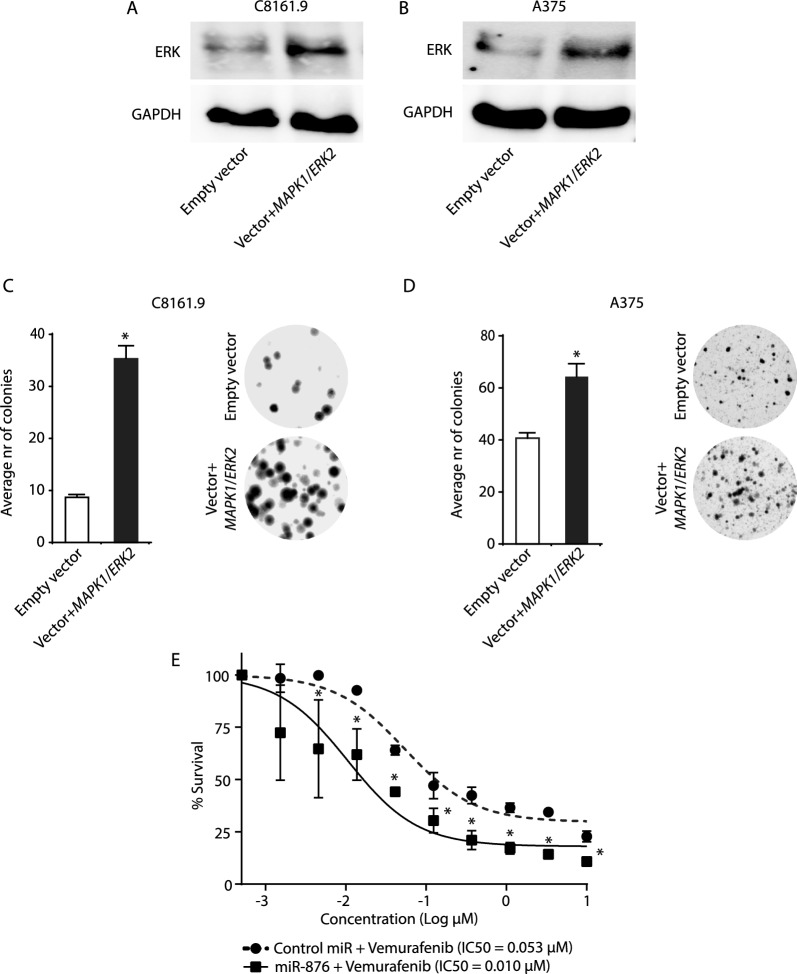
Results: miR-876 expression was significantly decreased in primary melanoma samples when compared with nevi, and in human melanoma cell lines when compared with human melanocytes. Analysis of the TCGA cohort revealed deletions in MIR876 in > 50% of melanomas. miR-876 overexpression resulted in decreased melanoma cell colony formation, migration, and invasion, which was accompanied by cell cycle arrest and increased apoptosis. Intra-tumoral injections of miR-876 significantly suppressed melanoma growth in vivo. RNA-Seq analysis of miR-876-treated tumors revealed downregulation of several growth-promoting genes, along with upregulation of tumor suppressor genes, which was confirmed by qRT-PCR analysis. Computational analyses identified MAPK1 (or ERK2) as a possible target of miR-876 action. Overexpression of miR-876 significantly suppressed luciferase expression driven by the MAPK1/ERK2 3' UTR, and resulted in decreased ERK protein expression in melanoma cells. MAPK1/ERK2 cDNA overexpression rescued the effects of miR-876 on melanoma colony formation. miR-876 overexpression sensitized melanoma cells to treatment with the BRAF inhibitor vemurafenib.

Conclusions: These studies identify miR-876 as a distinct tumor suppressor on 9p21 that is inactivated in melanoma and suggest miR-876 loss as an additional mechanism to activate ERK and the mitogen activated protein kinase (MAPK) pathway in melanoma. In addition, they suggest the therapeutic potential of combining miR-876 overexpression with BRAF inhibition as a rational therapeutic strategy for melanoma.

Keywords: Melanoma; Mitogen activated protein kinase; Tumor suppressor; microRNA-876.


Conflict of interest statement

The authors declare no competing interest.

Figures

Fig. 1
Fig. 1
miR-876 and CDKN2A expression and copy number in human melanoma samples and cell lines. A Relative expression by qRT-PCR of miR-876 in melanoma samples (n = 55) compared to nevi (n = 48). B Relative expression by qRT-PCR of miR-876 in a panel of melanoma cell lines normalized to a human melanocyte (HEM) cell line. C Analysis of miR-876 copy number variation in the TCGA melanoma cohort. D Relative expression by qRT-PCR of CDKN2A in a panel of human melanoma cell lines normalized to a human melanocyte (HEM) cell line. E, F Representative FISH images detecting the CDKN2A locus in red and CEP9 in green in HEM (panel E) and C8161.9 cells (panel F). Data are shown as the mean ± SEM. *P < 0.05 denotes statistically significant differences compared with control. Scale bar, 20 µm
Fig. 2
Fig. 2
Effects of miR-876 overexpression on melanoma colony formation and cell cycle progression. A, B Relative expression by qRT-PCR of miR-876 (panel A) in C8161.9 and A375 (panel B) stable transformants. C, D Bar graphs showing the average number of colonies in C8161.9 (panel C) and A375 (panel D) cells stably expressing miR-876 compared to a control-miR (termed cont. miR) sequence, along with representative bright field images of C8161.9 and A375 colonies in culture stably expressing miR-876 vs control-miR. Data are shown as the mean ± SEM. *P < 0.05 denotes statistically significant differences compared with control. E, F Cell cycle profile with accompanied analysis of distribution of C8161.9 (panel E) and A375 melanoma (panel F) cells stably expressing either miR-876 or control-miR in different phases of the cell cycle
Fig. 3
Fig. 3
Effects of miR-876 overexpression on apoptosis, migration and invasion in melanoma cells. A, B Analysis of apoptotic rates with representative dot plots and bar graphs indicating percentage of total apoptotic C8161.9 (panel A) and A375 cells (panel B) stably expressing miR-876 compared to control-miR. C, D Migratory capacity of C8161.9 (panel C) and A375 cells (panel D) stably expressing miR-876 compared to control-miR. E, F Invasive capacity of C8161.9 (panel E) and A375 cells (panel F) stably expressing miR-876 compared to control-miR. Data are shown as the mean ± SEM. *P < 0.05 denotes statistically significant differences compared with control
Fig. 4
Fig. 4
The effects of miR-876 overexpression on C8161.9 melanoma cells in vivo. A Tumor volume of C8161.9 cells subcutaneously injected in nude mice following intratumoral treatment with miR-876 or control-miR twice weekly for 24 days. B Relative miR-876 expression by qRT-PCR in C8161.9 in vivo tumors treated with miR-876 or control (N = 4 per group). C RNA-Seq followed by supervised hierarchical analysis of C8161.9 tumors treated in vivo with either miR-876 or control-miR (N = 3 per group). D Relative expression by qRT-PCR of differentially expressed genes in C8161.9 tumors (N = 3 per group). E Relative expression by qRT-PCR of differentially expressed genes in C8161.9 cells grown in culture following transfection with miR-876 or control miR. Data are shown as the mean ± SEM. *P < 0.05 denotes statistically significant differences compared with control
Fig. 5
Fig. 5
ERK as a potential target of miR-876. A The putative binding sites of miR-876 in the 3′UTR of MAPK1/ERK2. B, C Luciferase reporter assays verifying the regulation of miR-876 in the 3’UTR of MAPK1/ERK2 in C8161.9 (panel B) and A375 (panel C) cells transiently co-transfected with miR-876 (or control-miR) and a plasmid encoding the luciferase reporter gene and MAPK1/ERK2 3’ UTR. D, E Western blot analysis of ERK expression in C8161.9 (panel D) and A375 (panel E) expressing miR-876 vs control-miR
Fig. 6
Fig. 6
Effects of regulation of ERK by miR-876 on melanoma colony formation and drug sensitivity. A, B Western analysis of ERK expression in C8161.9 (panel A) and A375 (panel B) cells stably expressing miR-876 transfected with a plasmid encoding MAPK1/ERK2 cDNA or empty vector. C, D Bar graphs showing the average number of colonies in C8161.9 (panel C) and A375 (panel D) cells stably expressing miR-876 transfected with a plasmid encoding MAPK1/ERK2 cDNA or empty vector, with accompanying representative bright field images of C8161.9 and A375 colonies. E Viability of A375 cells expressing miR-876 or control miR following treatment with various concentrations of vemurafenib. Data are shown as the mean ± SEM. *P < 0.05 denotes statistically significant differences compared with control

References

    1. Siegel RL, Giaquinto AN, Jemal A. Cancer statistics, 2024. CA Cancer J Clin. 2024;74:12–49. 10.3322/caac.21820 - DOI - PubMed
    1. Cancer Genome Atlas N. Genomic classification of cutaneous melanoma. Cell. 2015;161:1681–96. 10.1016/j.cell.2015.05.044 - DOI - PMC - PubMed
    1. Holland EA, Beaton SC, Edwards BG, Kefford RF, Mann GJ. Loss of heterozygosity and homozygous deletions on 9p21-22 in melanoma. Oncogene. 1994;9:1361–5. - PubMed
    1. Coleman A, Fountain JW, Nobori T, Olopade OI, Robertson G, Housman DE, Lugo TG. Distinct deletions of chromosome 9p associated with melanoma versus glioma, lung cancer, and leukemia. Cancer Res. 1994;54:344–8. - PubMed
    1. Ranade K, Hussussian CJ, Sikorski RS, Varmus HE, Goldstein AM, Tucker MA, Serrano M, Hannon GJ, Beach D, Dracopoli NC. Mutations associated with familial melanoma impair p16INK4 function. Nat Genet. 1995;10:114–6. 10.1038/ng0595-114 - DOI - PubMed

Substances