. 2024 Dec;29(1):2391139.
doi: 10.1080/13510002.2024.2391139. Epub 2024 Aug 13.

Sigma-1 receptor exerts protective effects on ameliorating nephrolithiasis by modulating endoplasmic reticulum-mitochondrion association and inhibiting endoplasmic reticulum stress-induced apoptosis in renal tubular epithelial cells

Affiliations

Sigma-1 receptor exerts protective effects on ameliorating nephrolithiasis by modulating endoplasmic reticulum-mitochondrion association and inhibiting endoplasmic reticulum stress-induced apoptosis in renal tubular epithelial cells

Hu Ke et al. Redox Rep. 2024 Dec.

Abstract

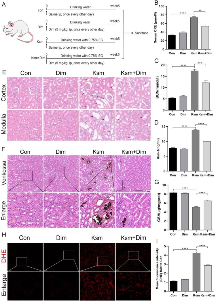
Oxalate-induced damage to renal tubular epithelial cells (RTECs) is an essential factor in the incident kidney stone, but the specific mechanism is unclear. Recent research has pinpointed interacting areas within the endoplasmic reticulum and mitochondria, called mitochondria-associated membranes (MAMs). These studies have linked endoplasmic reticulum stress (ERS) and oxidative imbalance to kidney disease development. The sigma-1 receptor (S1R), a specific protein found in MAMs, is involved in various physiological processes, but its role in oxalate-induced kidney stone formation remains unclear. In this study, we established cellular and rat models of oxalate-induced kidney stone formation to elucidate the S1R's effects against ERS and apoptosis and its mechanism in oxalate-induced RTEC injury. We found that oxalate downregulated S1R expression in RTECs and escalated oxidative stress and ERS, culminating in increased apoptosis. The S1R agonist dimemorfan up-regulated S1R expression and mitigated ERS and oxidative stress, thereby reducing apoptosis. This protective effect was mediated through S1R inhibition of the CHOP pathway. Animal experiments demonstrated that S1R's activation attenuated oxalate-induced kidney injury and alleviated kidney stone formation. This is the first study to establish the connection between S1R and kidney stones, suggesting S1R's protective role in inhibiting ERS-mediated apoptosis to ameliorate kidney stone formation.

Keywords: Sigmar-1 receptor; apoptosis; dimemorfan; endoplasmic reticulum stress; kidney stone; mitochondria-associated endoplasmic reticulum membrane; oxalate; oxidative stress.


Conflict of interest statement

No potential conflict of interest was reported by the author(s).

Figures

Figure 1.
Figure 1.
Activation of S1R attenuated oxidative stress and excessive ER-mitochondrial contact in oxalate-induced HK-2 cells. The concentration of oxalate (Ox) is 2 mM. NC group represented that HK-2 cells were incubated with the complete medium for 24 hours. The Dim group represented that HK-2 cells were incubated with the complete medium containing 10μM Dim for 24 hours. The Ox group represented that HK-2 cells were incubated with 2 mM oxalate for 24 hours. HK-2 cells in the Ox + Dim group were incubated with the complete medium containing 2 mM oxalate and 10μM Dim for 24 h. (A) HK-2 cells were treated with varying concentrations of Dim (0, 5,10, 20, and 40μM) and incubated for 24 h. The cell viability was then detected using CCK-8 assay. (B) HK-2 cells were treated with 2 mM oxalate in the presence of coincubation with different concentrations of Dim (0, 5, 10, 20, and 40μM) for 24 h, and then the viability of cells was determined. (C) The expression of S1R was detected in HK-2 cells treated with different concentrations of Dim. (D, E) 10μM Dim was selected as the optimal concentration to interfere with HK-2 cells. The images show intracellular ROS levels, with brighter green indicating higher cell ROS levels. Images were captured under a dark field (100x magnification), and the bar chart shows the mean fluorescence intensity. (F, G) Immunofluorescence of JC-1 was performed to determine HK-2 cell's mitochondrial membrane potential. Images were captured under a dark field (400x magnification), and the bar graph shows the red-to-green fluorescence ratio. The lower the ratio, the worse the mitochondrial membrane potential. (H) The content of GSH in each group was detected. (I) The connection between the endoplasmic reticulum and mitochondria in HK-2 cells was observed using a transmission electron microscope (Scale bar = 1μm, 500 nm). The red arrow indicates mitochondria-associated endoplasmic reticulum membrane. M: mitochondrion; ER: endoplasmic reticulum. Data are presented as the means ± SEM from three independent experiments. One set of representative images of three independent experiments is shown. *P < 0.05, **P < 0.01, ***P < 0.001, ****P < 0.0001. #P < 0.05, ##P < 0.01, ###P < 0.001, ####P < 0.0001 versus the Ox group; ns represents P>0.05 versus the NC group.
Figure 2.
Figure 2.
Activation of S1R reduced oxalate-induced ERS by inhibiting the PERK-ATF4-CHOP signal pathway in HK-2 cells. (A-F) In each group, western blot and quantitative analyses for ERS-related protein (GRP78, PERK, ATF4, and CHOP) and S1R expression were performed. (G, I) The immunofluorescence analysis illustrated the protein expressions of ATF4 (400x magnification). (H, J) The immunofluorescence analysis illustrated the protein expressions of CHOP (400x magnification). This table presents the means and standard errors from three independent experiments. *P < 0.05, 371, **P < 0.01, ***P < 0.001, ****P < 0.0001. ns represents P>0.05 versus the NC group.
Figure 3.
Figure 3.
Activation of S1R reduced oxalate-induced apoptosis in HK-2 cells. (A, B) TUNEL staining analysis of cell apoptosis in HK-2 cells. Images were captured under a dark field (200x magnification). (C, D) The immunofluorescence analysis illustrated the protein expressions of Bax (400x magnification). (E-G) Representative Western blot and quantitative analysis of apoptosis-related proteins (Bax and cleaved C3) in each group. This table presents the means and standard errors from three independent experiments. *P < 0.05, 371, **P < 0.01, ***P < 0.001, ****P < 0.0001. ns represents P>0.05 versus the NC group.
Figure 4.
Figure 4.
Knockdown of S1R reversed the protective effect of S1R activation against oxalate-induced HK-2 cell injury. (A) The cell model of S1R knockdown was verified. (B) The content of GSH was detected in four groups. (C, E) Images (100x magnification) show the levels of cellular ROS, and the bar graph shows the mean fluorescence intensity in each group. (D, F) Immunofluorescence of JC-1 was performed to determine HK-2 cell's mitochondrial membrane potential in each group. Images were captured under a dark field (400x magnification), and the bar graph shows the red-to-green fluorescence ratio. (G, H) Western blot and quantitative analyses for the ERS-related protein (GRP78, PERK, ATF4, and CHOP) expression in each group. (I, J) The immunofluorescence analysis illustrated the protein expressions of CHOP (400x magnification). This table presents the means and standard errors from three independent experiments. *P < 0.05, 371, **P < 0.01, ***P < 0.001, ****P < 0.0001.
Figure 5.
Figure 5.
S1R knockdown reversed the anti-apoptotic effect of Dim in oxalate-induced HK-2 cell injury. (A-B) Western blot and quantitative analysis of apoptosis-related proteins (Bax and cleaved C3) in each group. (C, D) The immunofluorescence analysis illustrated the protein expressions of Bax (400x magnification). (E, F) TUNEL staining analysis of cell apoptosis in HK-2 cells. Images were captured under a dark field (200x magnification). This table presents the means and standard errors from three independent experiments. *P < 0.05, **P < 0.01, ***P < 0.001, ****P < 0.0001.
Figure 6.
Figure 6.
S1R activation ameliorates renal impairment, the deposition of crystals, and oxidative stress in the rat kidney stone model. (A) This diagram represents the operation of animal experiments. (B-D) The serum CRE and BUN content in blood and Kim-1 in urine were detected in four groups. (E) Renal paraffin tissues were observed by H&E staining, and the black arrows indicate dilated renal tubules (200x magnification). (F) Renal paraffin tissues were observed by Von Kossa staining, and the brown block mass indicates calcium salt deposition (100x magnification). (F, G) The content of GSH in kidney tissues was detected in four groups. (H, I) DHE staining of renal frozen sections in four groups (200x magnification). This table presents the means and standard errors from five independent experiments. *P < 0.05, **P < 0.01, ***P < 0.001, ****P < 0.0001.
Figure 7.
Figure 7.
S1R activation alleviates oxalate-induced ERS in rats. (A-F) Western blot and quantitative analyses for ERS-related protein (GRP78, PERK, ATF4, and CHOP) and S1R expression in four groups. (G-J) Immunohistochemical staining and immunofluorescent staining of ATF4 and CHOP (200x magnification). This table presents the means and standard errors from five independent experiments. *P < 0.05, **P < 0.01, ***P < 0.001, ****P < 0.0001.
Figure 8.
Figure 8.
(A) TUNEL staining was used to observe the renal apoptosis of rats in each group. (B-D) Western blot was used to detect the expression of apoptosis-related proteins (Bax and cleaved C3). (E-H) Immunohistochemical staining and immunofluorescent staining of Bax (200x magnification). This table presents the means and standard errors from five independent experiments. *P < 0.05, **P < 0.01, ***P < 0.001, ****P < 0.0001.

References

    1. Khan SR, et al. . Kidney stones. Nat Rev. Dis Primers. 2016;2:16008. doi:10.1038/nrdp.2016.8 - DOI - PMC - PubMed
    1. Scales CD, Jr., Smith AC, Hanley JM, et al. . Prevalence of kidney stones in the United States. Eur Urol. 2012;62:160–165. doi:10.1016/j.eururo.2012.03.052 - DOI - PMC - PubMed
    1. Zhu C, et al. . Epidemiological trends of urinary tract infections, urolithiasis and benign prostatic hyperplasia in 203 countries and territories from 1990 to 2019. Mil Med Res. 2021;8:64. doi:10.1186/s40779-021-00359-8 - DOI - PMC - PubMed
    1. Wigner P, Bijak M, Saluk-Bijak J.. Probiotics in the prevention of the calcium oxalate urolithiasis. Cells. 2022;11:284; doi:10.3390/cells11020284 - DOI - PMC - PubMed
    1. Witting C, et al. . Pathophysiology and treatment of enteric hyperoxaluria. Clin J Am Soc Nephrol 2021;16:487–495. doi:10.2215/cjn.08000520 - DOI - PMC - PubMed

LinkOut - more resources