Review
. 2024 Aug 21;138(16):991-1007.
doi: 10.1042/CS20240717.

To target cellular senescence in diabetic kidney disease: the known and the unknown

Affiliations
Review

To target cellular senescence in diabetic kidney disease: the known and the unknown

Yuehan Wei et al. Clin Sci (Lond). .

Abstract

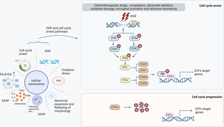
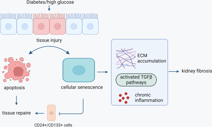
Cellular senescence represents a condition of irreversible cell cycle arrest, characterized by heightened senescence-associated beta-galactosidase (SA-β-Gal) activity, senescence-associated secretory phenotype (SASP), and activation of the DNA damage response (DDR). Diabetic kidney disease (DKD) is a significant contributor to end-stage renal disease (ESRD) globally, with ongoing unmet needs in terms of current treatments. The role of senescence in the pathogenesis of DKD has attracted substantial attention with evidence of premature senescence in this condition. The process of cellular senescence in DKD appears to be associated with mitochondrial redox pathways, autophagy, and endoplasmic reticulum (ER) stress. Increasing accumulation of senescent cells in the diabetic kidney not only leads to an impaired capacity for repair of renal injury, but also the secretion of pro-inflammatory and profibrotic cytokines and growth factors causing inflammation and fibrosis. Current treatments for diabetes exhibit varying degrees of renoprotection, potentially via mitigation of senescence in the diabetic kidney. Targeting senescent cell clearance through pharmaceutical interventions could emerge as a promising strategy for preventing and treating DKD. In this paper, we review the current understanding of senescence in DKD and summarize the possible therapeutic interventions relevant to senescence in this field.

Keywords: Cellular Senescence; diabetic nephropathy; inlfammation; renal fibrosis.


Conflict of interest statement

The authors declare that there are no competing interests associated with the manuscript.

Figures

Figure 1
Figure 1. Characteristics of cellular senescence
Left panel: Cellular senescence is characterized by increased activity of SA-β-gal, SASP, cell cycle arrest, DDR, oxidative stress, abnormal expansion and flattening of cell in morphology as well as SAHF. SASP can be identified by detection of secreted chemokines, ECMs, TGFβ, and inflammatory cytokines such as IL-1α, IL-6, and IL-8. SAHF is presented as DNA foci in the nucleus that are colocalized with enriched H3K9me3, macroH2A, and HP1. Oxidative stress is driven by excessive ROS accumulation. There is an association between DDR and cell cycle arrest. Right and upper panel: Exogenous or endogenous stress causes DNA damage, accompanied by up-regulation of γH2AX. ATR and ATM are DNA damage sensors, that can phosphorylate CHK1 and CHK2, respectively. Activated CHK1/2 phosphorylate p53, resulting in p53 up-regulation, which in turn up-regulates p21, the CDK inhibitor. The inhibition of CDK1/2 by p21 and the inhibition of CDK 4/6 by p16 result in hypo- or mono-phosphorylation of RB, promoting RB-E2F complex formation, leading to inhibition of the proliferation-promoting function of E2F and ultimately cell cycle arrest. Right lower panel: CDKs promote hyper-phosphorylation of RB, supporting RB dissociation from E2F and cell cycle progression.
Figure 2
Figure 2. Model of CDA1’s action in promoting cellular senescence in diabetic kidney via enhancing TGFβ signaling and activating p53/p21 pathways
CDA1 has been shown to synergistically enhance TGFβ signaling in the context of diabetes [130,124] and to induce p53 in DNA damage response [127,128]. Indeed, CDA1 overexpression leads to arrest of cell cycle, as a result of CDA1-induced p21 up-regulation, which can be inhibited by p53 siRNA knockdown, ERK MAPK inhibitors or TGFβ type I receptor inhibitor. Genetic deletion of CDA1 or pharmacological inhibition of CDA1 in diabetic mouse has been shown to attenuate renal inflammation and fibrosis, the key features of DKD [129,131]. These findings support the view that CDA1 synergistically enhances profibrotic TGFβ signaling and induces p53 leading to cellular senescence in diabetic kidney, which further stimulates inflammation and fibrosis via SASP.
Figure 3
Figure 3. Impacts of cellular senescence in DKD
Diabetic or high glucose environment promotes tissue injury, resulting in cellular senescence and cell death such as apoptosis. Renal CD24+/CD133+ cells with self-renewal potential participate in the repair process upon injury. However, in diabetes, increased cellular senescence compromises their proliferation and senescent cells secret proinflammatory and profibrotic molecules in the kidney, leading to maladaptive repair and increasing loss of functional kidney tissue. Furthermore, senescent cells secrete pro-inflammatory cytokines and pro-fibrotic molecules, which further damage kidney tissue accompanied by sustained inflammation and fibrosis, ultimately leading to loss of renal function or kidney failure as a result of increasingly accumulated unrepaired tissue damage.

Similar articles

Cited by

References

    1. Minot C.S. (1891) Senescence and rejuvenation. J. Physiol. 12, 97.113–192.113 10.1113/jphysiol.1891.sp000369 - DOI - PMC - PubMed
    1. Hayflick L. and Moorhead P.S. (1961) The serial cultivation of human diploid cell strains. Exp. Cell. Res. 25, 585–621 10.1016/0014-4827(61)90192-6 - DOI - PubMed
    1. Sharpless N.E. and Sherr C.J. (2015) Forging a signature of in vivo senescence. Nat. Rev. Cancer 15, 397–408 10.1038/nrc3960 - DOI - PubMed
    1. Calcinotto A., Kohli J., Zagato E., Pellegrini L., Demaria M. and Alimonti A. (2019) Cellular senescence: aging, cancer, and injury. Physiol. Rev. 99, 1047–1078 10.1152/physrev.00020.2018 - DOI - PubMed
    1. Hernandez-Segura A., Nehme J. and Demaria M. (2018) Hallmarks of cellular senescence. Trends Cell Biol. 28, 436–453 10.1016/j.tcb.2018.02.001 - DOI - PubMed