

Dermoscopy of skin metastases in advanced cancer-systemic (visceral, hematologic) and cutaneous
- PMID: 39139791
- PMCID: PMC11319161
- DOI: 10.3389/fmed.2024.1445811
Dermoscopy of skin metastases in advanced cancer-systemic (visceral, hematologic) and cutaneous
Abstract
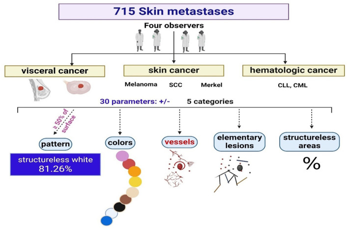
Introduction: Skin metastases arise in 10% of cancer patients, but standardized dermoscopy diagnostic criteria for skin metastases remain poor. This study's objective was to analyze the dermoscopy features of skin metastases from advanced systemic and cutaneous cancers.
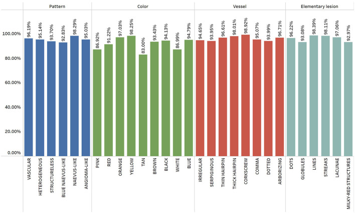
Methods: A retrospective study on 715 dermoscopy images of skin metastases from 33 patients with various primary cancers (breast, ovary, melanoma, non-melanoma skin cancer, and chronic leukemia) attending two academic centers between 2013 and 2023 was performed. Four independent observers blindly analyzed patterns, colors, vessels, and elementary lesions for each metastasis (30 parameters in total).
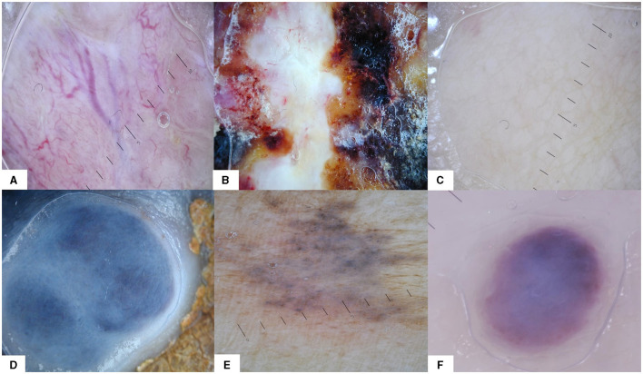
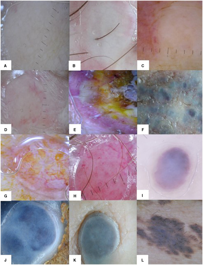
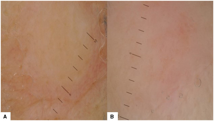
Results: The structureless white pattern was the most prominent indicator of cutaneous metastasis (81.26%, p < 0.001). Regardless of the primary tumor, colors pink, red, white, and tan were identified. Elementary lesions were infrequent, except for melanoma metastases that displayed dots (13.23%) and globules (11.11%). Breast cancer metastases presented: blue (41.48%) and red (34.32%) colors, irregular vessels (13.58%), and a blue-naevus pattern (22.22%). Melanoma metastases displayed: a blue-naevus pattern (61.38%), a blue color (85.71%), and a structureless-blue combination pattern (79.37%). Non-melanoma skin cancer metastases were characterized by vascular (42.11%) and angioma-like (31.58%) patterns, pink (57.89%) and red (57.89%) colors, irregular (57.89%), thin hairpin (47.37%), comma (47, 37%), and thick hairpin (26, 32%) vessels and a red, white and irregular vessels combination pattern (52, 63%). A pink structureless combination pattern was frequent (61.05%) in chronic leukemia metastases. Ovarian cancer metastases displayed a white and tan structureless combination pattern (100%) and frequently had dotted vessels (42.85%).
Conclusion: Papules and nodules with a white structureless pattern suggest skin metastases, regardless of the primary tumor. A blue structureless lesion is indicative of melanoma metastasis and a vascular pattern with irregular vessels indicates a non-melanoma skin cancer metastasis. Dermoscopy stands as a reliable non-invasive diagnostic method for suspected cutaneous metastases in patients with a known cancer history.
Keywords: cutaneous metastases; dermoscopy; diagnosis; metastases; skin cancer; skin metastases.
Copyright © 2024 Simionescu, Petrică, Avram, Costache, Scurtu, Tudorache, Iorga and Grigore.
Conflict of interest statement
The authors declare that the research was conducted in the absence of any commercial or financial relationships that could be construed as a potential conflict of interest.
Figures
Similar articles
-
Clinical and dermoscopic features of cutaneous BAP1-inactivated melanocytic tumors: Results of a multicenter case-control study by the International Dermoscopy Society.J Am Acad Dermatol. 2019 Jun;80(6):1585-1593. doi: 10.1016/j.jaad.2018.09.014. Epub 2018 Sep 20. J Am Acad Dermatol. 2019. PMID: 30244062 Free PMC article.
-
Amelanotic/hypomelanotic melanoma: clinical and dermoscopic features.Br J Dermatol. 2004 Jun;150(6):1117-24. doi: 10.1111/j.1365-2133.2004.05928.x. Br J Dermatol. 2004. PMID: 15214897
-
Dermoscopy of Pilonidal Cyst Disease: A Case-series.Acta Dermatovenerol Croat. 2022 Nov;30(3):194-196. Acta Dermatovenerol Croat. 2022. PMID: 36812282
-
Eccrine porocarcinoma shares dermoscopic characteristics with eccrine poroma: A report of three cases and review of the published work.J Dermatol. 2016 Mar;43(3):332-5. doi: 10.1111/1346-8138.13082. Epub 2015 Sep 1. J Dermatol. 2016. PMID: 26333057 Review.
-
Assessment of Diagnostic Accuracy of Dermoscopic Structures and Patterns Used in Melanoma Detection: A Systematic Review and Meta-analysis.JAMA Dermatol. 2021 Sep 1;157(9):1078-1088. doi: 10.1001/jamadermatol.2021.2845. JAMA Dermatol. 2021. PMID: 34347005 Free PMC article.
References
LinkOut - more resources
Full Text Sources